In vitro
evaluation of osteoblastic cell
adhesion on machined osseointegrated
implants
Abstract: At present the major consideration in planning an implant de-sign is to seek biocompatible surfaces that promote a favorable response from both cells and host tissues. Different treatments of implant surfaces may modulate the adhesion, proliferation and phenotypic expression of osteoblastic cells. For this reason, the aim of the present study was to evaluate the biocompatibility of an implant surface, observing adhesion, cell morphology and proliferation of osteoblast-like cells cultivated on a commercially available titanium dental implant (Titamax Liso,
Neo-dent, Curitiba, PR, Brazil). The implant samples were immersed into an osteoblast-like cell (Osteo-1) suspension for a period of 24, 48 and 72 hours. After seeding the cells, the samples were prepared for analyses through scanning electron microscopy. Based on the surface analysis, the osteoblastic cells adhered to the machined surface after 24 hours in cul-ture. In 48 hours, the cells spread over the implant surface, and after 72 hours a proliferation of cells with large and lat bodies was observed over the machined implant surface. These results demonstrate that the ma-chined titanium surface studied is biocompatible since it allowed adhe-sion and proliferation of the osteoblast-like cells, in addition to preserv-ing cell integrity and the morphologic characteristics of cells durpreserv-ing the studied period.
Descriptors: Titanium; Dental implants; Osteoblasts; Osseointegration; Scanning electron microscopy.
Sandra Fabiano Alves(a) Thomaz Wassall(b)
(a) Coordinator, Master’s Course in Implant
Dentistry; (b)Coordinator, Graduate Course
– São Leopoldo Mandic Dental Research Center, Campinas, SP, Brazil.
Corresponding author:
Thomaz Wassall
Rua José Rocha Junqueira, 13, Swift Campinas - SP - Brazil
CEP: 13045-755
E-mail: [email protected]
Introduction
Studies in the Implant Dentistry area have ex-panded rapidly over the last 40 years. The recog-nition that oral implants could attain high success rates provided the basis for this large number of studies.1-5
The inding that titanium is a biocompatible ma-terial led some authors6,7 to study its surface
proper-ties, such as chemical composition, micro- and mac-rostructure, contamination, cleanliness and surface properties of interaction with biomolecules.
As a biocompatible material, the applicability of titanium was conirmed mainly because of two fac-tors: (1) its excellent resistance to corrosion, limiting the quantity of titanium ions released into the tis-sues; (2) its biological inactivity, in which signs of the presence of the metal appear not to inluence the tissues.8,9
With regard to titanium biocompatibility, the surface of the implant must be chemically and me-chanically cleaned to remove any strange particle or contaminative material to preserve the integrity of the receptor bed cells.10 Irrespective of the material
chosen for making the implants, it is the material’s surface that comes into contact with the bone, and, in the case of titanium, the surface is covered with titanium oxide, which is formed as soon as the im-plant comes into contact with the oxygen molecules in the environment. Titanium can form numerous oxides, titanium dioxide being of major relevance because of its high dielectric constant.11
Titanium biocompatibility, as regards cellular ad-hesion and proliferation, has been assessed in
vari-ous in vitro studies that have shown that osteoblasts
appear to adhere more rapidly to titanium surfaces with a rougher microtopography.12-17
Some studies have also demonstrated the capac-ity of machined titanium surfaces to promote cel-lular adhesion and proliferation.18-20 In addition,
rough surfaces enhance osteoblastic phenotype dif-ferentiation and the capacity of osteoblasts to syn-thesize bone matrix.21-23
With regard to cellular morphology related to surfaces with different textures, the related litera-ture has shown that cells spread themselves over a larger area on machined surfaces than they do on
rough surfaces. On machined surfaces, cells pres-ent a compact and lattened morphology, and must make a big lateral effort to spread. According to an author,24 this could be because they do not ind a
three dimensional structure as they do on rough substrates, which allows cellular accommodation and a larger contact surface to accommodate them-selves. The cells on rough substrates present a more polygonal shape.25,26
Some studies have shown that, in implants with machined surfaces, cells have a tendency to follow an orientation parallel to the scratches arising from the machining of the titanium surface (anisotropic characteristic) as opposed to rough surfaces, where the cells spread throughout the entire extent of the surface (isotropic characteristic).27-29
In view of the foregoing explanations, the aim of the present in vitro study was to analyze the bio-compatibility of an implant surface, observing the adhesion, cell morphology and proliferation of an osteoblastic cell lineage30 cultivated on a
commer-cially available dental implant (Titamax Liso,
Neo-dent, Curitiba, PR, Brazil) through scanning elec-tron microscopy (SEM).
Material and Methods
For this experiment Titamax Liso implants
(Neodent, Curitiba, PR, Brazil) were used, mea-suring 3.75 mm in diameter and 13 mm in length. These implants have a cylindrical design with pyra-midal threads and pitch varying with implant diam-eter (macro surface). They have a smooth surface, resulting from machining in an automatic CNC lathe (TNL 12, Index Traub, Esslingen, Baden-Württemberg, Germany). The raw material used for manufacturing the implants was Grade II Titanium (ASTMF67). The cellular lineage used was origi-nated from the parietal bone tissue of newborn rats (Osteo-1).15 The cells were stored in liquid nitrogen,
protected by dimethyl-sulphoxide, and defrosted in
a bain-marie at 37°C for 2 minutes. The cells in
bovine fetal serum (Cultilab Ltda., Campinas, SP, Brazil). The cells were maintained in an incubator at 37°C, in a humidiied 5% CO2 atmosphere. Cell culture development was assessed under an inverted phase microscope. After the surface was colonized, the culture medium was removed, the plates were washed in PBS and the cells were enzymatically re-leased. The enzyme was inactivated with culture medium and the cells in suspension were centrifuged at 3,000 rpm for ive minutes. After the supernatant had been aspired, aliquots were distributed onto new plates. After cultivation, a cell suspension of 109 cells per mm2 was prepared, and this suspension
was plated onto three samples of the implant. The receptacles with the samples were incubated at 37°C in a humidiied atmosphere with 5% of CO2. After 24, 48 and 72 hours of plating, the samples were ixed in 2% glutaraldehyde in 0.1 M phosphate buf-fer and post ixed in 1% osmium tetroxide in the same buffer. The samples were then dehydrated in 100% ethanol and submitted to chemical drying in hexamethyldisilane (HMDS – Electron Microscopy Sciences, Fort Washington, PA, USA). The samples were then sputter-coated with gold (Sputtering SCD 020, Bal-Tec, Balzers, Liechtenstein) and studied by means of scanning electron microscopy (Leo 430 SEM, Leo Ldta., Cambridge, UK).
Results
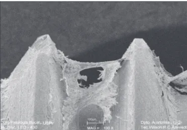
A surface topography of clean titanium was ob-served, without the presence of foreign particles and
with scratches arising from the machining process (Figure 1).
After 24 hours of plating (Figure 2), cells ad-hered on part of the machine surface of the implant were observed. The cells had an anisotropic orienta-tion, with afinity for the scratches of the implant machining process.
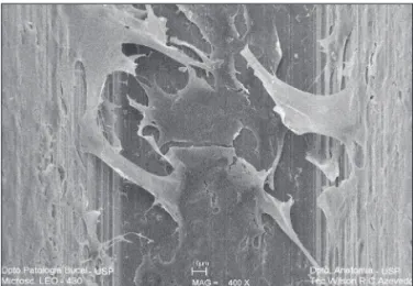
After 48 hours, when analyzing a photomicro-graph at 400 X magniication (Figure 3), it could be observed that the cells presented a more deined morphologic characteristic, with a large and lat body (lattened) and with short cytoplasmatic pro-longations.
Analysis after 72 hours (Figures 4 and 5) also re-vealed a favorable cell behavior response by spread-ing and proliferation of cells over the machined ti-tanium surface, with large and lat cells with short cytoplasmatic prolongations accompanying the im-plant machining scratches.
Discussion
In the related literature, many publications re-porting a superiority of rough surfaces submitted to different treatment procedures in comparison with machined surfaces as regards osseointegration in terms of cellular adhesion and proliferation can be found.12,14,15,17,19 However, the present in vitro
ex-periment demonstrated that the machined surfaces promoted a favorable cell behavior response, as is also shown in the related literature.12,14,18,20
Irrespective of the type of surface treatment to
which an implant is submitted, in the case of tita-nium implants, it is their supericial layer of oxide which comes into contact with bone and provides the basis for their exceptional biocompatibility.11
Moreover, in order for a material to be biocompati-ble, it must not be cytotoxic.9,10 In the present study,
the samples used and analyzed by SEM were shown to be biocompatible for their capacity of preserving the integrity of the Osteo-1 cells for a period of 24 to 72 hours.
Our observations allowed us to conclude that the machined surfaces analyzed promoted cell adhesion observed by SEM at 24 hours, and that, at 48 hours, the cells were adhered to the implant surface, with morphologic characteristics already deined. After
72 hours, our SEM observation revealed spreading and proliferation of the Osteo-1 cells over the ana-lyzed substrate.
As regards the morphologic characteristics of the cells submitted to culture, the related literature has shown some differences with regard to cellular shape among surfaces with different textures. This could be due to the three dimensional structure of rough substrates, where cells conforming to the sur-face roughness attained a contact sursur-face without the need for a greater lateral effort to spread out. In the present experiment, when analyzed by SEM, the cells were shown to have a large lattened body with short cellular prolongations, in agreement with what has been observed in the related literature with respect to cellular interaction and smooth surfaces21
In contrast, cells on rough surfaces present a more elongated or polygonal shape, and longer cellular extensions.25,26
We also observed that the cells analyzed followed a direction parallel to the machining scratches. The literature relates this tendency in machined implants as opposed to rough surfaces, where the cells prolif-erate over the entire surface without any predomi-nant direction.27-29
In the present experiment, and in agreement with the related literature, it was possible to observe that adhesion and an initial interaction between cell and substrate occurred irrespective of the treatment used on the titanium surface.
Figure 5 - SEM - 72 hours - Cellular proliferation over the machined titanium surface (100 X).
Figure 4 - SEM - 72 hours - Cells with large flat bodies, spread over the machined titanium surface (400 X).
Conclusion
Based on the indings of the present study, we concluded that the studied machined implant sur-face is biocompatible since it preserved the integrity
of the cultivated osteoblast-like cells (Osteo-1) for a period of 24 to 72 hours, allowing their adhesion and proliferation, and maintaining their morpho-logic characteristics.
References
1. Adell R, Lekholm U, Rockler B, Brånemark PI. A 15-year study of osseointegrated implants in the treatment of the eden-tulous jaw. Int J Oral Surg. 1981;10(6):387-416.
2. Albrektsson T, Brånemark PI, Hansson HA, Lindström J. Os-seointegrated titanium implants. Requirements for ensuring a long-lasting, direct bone-to-implant anchorage in man. Acta Orthop Scand. 1981;52(2):155-70.
3. Albrektsson T, Zarb G, Worthington P, Eriksson AR. The long-term efficacy of currently used dental implants: a re-view and proposed criteria of success. Int J Oral Maxillofac Implants. 1986;1(1):11-25.
4. Brånemark PI. Vital microscopy of bone marrow in rabbit. Scand J Clin Lab Invest. 1959;11(Supp 38):1-82.
5. Branemark PI, Hansson BO, Adell R, Breine U, Lindstrom J, Hallen O et al. Osseointegrated implants in the treatment of the edentulous jaw. Experience from a 10-year period. Scand J Plast Reconstr Surg Suppl. 1977;16:1-132.
6. Albrektsson T. The response of bone to titanium implants. Crit Rev Biocompat. 1985;1:53-84.
7. Wataha JC. Biocompatibility of dental casting alloys: a review. J Prosthet Dent. 2000 Feb;83(2):223-34.
8. Williams DF. Implants in dental and maxillofacial surgery. Biomaterials. 1981 Jul;2(3):133-46.
9. Williams DF. Titanium: epitome of biocompatibility or cause for concern. J Bone Joint Surg Br. 1994 May;76(3):348-9. 10. Kasemo B, Lausmaa J. Biomaterial and implant surfaces: on
the role of cleanliness, contamination, and preparation proce-dures. J Biomed Mater Res. 1988 Aug;22(A2 Suppl):145-58. 11. Kasemo B, Lausmaa J. Metal selection and surface character-istics. In: Brånemark PI, Zarb GA, Albrektsson T. Osseoin-tegration in clinical dentistry. Chicago: Quintessence; 1995. p. 99-116.
12. Anselme K. Osteoblast adhesion on biomaterials. Biomateri-als. 2000;21(7):667-81.
13. Att W, Tsukimura N, Suzuki T, Ogawa T. Effect of suprami-cron roughness characteristics produced by 1- and 2-step acid etching on the osseointegration capability of titanium. Int J Oral Maxillofac Implants. 2007 Sep-Oct;22(5):719-28. 14. Deligianni DD, Katsala N, Ladas S, Sotiropoulou D, Amedee
J, Missirlis YF. Effect of surface roughness of the titanium alloy Ti-6Al-4V on human bone marrow cell response and on protein adsorption. Biomaterials. 2001 Jun;22(11):1241-51. 15. Guizzardi S, Galli C, Martini D, Belletti S, Tinti A, Raspanti
M et al. Different titanium surface treatment influences
hu-man osteoblast response. J Periodontol. 2004 Feb;75(2):273-82.
16. Nishimoto SK, Nishimoto M, Park SW, Lee KM, Kim HS, Koh JT et al. The effect of titanium surface roughening on protein absorption, cell attachment, and cell spreading. Int J Oral Maxillofac Implants. 2008 Jul;23(4):675-80.
17. Rouahi M, Champion E, Hardouin P, Anselme K. Quantita-tive kinetic analysis of gene expression during human osteo-blastic adhesion on orthopaedic materials. Biomaterials. 2006 May;27(14):2829-44.
18. Martin JY, Schwartz Z, Hummert TW, Schraub DM, Simpson J, Lankford J Jr et al. Effect of titanium surface roughness on proliferation, differentiation, and protein synthesis of hu-man osteoblast-like cells (MG63). J Biomed Mater Res. 1995 Mar;29(3):389-401.
19. Pebé P, Barbot R, Trinidad J, Pesquera A, Lucente J, Nishimura R et al. Countertorque testing and histomorphometric analysis of various implant surfaces in canines: a pilot study. Implant Dent. 1997;6(4):259-65.
20. Postiglione L, Di Domenico G, Ramaglia L, Montagnani S, Salzano S, Di Meglio F et al. Behavior of SaOS-2 cells cultured on different titanium surfaces. J Dent Res. 2003 Sep;82(9):692-6.
21. Protivinsky J, Appleford M, Strnad J, Helebrant A, Ong JL. Effect of chemically modified titanium surfaces on protein adsorption and osteoblast precursor cell behavior. Int J Oral Maxillofac Implants. 2007 Jul-Aug;22(4):542-50.
22. Scheideler L, Geis-Gerstorfer J, Kern D, Pfeiffer F, Rupp F, Weber H et al. Investigation of cell reactions to microstruc-tured implant surfaces. Mater Sci Eng. 2003;23:455-9. 23. Zhu X, Chen J, Scheideler L, Altebaeumer T, Geis-Gerstorfer
J, Kern D. Cellular reactions of osteoblasts to micron- and submicrom-scale porous structures of titanium surfaces. Cells Tissues Organs. 2004;178(1):13-22.
24. ter Brugge PJ, Jansen JA. Initial interaction of rat bone marrow cells with non-coated and calcium phosphate coated titanium substrates. Biomaterials. 2002 Aug;23(15):3269-77. 25. Araújo NS, Jaeger RG, Todescan FF, Jaeger MMM, Groll W.
Teste de cultura celular para avaliar a adesão e proliferação sobre implantes de titânio com superfícies modificadas. RPG Rev Pós Grad. 2001 abr-jun;8(2):103-9.
27. Anselme K, Bigerelle M, Noel B, Dufresne E, Judas D, Iost A et al. Qualitative and quantitative study of human osteoblast adhesion on materials with various surface roughnesses. J Biomed Mater Res. 2000 Feb;49(2):155-66.
28. Elias CN, Lima JHC, Figueira DC. Implantes dentários com superfícies anisotrópicas e isotrópicas. Rev Bras Implant. 2005;11(1):9-12.
29. Lindhe J. Topography and titanium surfaces. In: Lindhe J, Karring T, Lang NP. Clinical periodontology and implant dentistry. Copenhagen: Blackwell Munksgaard; 2003. p. 799-806.