Effect of perillyl alcohol on gene expression of human
pulmonary adenocarcinoma cells*
JULIANA DE SALDANHA DA GAMA FISCHER1, MARCELO SOARES DA MOTA E SILVA2, MARCOS EDUARDO PASCHOAL3, CERLI ROCHA GATTASS4, PAULO COSTA CARVALHO5,
MARIA DA GLORIA DA COSTA CARVALHO4
Keywords: Adenocarcinoma; Lung neoplasms; Monoterpenes; Cell culture; Lung/cytology
*Study carried out in the Laboratory of Molecular Oncology at the Institute for Thoracic Diseases of the Clementino Fraga University Hospital - Universidade Federal do Rio de Janeiro (UFRJ, Federal University of Rio de Janeiro), Rio de Janeiro, Rio de Janeiro, Brazil
Source of funding: Capes and CNPq - Grant number: 5506532002-3
1. Masters in Medicine from the Universidade Federal do Rio de Janeiro (UFRJ, Federal University of Rio de Janeiro) School of Medicine, Rio de Janeiro, Rio de Janeiro, Brazil
2. Basic research student at the Clementino Fraga University Hospital - Universidade Federal do Rio de Janeiro (UFRJ, Federal University of Rio de Janeiro), Rio de Janeiro, Rio de Janeiro, Brazil
3. Physician at the Tuberculosis Clinic of the Clementino Fraga Filho University Hospital - Universidade Federal do Rio de Janeiro (UFRJ, Federal University of Rio de Janeiro), Rio de Janeiro, Rio de Janeiro, Brazil
4. Adjutant Professor and Chief of the Gene Expression Laboratory at the Carlos Chagas Filho Institute of Biophysics -Universidade Federal do Rio de Janeiro (UFRJ, Federal University of Rio de Janeiro), Rio de Janeiro, Rio de Janeiro, Brazil 5. Masters student at the Instituto Oswaldo Cruz (IOC, Oswaldo Cruz Institute), Rio de Janeiro, Rio de Janeiro, Brazil Correspondence to: Maria da Gloria da Costa Carvalho. Rua Nascimento Silva, 241/501, Ipanema, Rio de Janerio - RJ. CEP:22421-020. Phone: (21) 22759789. E-mail: [email protected]
Submitted: 9 March 2005. Accepted, after review: 20 May 2005.
ABSTRACT
INTRODUCTION
Patients with lung cancer have an extremely low survival rate, which reflects their high mortality rate. The main reasons for this are late diagnosis of the disease and the inefficacy of the adopted treatments. For these reasons, it is necessary to seek new chemotherapy treatments.
Perillyl alcohol (PA), which is a monoterpene isolated from the essential oils in mint, cherries and celery seeds, as well as from those found in other plants,(1) has been studied as an alternative treatment
for solid tumors. Studies in animals have shown that PA is an efficacious chemotherapeutic agent for the regression of breast,(2) pancreas,(3) liver(4) and prostate
tumors,(5) and that it is a chemopreventive agent for
colon tumors,(6) melanomas(7) and neuroblastomas.(8)
Active in the induction of apoptosis in tumor cells, PA does not affect normal cells and might even revert tumor cells to a differentiated stage.(1) The
PA-induced inhibition of signal transduction through the membrane prevents the anchorage of Ras proteins through the inhibition of Ras isoprenylation. In lung adenocarcinoma, Ras protein mutates in 19% of cases; hence it is an important therapeutic target.(9)
Based on these problems, the objective of the present study was to evaluate the effect of PA on the gene expression of human pulmonary adenocarcinoma cells in order to better understand its mechanism of action as a chemotherapeutic agent.
METHODS
Monolayers of pulmonary adenocarcinoma cells (A549; American Type Culture Collection, Manassas, VA, USA) were cultivated in RPMI 1640 culture medium (Gibco BRL, Gaithersburg, MD, U S A ) . F o r c e l l s u b c u l t u r e , t h e c o n f l u e n t monolayers (1 x 10⁷ cells/bottle) were washed with phosphate-buffered saline (PBS), pH 7.2, with
0.8% NaCl, 0.02% KCl, 0.15% Na2HPO4 and 0.02%
KH2PO4.H2O, followed by rapid treatment with
trypsin solution (0.25% in PBS), and resuspended, with mild agitation, in fresh culture medium. The PA (96%; Sigma-Aldrich, St. Louis, MO, USA) was added to the semiconfluent cell monolayers in various dilutions (0.03%, 0.003 % and 0.0003%), and the cells were incubated in a carbon dioxide incubator at 37ºC for 48 hours.
Cell viability was quantified using [3-(4,5-dimethylthiazol-2-yl)-2,5 diphenyltetrazolium bromide] assays (MTT method) as described by
Mosmann.(10) In summary, 104 cells/well were
grown in 96-well plates, which, 24 hours later, were incubated with perillyl alcohol at the various dilutions previously cited. After 24 hours of incubation, 20 µL of MTT (5µg/mL) were added to each well, and cells were incubated in total darkness at 37°C for 4 hours. The supernatant was then discarded and the precipitate was suspended in 150 µL of dimethyl sulfoxide. The microplates were read with a BenchMark ELISA reader (BioRad, Hercules, CA, USA) at 570-nm using a 630-nm reference filter.
Semiconfluent cell monolayers were incubated in PA dilutions of 0.03%, 0.003% and 0.0003%. After 48 hours, the monolayers were removed from the culture medium and immersed in serum-f r e e E a g l e ' s M i n i m u m E s s e n t i a l M e d i u m containing 40 µCi/mL of methionine for 120 min. After incorporation, cell monolayers were treated with a buffer solution: 62.5 mM of Tris-HCl, pH 6.8; 2% sodium dodecyl sulfate (SDS); 10% glycerol; 5% 2-mercaptoethanol; and 0.001% bromophenol blue. Radiolabeled proteins were then analyzed using autoradiography.
The fractionated proteins were analyzed using electrophoresis on a 12% polyacrylamide gel in accordance with the technique described by
Laemmli in 1970.(11) The molecular weight of the
proteins was determined using co-electrophoresis of proteins whose molecular weight was known (Life Technology, Inc., Gaithersburg, MD, USA): myosin (200 kDa); phosphorylase B (97.4 kDa); bovine serum albumin (68 kDa); beta- lactoglobulin (18.4 kDa); and lysozyme (14.3 kDa). Gel samples were stained with Coomassie blue, dried and exposed to Kodak X-Omat YAR-S radiographic film (Eastman Kodak, Rochester, NY, USA).
The exposed film was scanned using an LKB 2202 Ultroscan laser densitometer (Pharmacia LKB, Uppsala Sweden). The densitometric tracing presented the protein profile of the control cells as well as that of those incubated with perillyl alcohol for 48 hours.
Western blots were performed for the following proteins: p44/42 kinases (ERK1 and ERK2), -tubulin and p53. The proteins were fractionated
nitrocellulose membrane and submitted to separate immunodetection reactions for each antibody. The nitrocellulose membrane was blocked using a 5% w/v skimmed milk powder in TBST (10 mM Tris, pH 8.3; 150 mM NaCl; 0.15% Tween 20).
After three 15-min rinses in TBST, membranes were separately incubated for 24 hours with the following monoclonal antibodies: anti-p53 (Mab 1801, Gibco); anti tubulin (monoclonal anti -tubulin clone B-5-1-2; Sigma, St. Louis, MO, USA); and anti-ERK1/2 [Phospho-p44/42 MAPK (Thr 202/ Try 204) E10 monoclonal antibody; Cell Signaling Technology, Beverly, MA, USA], all at the dilutions recommended by the manufacturers. Subsequently, membranes were reincubated with peroxidase-conjugated mouse secondary anti-IgG antibody (Amersham Biosciences, Piscataway, NJ, USA) at a dilution of 1:1.000. The complexes were visualized using the Amersham Enhanced Chemiluminescence System development kit (Amersham) with exposure to X-Omat YAR-S radiographic film (Kodak).
Three independent experiments were carried out in triplicate. Statistical significance was analyzed using the unpaired Student's t-test. Values of p < 0.05 were considered statistically significant.(12-13)
RESULTS
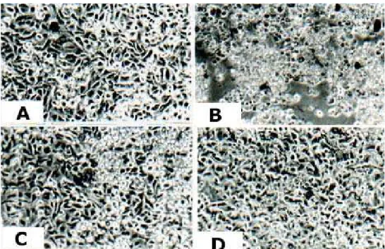
We studied the alterations in cell morphology at PA dilutions ranging from 0.03% to 0.0003%. Figure 1A shows the morphology of cells not incubated with PA. Figure 1B shows that, in comparison with control cells (Figure 1A), the 0.03% dilution provoked intense cell lysis, which was slight at the 0.003% and 0.0003% dilutions (Figures 1C and 1D). The inhibition of cellular viability, quantified using the MTT method, was 60.17% (p < 0.001), 15.62% (p < 0.001) and 11.53% (p < 0.05), respectively, at PA dilutions of 0.03%, 0.003% and 0.0003% (Figure 2). These results are in accordance with the alterations in cell morphology shown in Figure 1.
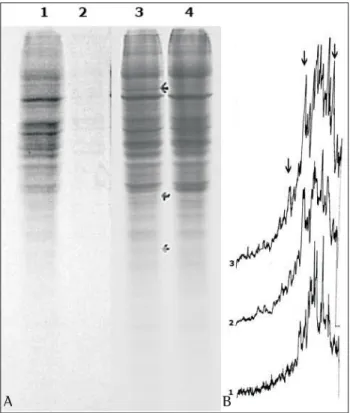
With the objective of determining whether the altered control of gene expression at the protein synthesis level was associated with the results shown in Figures 1 and 2, the protein pattern was analyzed through the incorporation of a radioactive precursor (Figure 3). The results showed that treatment with 0.003% PA led to the induction of 110-kDa, 42-kDa and 28-kDa proteins. However, this was not observed at the 0.0003% dilution, as indicated by the arrows in the autoradiogram (Figure 3A) and in the densitometric tracing (Figure
3B). Since PA led to a decrease in cell viability (Figure 2), we also analyzed the possible involvement of proteins present in the proliferation and apoptosis pathways (Figure 4). The expression of p53 and p44/42 proteins was determined using the Western blot method. As an internal control,
-tubulin was used. The same quantity of --tubulin was detected for all dilutions (Figure 4A), except for the 0.03% PA dilution, in which cell viability was found to be reduced (Figure 2). Figure 4B shows that there was no statistically significant difference for p53. In comparison with -tubulin expression, the 0.003% PA dilution provoked a marked decrease in p44 phosphorylation and an increase in p42 phosphorylation (Figure 4C).
DISCUSSION
The understanding of the molecular profile of human tumors is essential for the effective use of chemotherapeutic agents. The cellular alterations demonstrated in this study suggest that PA is a potential chemotherapeutic agent for lung cancer. These results show a new mechanism of action of this agent, through alteration of the pathways that regulate cell growth and differentiation.
The p42 and p44 kinase proteins are present in the cascade of kinases that regulate cell growth and differentiation. The mitogen-activated protein kinases are activated by a variety of extracellular factors, including growth factors, hormones and
neurotransmitters.(14-16) The activation of the
mitogen-activated protein kinases occurs through the phosphorylation of tyrosine and threonine (202 and 204 of human ERK1, or 183 and 185 of mouse
Figure 2 - Cell viability after 48 hours of treatment with perillyl alcohol
Figure 3 - A - Autoradiogram on a polyacrylamide gel: channel 1 - control A549 cells; channels 2 to 4 - A549 cells incubated with 0.03%, 0.003% and 0.0003% perillyl alcohol, respectively, for 48 h. B - Densitometry of the autoradiogram: 1 - control A549 cells; 2 - A549 cells with 0.0003% perillyl alcohol; 3 - cells with 0.003% perillyl alcohol. Arrows show the alterations due to the effect of perillyl alcohol
A B
Figure 4 - Western Blot: A - α-tubulin, B - p53 and C - p44/ 42. Channel 1: control A549 cells; Channels 2 to 4: A549 cells incubated with perillyl alcohol at dilutions of 0.03%, 0.003% and 0.0003%, respectively, for 48 hours
ERK2) in the threonine and tyrosine amino acid sequence by one single kinase.(17-18) The results
indicated that PA was able to induce p42 protein phosphorylation at the 0.003% and 0.0003% dilutions within 48 hours (Figure 4C) and to inhibit p44 phosphorylation. The physiologic meaning of this in vitro activation/inhibition has not been well established.
The results show, for the first time, that ERK1/2 was one of the molecular targets of the modulation of the response to PA in human pulmonary adenocarcinoma cells. The effect of PA is not limited to inhibition of Ras farnesylation but also directly affects the kinase phosphorylation state regulated by extracellular signals (ERK).
ACKNOWLEGMENTS
We would like to thank Professor Maria Christina Soares Rebello for her collaboration in writing the Discussion.
REFERENCES
1. Belanger JT. Perillyl alcohol: applications in oncology. Altern Med Rev. 1998;3(6):448-57.
2. Haag JD, Lindstrom MJ, Gould MN. Limonene-induced regression of mammary carcinomas. Cancer Res. 1992;52(14):4021-6.
3. Stark MJ, Burke YD, McKinzie JH, Ayuobi AS, Crowell PL. Chemotherapy of pancreatic cancer with the monoterpene perillyl alcohol. Cancer Lett. 1995;96(1):15-21. 4. Mills JJ, Chari RS, Boyer IJ, Gould MN, Jirtle RL. Induction
of apoptosis in liver tumors by the monoterpene perillyl
alcohol. Cancer Res.1995;55(5):979-83.
5. Jeffers L, Church D, Gould M, Wilding G. The effect of perillyl alcohol on the proliferation of human prostatic cell lines. Proc Am Assoc Cancer Res. 1995;36:303. 6. Reddy BS, Wang CX, Samaha H, Lubet T, Steele VE,
K e l l o f f G J , e t a l . C h e m o p r e v e n t i o n o f c o l o n carcinogesis by dietary perillyl alcohol. Cancer Res. 1997;57(3):420-5.
7. He L, Mo H, Hadisusilo S, Oureshi AA, Elson CE. Isoprenoids suppress the growth of murine B16 melanomas in vitro and in vivo. J Nutr. 1997;127(5):668-74.
8. Shi W, Gould MN. Induction of differentiation in neuro-2A cells by the monoterpene perillyl alcohol. Cancer Lett. 1995;95(1-2):1-6.
9. Kawamura MT. Estudo das alterações moleculares em soros de pacientes com câncer de pulmão[tese]. Rio de Janeiro: Universidade Federal do Rio de Janeiro; 2002. 98p. 10. Mosmann T. Rapid colorimetric assay for cellular growth
and survival: application to proliferation and cytoxicity assays. J Immunol Methods. 1983;65(1-2):55-63. 11 . Laemmli UK. Cleavage of structural proteins during
the assembly of the head of bacteriophage T4. Nature. 1970;227(5259):680-5.
1 2 . Kanji GK. 100 statistical tests. London: Sage;1983. 1 3 . Medronho RA, Carvalho DM, Bloch KV, Luiz RR, Werneck
GL. Epidemiologia. São Paulo: Atheneu; 2003. 1 4 . Marshall CJ. Specificity of receptor tyrosine kinase
signaling: transient versus sustained extracellular signal-regulated kinase activation. Cell. 1995;80(2):179-85. 1 5 . Hunter T. Protein kinase and phosphatases: the yin and y a n g o f p r o t e i n p h o s p h o r y l a t i o n a n d s i g n a l i n g . Cell.1995;80(2):225-36.
1 6 . Hill CS, Treisman R. Transcriptional regulation by extracellular signals: mechanism and specificity. Cell. 1995;80(2):199-211.
1 7 . C o b b M H , G o l d s m i t h E J . H o w M A P k i n a s e s a r e regulated. J Biol Chem.1995;270(25):14843-6. 1 8 . B u ra c k W R , S t u rg i l l T W. T h e a c t i va t i n g d u a l