D e p a rtment of Surgical Pathology - Universidade de Brasília - Brazil, and Faculty of Medicine - University of O Porto - Port u g a l :
1Pathologist - Secretaria de Estado da Saúde do Distrito Federal, Brasília, Brazil;2Pathologist - Universidade de Brasília, Brazil; 3N e u ro s u rgeon - Hospital Universitário de Brasília, Brazil;4P rofessor of Pathology and Senior Researcher at Medical Faculty of Port o
University and IPATIMUP, Portugal;5Endocrinologist - Hospital Universitário de Brasília, Brazil.
Received 9 August 2005, received in final form 10 October 2005. Accepted 12 October 2005.
Dr. Carlos Henrique A. Botelho - SQSW 105 - Bloco B / Apto. 510 - 70670-420 Brasília DF - Brasil. [email protected]
EXPRESSION OF P53, KI-67 AND C-ERB B2
IN GROWTH HORMONE- AND/OR
PROLACTIN-SECRETING PITUITARY ADENOMAS
Carlos Henrique A. Botelho
1, Albino Verçosa de Magalhães
2,
Paulo Andrade de Mello
3, Fernando C. Schmitt
4, Luiz Augusto Casulari
5ABSTRACT - The subcellular events implicated on the formation and behavior of pituitary adenomas are not fully understood. In this study we investigated the presence of p53, Ki-67 and c-erb B2 in 38 pituitary adenomas with immunohistochemical positivity for GH and prolactin (n=26; 68.4%), for prolactin (n=9; 23.7%) and for GH (n=3. 7.8%). The analyses revealed the following results: 24 (63.2%) tumors expressed variable positivity for c-erb B2, 11 (28.9%) expressed p53 positivity and 11 (28.9%) tumors were variably positive for Ki-67. Our results demonstrated a high percentage of GH/prolactin-, prolactin- and GH-secre t-ing tumors with immunohistochemical positivity for c-erb B2. Once this membrane receptor is related to g rowth factors EGF and TGFand both have a definite effect on tumor growth, our data suggest a possi-ble role for c-erb B2 on the evolution of these tumors.
KEY WORDS:p i t u i t a ry adenomas, growth hormone, prolactin, p53, Ki-67, c-erb B2, immunohistochemistry.
E x p ressão de p53, Ki-67 e c-erb B2 em adenomas hipofisários secre t o res de prolactina e/ou hor-mônio de crescimento
RESUMO - Os eventos subcelulares implicados na formação e comportamento dos adenomas hipofisários não são completamente compreendidos. Neste estudo nós investigamos a presença de p53, Ki-67 e c-erb B2 em 38 adenomas hipofisários com positividade imuno-histoquímica para GH e prolactina (n=26, 68,4%), para prolactina (n=9, 23,7%) e para GH (n=3, 7,8%). A análise revelou os seguintes resultados: 24 tumore s (63,2%) expressaram positividade variável para c-erb B2, 11 (28,9%) expressaram positividade para p53 e 11 tumores (28,9%) foram variavelmente positivos para Ki-67. Nossos resultados demonstraram elevada p e rcentagem de tumores secre t o res de GH/prolactina, prolactina e GH com positividade imuno-histoquími-ca para c-erb B2. Desde que este receptor de membrana está relacionado aos fatores de crescimento EGF e TGFe ambos têm efeito definido no crescimento tumoral, nossos dados sugerem possível função para o c-erb B2 na evolução destes tumores.
PA L AV R A S - C H AV E : adenomas hipofisários, hormônio de crescimento, prolactina, p53, Ki-67, c-erb B2, imuno-histoquímica.
Human pituitary originates from Rathke’s pouch during the initial stage of embryonal development. Its ventral epithelium differentiates in the direction of anterior pituitary, while dorsal epithelium origi-nates the intermediate lobe. A process of cellular dif-f e rentiation gives rise to didif-fdif-f e rent cells: acidophil cells, that are the progenitor cells for GH- and pro-l a c t i n - s e c reting cepro-lpro-ls, and basophipro-lic cepro-lpro-ls, originat-ing ACTH-, TSH-, LH- and FSH-secretoriginat-ing cells1. There
a re experimental evidences that most of the
lac-t o lac-t rophic cells come from somalac-tolac-trophic cells. Des-t rucDes-tion of somaDes-toDes-trophic cells resulDes-ts on Des-the elimi-nation of most lactotrophs, despite persistence of a small percentage of these cells1. Besides that, most
p rolactin is secreted by lactotrophs in normal sub-jects, but there is a small fraction that is secreted by m a m o s o m a t o t rophic cells, also capable of GH secre-t i o n2. Among human foetal cells, between 18 and 22
GH- and pro l a c t i n - s e c reting is related to Pit 1, a tran-scription factor expressed in lactotrophs and soma-totrophs (and also thyrotrophs) that is a critical ele-ment for activating the expression of prolactin, GH and TSH genes4. Reinforcing this idea, acro m e g a l i c
patients disclose adenomas that secrete GH or mixed adenomas, composed of two diff e rent types of cells, GH- and pro l a c t i n - s e c reting cells, or adenomas secre t-ing both, GH and prolactin, in the same cell5. Pituitary
adenomas are monoclonal. An intrinsic genetic alter-ation gives rise to a clonal expansion of only one cell, resulting in adenoma form a t i o n6. Point mutations
on thechain of stimulating G protein gene, the G s p oncogene, are present in 40% of the GH-secre t i n g a d e n o m a s7. Also associated with this group of tumors
a re the phosphorilation of the c-AMP binding pro-tein (with subsequent increased secretion of GH and i n c reased somatotrophic proliferation) and the in-c reased expression of pituitary tumor transform i n g gene (PTTG), with its cell transforming effect. Fibro-blast growth factor 2 (FGF-2) is abundantly found in n o rmal pituitary, stimulating prolactin secretion by normal and adenomatous cells8.
The sub-cellular events involved in the form a t i o n and evolution of pituitary adenomas are not fully understood. Proto-oncogene c-erb B2 codifies a trans-membrane protein and is a receptor for the epider-mal growth factor (EGF). This growth factor has an i m p o rtant role on the evolution of pituitary tumors8.
Despite this fact, we did not find references to pos-sible associations between c-erb B2 and pituitary tu-mors. Tumor suppressor gene p53 controls entry of the cells on the cell cycle, on phase G1, and mutat i o n s on this gene permit pro g ression of tumor cells in the same cell cycle. The presence of p53 mutations in pi-t u i pi-t a ry pi-tumors is rarely described9 , 1 0, but some
au-thors found a relationship with proliferative states1 1 , 1 2
or mediating bromocriptine action in pro l a c t i n o m a s1 3.
The nuclear antigen Ki-67 has not been demonstraed to be associatdemonstraed with GH- and/or pro l a c t i n - s e c re t-ing tumors1 4, but may have some relationship with
invasiveness of pituitary tumors1 5or their pro l i f e r
a-tive potential1 6, observations contested by Mastro n a
r-di et al.17. Pituitary adenomas are almost always
be-nign. They grow slowly, may be confined to the sel-lar compartment or extend superiorly through sell-ar diaphragm, compressing optic quiasm and invad-ing cavernous as well as sphenoidal sinuses. In up to 30% of cases they infiltrate adjacent bone and, more r a re l y, the brain, characterizing an invasive adeno-ma18. Due to this particular behavior, the search for
cell proliferation markers could be useful for
under-standing their clinical evolution. Up to this time, no parameters are available as dependable tools for identifying aggressive tumors on risk of re c u rre n c e1 9.
Due to the common origin of GH- and prolactin-s e c reting cellprolactin-s, the preprolactin-sence of leprolactin-sionprolactin-s prolactin-secreting both h o rmones and the need for understanding the intrin-sic cell factors that may be involved in cell pro l i f e r a-tion of these tumors, we studied the presence of tu-mor markers (p53, Ki-67 and c-erb B2) in a series of 38 GH- and prolactin-secreting tumors.
METHOD
T h i rtyeight patients presenting with GH and pro l a c t i n -s e c reting pituitary adenoma-s (13 male - 34.2% - and 25 fe-male - 65.8%) were selected for this study based on the availability of clinical information and of histological sam-ples. In the male populat ion the mean age was 37 years, v a rying from 18 to 60 years (median: 39 years); in the female population the mean age was 40 years, varying from 18 to 62 years (median: 41 years). All patients were submitted to adenomectomy at the Hospital Universitário de Brasília.
Nine patients were re-operated, for periods that last-ed from 1 to 95 months after the first operation. Four male patients (44.5%) presented with tumors from 1 to 95 months after the first surgery (mean: 30 months; median: 12 months). One of the patients (case 27) was re - o p e r a t e d twice (7 and 20 months, re s p e c t i v e l y, after the initial pro-cedure). He was recently seen and considered inoperable, waiting for complementary radiotherapy. Five female pa-tients (55.5%) with tumors detected from 2 to 96 months, re s p e c t i v e l y, from the first intervention (mean: 28 months; median: 14 months).
In this study we used paraffin block-embedded mate-rial stored at the archives of the Department of Pathology, Universidade de Brasília. The specimens were obtained dur-ing operative pro c e d u res intended to cure or limit the extension of the disorders, for which informed consent was
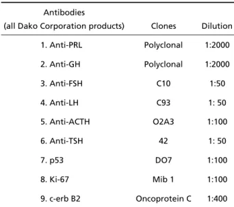
Table 1. Used antibodies, clones and dilutions.
Antibodies
(all Dako Corporation products) Clones Dilution
1. Anti-PRL Polyclonal 1:2000
2. Anti-GH Polyclonal 1:2000
3. Anti-FSH C10 1:50
4. Anti-LH C93 1: 50
5. Anti-ACTH O2A3 1:100
6. Anti-TSH 42 1: 50
7. p53 DO7 1:100
8. Ki-67 Mib 1 1:100
obtained from all patients. Approval by the Committee on Ethics in Research of the Univ ersidade de Br asília was an o b l i g a t o ry re q u i rement for this study, being obtained after fulfilling extensive pre requisites. Techniques were per-f o rmed both at the Department oper-f Pathology, Universidade de Brasília, Brazil, and IPATIMUP, O Porto, Portugal.
Specimens were fixed in 10% formalin and submitted to embedding in paraffin according to standard histolog-ical pro c e d u res. Hematoxylin and eosin staining was perf o r-med in all sections. Immunohistochemical evaluation (using S t reptavidin-Biotin systems) included hormonal as well as “ p rognostic” profiles. The used antibodies, clones and re s-pective dilutions are depicted in Table 1. Reactions were developed with diaminobenzidine (DAB) and counter-stained with hematoxylin20.
I m m u n o h i s t o c h e m i s t ryresults were interpreted accord-ing to Table 2.
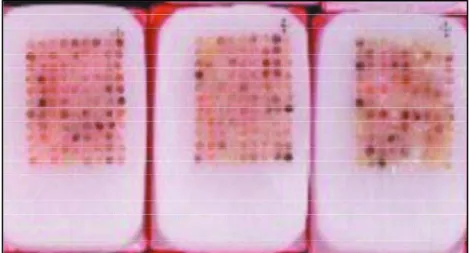
Immunohistochemical evaluation was done accord i n g to standard techniques and by “tissue microarray”. In this last modality, only the most re p resentative areas of the lesions were selected and extracted from the original paraf-fin block using specially design ed syringes. Samples fro m d i ff e rent cases were then joined in new paraffin blocks, p e rmitting simultaneous analysis of many surgical speci-mens (Figs 1 and 2).
RESULTS
Data from immunohistochemical evaluation
con-Table 2. Immunohistochemistry. Data analyses.
A. GH, PRL, FSH, LH, TSH and ACTH antibodies (positive: cytoplasmic staining):
1 less than 10% of cells with positive staining 2 between 10 and 20% of cells with positive reaction 3 between 21 and 50% of cells with positive reaction 4 more than 51% of cells with positive reaction
Note: We considered, as secreting, tumors with any staining intensity.
B. c-erb B2 (positive:membrane staining):
0 lack of positivity in more than 90% of tumor cells 1 d i s c rete immunopositivity in more than 10% of tumor cells 2 d i s c rete to moderate positivity in more than 10% of tumor
cells
3 intense and complete immunoreactivity in more than 10% of tumor cells
C. Ki-67 and p53 (positive:nuclear staining): 0 complete lack of positivity in tumor cell
1 immunoexpression in less than 10% of tumor cells 2 immunoexpression between 10 and 25% of tumor cells 3 immunoexpression between 25 and 50% of tumor cells 4 immunoexpression in more than 50% of tumor cells
Note: Lesions with less than 10% of nuclear staining for Ki-67 were con-s i d e red of low proliferative potential; lecon-sioncon-s with more than 25% of nuclear staining as highly proliferative, and intermediate values as mod-erate or intermediate proliferative potential.
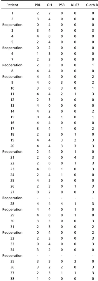
Table 3. Immunohistochemical semi-quantitative analysis of p i t u i t a ry hormones PRL and GH and associated Ki-67, p53 and c-erb B2 expression.
Patient PRL GH P53 Ki 67 C-erb B2
1 2 2 0 0 0
2 3 4 0 0 0
Reoperation 0 4 0 0 0
3 3 4 0 0 3
4 4 0 0 0 0
5 2 4 0 0 0
Reoperation 0 2 0 0 0
6 1 3 0 0 0
7 2 3 0 0 0
Reoperation 2 3 0 0 1
8 4 4 0 0 0
Reoperation 4 4 0 0 2
9 4 0 3 0 2
10 3 0 3 0 1
11 4 4 2 1 3
12 2 3 0 0 0
13 4 0 0 0 0
14 4 2 0 0 2
15 0 4 1 0 1
16 4 4 0 0 0
17 3 4 1 0 2
18 2 3 0 1 0
19 4 4 3 2 3
20 4 4 3 3 3
Reoperation 2 4 0 1 0
21 2 0 0 4 3
22 2 0 0 1 3
23 4 0 1 0 3
24 2 4 1 0 0
25 4 2 0 0 0
26 2 3 0 1 3
27 0 2 0 0 3
Reoperation - - - -
-28 4 4 4 1 3
Reoperation 4 4 0 1 0
29 4 0 0 1 0
30 3 3 0 0 3
31 2 3 0 0 2
Reoperation 0 4 0 0 2
32 2 3 0 0 3
33 0 4 0 0 3
34 3 2 0 0 0
Reoperation - - - -
-35 3 3 0 3 0
36 3 2 2 0 3
37 2 3 1 1 3
c e rning hormonal as well as prognostic profiles are presented in Table 3.
Immunohistochemical analysis of the lesions obtai-ned from the first operation revealed that 26 (68.4%) w e re positive for GH and prolactin, nine (23.7%) were positive for prolactin and three (7.8%) for GH. Immu-nohistochemical profiles of the tumors operated for the second time disclosed concordant results with first operation in four cases; in two cases analysis was not done (one of them because of lack of tumoral tissue at reoperation - case 34), and in three cases re-sults were discordant (originally GH- and PRL-posi-tive, and only GH positivity in the second evaluation). Two of the GH- and prolactin-positive cases showed also LH- and TSH-positivity; other case disclosed simul-taneous positivity for ACTH. A PRL-positive case and a GH-positive case disclosed also positivity for LH.
Among the cases operated on for the first time, 20 (52.6%) showed c-erb B2 positivity from 2 to 3, and two were 1 (Fig 3). Considering the horm o n a l e x p ression, the distribution was 14/26 (53.8%) GH/
PRL-positive, 6/9 (66.6%) PRL-positive (Fig 4) and 1/3 (33.3%) GH-positive (Fig 5). These diff e rences were not statistically significant (Fischer test p=0.394 GH/ PRL and prolactin; p=0.363 PRL and GH). From seven tumors analysed for c-erb B2 after the second oper-ation, three were concordant with the first pro f i l e , two were not analyzed and four were discord a n t ; the discordant ones were two negatives on first and 1 or 2 on the second intervention, and two 3 on the first and negative on the second.
C o n c e rning p53, 11 (28.9%) tumors showed ex-pression, but only six (15.9%) from 2 to 3 and five 1 (Fig 6). The distribution, according to the histoche-mical type of tumor, was similar: 8/26 (26.9%) GH-/ P R L - s e c reting, 3GH-/9 (33.3%) PRL-secreting and 1GH-/3 (33.3%) GH-secreting. For Ki-67 we observed 11 pos-itive cases (28.4%), from which four (10.5%) from 2 to 4, and seven 1 (Fig 7). The distribution according to tumor type was 8/26 of GH-/PRL-secreting (30.7%) and 3/9 (33.3%) of PRL-secreting type. None of the isolated GH-secreting tumors was positive for Ki-67.
Fig 1. Microarray prepared paraffin blocks.
DISCUSSION
A c romegaly is due, in more than 95% of cases, to G H - s e c reting pituitary adenomas, that may be of dif-f e rent cell types. Exclusively GH-secreting adenomas a re identified in 60% of acromegalic patients5. In our
s t u d y, however, these adenomas re p resented only 7.8% of cases. Mixed adenomas (composed of two d i ff e rent populations of GH- and PRL-secreting cells) cause acromegaly with equally high prolactin levels. These are fast-growing, aggressive lesions. These
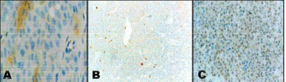
Fig 4. Intensity of cytoplasmic staining for prolactin: A:(0), 100X; B:(2), 200X; C:(4), 200X.
Fig 7. Intensity of nuclear staining for Ki-67. A: negative (0), 400X; B:(1), 100X; C:(1), 200X. Fig 5. Intensity of cytoplasmic staining for GH. A: negative (0),
200X; B: positive (4), 400X.
w e re the most common adenomas among our cases (68.4%), a finding not confirmed by other authors5.
Adenomas expressing both, GH and PRL, in the same cell, present with, generally, normal or lowly incre a-sed prolactin levels5.
A mutation in the somatotrophic cell is a pre re q-uisite for the development of a GH-secreting tumor. All adenomas composed exclusively of GH-secre t i n g cells are of monoclonal origin6. Many alterations were
described in the GH-secreting adenomas, such as irre g-ularities of the Gs p rotein (gsp) that simulates the function of GHRH. Patients carrying these adenomas have smaller tumors and keep GH suppressibility to glucose overc h a rg e2 1. Despite the evidences that a
s o m a t o t rophic cell may originate GH-secreting ade-nomas, the events leading to this tumoral expansion a re not clear. The presence of an activating oncogene may be necessary for initiating these adenomas.
The pathogenesis of prolactinomas is not equal-ly understood. Due to the inhibitory influence of hy-pothalamus (through dopamine) on PRL secretion, it has been considered that the decreased dopaminer-gic tonus should participate in this process. It has been described that prolactinomas are monoclonal, but that hypothalamus would exert a facilitating action on the clonal expansion of a genomically alte-red cell6. It was recently described that tubero i n f u
n-dibular dopaminergic (TIDA) neuronal lesion by estro-gen taken as a contraceptive method was not asso-ciated with a pituitary tumor, despite assoasso-ciated high PRL levels, probably for not being carrier of a mutant lactotrophic cell22.
P rotoncogene c-erb B2 is localized in chro m o-some 17, which encodes a transmembrane receptor p rotein with tyrosine kinase activity belonging to the e p i d e rmal growth factor receptor (EGFR) family. A positive membrane staining for the c-erb B2 pro d u c t is in most cases associated with real gene amplifica-tion, such as demonstrated by molecular biology tech-n i q u e s2 0. The pro p o rtion of positivity for c-erb B2 was
significative in the GH/PRL-, PRL- and GH-secre t i n g tumors included in our study. This finding was not paralleled in the consulted literature. We did not find papers that described the presence of c-erb B2 in pitu-i t a ry tumors. However, pitu-it pitu-is posspitu-ible that the detec-tion of c-erb B2 in the selected cases might have some influence on their evolution, considering the intima-te relationship between c-erb B2 and epiderm a l g rowth factor (EGF), as well as the fact that most pi-t u i pi-t a ryadenomas disclose EGF receppi-tors (pi-the expre s-sion being higher in non-functioning tumors as com-p a red to functioning ones). High levels of EGF re c e com-
p-tor in pituitary tumors suggest that they are involved in their pro g re s s i o n2 3. Besides that, serum levels of
EGF receptor were higher in patients presenting with macroadenomas and giant adenomas, as compared to pituitary hyperplasia, Rathke’s cyst and norm a l c o n t rols. There was a positive correlation between levels of EGF receptors and tumor size24.
Other growth factor related to EGF, the transform-ing growth factor alfa (TGF), that exerts biological e ffects on EGF re c e p t o r, has also been localized in prolactin-secreting cells. Both functioning and non-functioning adenomas, as well as normal pituitary, e x p ress this growh factor, suggesting that it might have some role in the pathogenesis of these tumors8.
In our cases, we identified p53 positivity in only 28.9% of tumors, with similar pro p o rtions among the three types of lesions (GH/PRL, PRL and GH). Be-sides that, only 6 (15.7%) expressed positivity high-er than 2. This suggests that p53 would not have an important role in our patients’ tumor evolution.
This finding was confirmed by other authors. Levy et al.9did not find p53 mutations on secreting or
non-s e c reting adenomanon-s, non-suggenon-sting that thinon-s gene would not have a role in the development of pituitary tu-mors. The high p53 protein levels identified thro u g h i m m u n o h i s t o c h e m i s t ry in pituitary tumors could be a consequence of binding to other cell proteins in these tumors9. Oliveira et al.1 0also observed low
pos-itivity frequency (1.3%) for p53 among their cases. The tumors studied by these authors were highly in-vasive, implying that p53 alterations would not be implicated in pituitary tumors aggressiveness. Howe-v e r, other authors identified p53 expression in 61% of pituitary adenomas, associating these finding with the proliferative status of these tumors, but not with their invasiveness or volume1 1. Alterations on p53
w e re identified in higher pro p o rtions of aggre s s i v e1 2
or invasive25neoplasms.
Ki-67 is a nuclear antigen expressed in phases G1, S, G2 and M of the cell cycle and recognized by the c o m m e rcially available antibody MIB-1. It has been re f e rred that MIB 1 values are not related neither to s e rum levels of prolactin and GH in patients with pro-lactinoma and acro m e g a l y, re s p e c t i v e l y, nor to inva-siveness of GH-secreting tumors. However, there is an association with positivity in staining for pro l a c t i n and growth hormone14.
Other studies showed greater MIB-1 positivity in tumors with dural invasion, compared to those in which no invasion was verified1 5. In non-functioning
ones) high levels of Ki-67 expression were detected, as well as a positive relationship between p53 and Ki-67 with invasiveness and tumor re c u rre n c e2 5. It
was also suggested that invasive adenomas would have a higher proliferation rate, compared to non-invasive, and would express greater amounts of Ki-6 72 6.In vitroKi-67 positivity was detected in ten out
of twelve pituitary adenomas and, in primary cultu-res of these tumors, in eleven out of twelve adeno-m a s1 6. The expression of cyclin A and the presence of
Ki-67 were significant survival and pro g ression fac-tors in pituitary adenomas27.
The low expression frequency observed in our cas-es suggcas-est that Ki-67 would not be involved in the development of GH/PRL, GH and PRL adenomas. Other authors did not found a significant re l a t i o n-ship between the presence of Ki-67 and pituitary t u m o r s2 8. These authors were not able to find a
pos-itive relationship between Ki-67 and tumor size and its invasiveness potential in neighboring stru c t u re s , but verified that tumors of older patients had gre a t e r Ki-67 positivity than tumors of younger ones17.
In conclusion, we found a high percentage of GH/ PRL-, GH- and PRL-secreting adenomas with immuno-histochemical expression for c-erb B2 not pre v i o u s l y identified in the literature. Once this membrane re-ceptor is associated to growth factors EGF and TGF
, that have a well-known effect on tumor growth, we suggest that c-erb B2 might have a role in the evolution of these lesions. Complementary studies are needed to confirm this hypothesis.
REFERENCES
1. Herman-Bonert VS, Prager D, Melmed S. Growth hormone. In Melmed S (ed). The pituitary. Cambridge: Blackwell Science, 1995:98-135. 2. Zurschmiede C, Ladolt AM. Distribution of growth hormone and
pro-lactin in secretory granules of the normal and neoplastic human ade-nohypophysis. Virchows Arch B 1987;53:308-315.
3. Mulchahey JJ, Jaffe RB. Detection of a potential progenitor cell in the human fetal pituitary that secretes both growth hormone and pro l a c t i n . J Clin Endocrinol Metab 1988;66:24-32.
4. Davis JRE. Tissue-specific regulation of prolactin gene expression. J Endocrinol 1990;125:171-173.
5. Kovacs K, Horvath E. Pathology of growth hormone-producing tumors of the human pituitary. Semin Diag Path 1986;3:18-33.
6. Herman V, Fagin J, Gonsky R, Kovacs K, Melmed S. Clonal origin of pituitary adenomas. J Clin Endocrinol Metab 1990;71:1427-1433.
7. Faglia G, A rosio M, Spada A. GS protein mutations and pituitary tu-mors: functional correlates and possible therapeutic implications. Metabolism 1996;45:117-119.
8. Melmed S, Shimon I. Genetical basis of endocrine disease: pituitary tumor pathogenesis. J Clin Endocrinol Metab 1997;82:1675-1681. 9. Levy A, Hall L, Yeudall WA, Lightman SL. P53 gene mutations in
pitu-itary adenomas: a rare events. Clin Endocrinol (Oxf) 1994;41:809-814. 10. Oliveira MC, Marroni CP, Pizarro CB, Lima JFP, Coutinho LMB. Ex-p ression of Ex-p53 Ex-protein in Ex-pituitary adenomas. Braz J Med Biol Res 2002;35:561-565.
11. Suliman M, Royds J, Cullen D, et al. Mdm2 and the p53 pathway in human pituitary adenomas. Clin Endocrinol 2001;54:317-325. 12. S u h a rdja A, Kovacs K, Rutka J. Genetic basis of pituitary adenomas
invasiveness: a review. J Neurooncol 2001;52:195-204.
13. Yin D, Tamaki N, Kokunai T, Yasuo K, Yonezawa K. Bro m o c r i p t i n e -induced apoptosis in pituitary adenoma cells: relationship to p53 and bcl-2 expression. J Clin Neurosci 1999;6:326-331.
14. Asano K, Tajika Y, Huang MC, Takakura K. The relation between cell-p roliferation activity and secretion in cell-pituitary adenoma: a review of 63 cases. No To Shinkei 1996;48:534-539.
15. Daita G, Yonemasu Y. Dural invasion and proliferative potential of pitu-itary adenomas. Neurol Med Chir 1996;36:211-214.
16. Atkin SL, Green VL, Hipin LJ, Landolt AM, Foy PM, Jeff reys RV, White MC. A comparison of proliferation indices in human anterior pituitary adenomas using formalin-fixed and in vitro cell culture. J Neuro s u rg 1997;87:85-88.
17. M a s t ro n a rdi L, Guiducci A, Puzzilli F, Maira G. Anterior pituitary ade-nomas in patients aged more than 65 years: analysis of growth fraction (using the MIB-1 monoclonal antibody) and of clinical features in com-parison to younger patients. Clin Neurol Neurosurg 2002;104:44-48. 18. Kumar V, Abbas AK, Fausto N. In Robbins & Cotran (eds). Pathologic
basis of disease. 7.ed. Philadelphia: Elsevier Saunders, 2005. 19. Penar PL, Nathan DJ, Nathan MH, Salsali A . Pituitary tumor
diagno-sis and treatment. Curr Neurol Neurosci Rep 2002;2:236-245. 20. Alves VA F, Bacchi CE, Vassallo J. Manual de Imuno-histoquímica.
Sociedade Brasileira de Patologia, 1999:35-37.
21. Landis CA, Harsh G, Lyons J, Davis FM, Bourne HR. Clinical charac-teristics of acromegalic patients whose pituitary tumors contain mutant Gs protein. J Clin Endocrinol Metab 1990;71:1416-1420.
22. Casulari LA, Celotti F, Naves LA, Domingues L, Papadia C. Case re p o r t : persistence of hyperprolactinemia after treatment of primary hypothy-roidism and withdrawal of the long term use of estrogen. A re the t u b e roinfundibular dopaminergic neurons permanently lesioned? A rq Bras Endocrinol Metab 2005, in press.
23. O n g u ru O, Scheithauer BW, Kovacs K, et al. Analysis of epidermal g rowth factor receptor and activated epidermal growth factor re c e p-tor expression in pituitary adenomas and carcinomas. Mod Pathol 2004;17:772-780.
24. Kong Y, Ren Z, Su C, Wang R. Detection of serum epidermal gro w t h factor receptor in the diagnosis of proliferation of pituitary adenoma. Zhonhua Yi Xue Za Zhi 2002;82:527-529.
25. S c h reiber S, Seager W, Ludecke DK. Proliferation markers in diff e re n t types of clinically non-secreting pituitary adenomas. Pituitary 1999;1:213-220.
26. Blevins LS Jr, Verity DK, Allen G. A g g ressive pituitary tumors. Oncology 1998;12:1307-1312.
27. Nakabayashi H, Sunada I, Hara M. Immunohistochemical analyses of cell cycle-related proteins, apoptosis, and proliferation in pituitary ade-nomas. J Histochem Cytochem 2001;49:1193-1194.