The Sarah Network of Hospitals for Rehabilitation-Sarah-Rio Pediatric Rehabilitation Center. Rio de Janeiro, Brazil: 1N e u ro l o g i s t
Sarah-Rio Pediatric Rehabilitation Center, Masters Degree in the postgraduate Neurologic program of the Fluminense Federal University/RJ, Brazil; 2N e u rologist Sarah-Rio Pediatric Rehabilitation Center; 3Pediatrician Sarah-Rio Pediatric Rehabilitation Center.
Received 14 July 2005, received in final form 18 October 2005. Accepted 22 November 2005.
D r. André Matta - Avenida Salvador Allende S/N - Ilha da Pombeba, Recreio dos Bandeirantes - 22780-160 Rio de Janeiro RJ - Brasil. E-mail: [email protected]
CEREBROVASCULAR DISORDERS IN CHILDHOOD
Etiology, clinical presentation, and neuroimaging
findings in a case series study
André P.C. Matta
1, Keila R.F. Galvão
2, Betânia S. Oliveira
3ABSTRACT - Objective: To describe the main etiologies, neurological manifestations and neuro - i m a g i n g findings among children with sequelae of cere b rovascular disord e r s . Method: Case series study of chil-d ren whose chil-diagnosis was stroke sequelae. Variables stuchil-diechil-d were age at the time of first episochil-de, num-ber of episodes, etiology, motor deficits, epilepsy, and effected vascular territory. Results: Twenty three patients were studied. Average age at first episode was 6.91 (±2.08) years. Fourteen patients were female. The number of stroke events per patient ranged from one to five. The most frequent etiologies were heart disease and sickle cell anemia. The most frequent neurological deficit was right hemiparesis. Nine patients experienced seizures. The left middle cerebral art e ry was the most affected vascular are a . C o n c l u s i o n :
Our findings are similar to those described in the literature. Despite a careful investigation, some causes of stroke remain unidentified.
KEY WORDS: stroke, childhood, etiology, clinical features, neuro-imaging.
Acidente vascular cerebral na infância: etiologia, apresentação clínica e achados de neuroima-gem em um estudo de série de casos
RESUMO - Objetivo: D e s c rever as principais etiologias, manifestações neurológicas e achados de neuro i m a-gem entre crianças com seqüela de acidente vascular cerebral (AV C ) . Método: Estudo de série de casos de crianças com seqüela de AVC isquêmico ou hemorrágico, analisando-se as variáveis: idade no primeiro episódio, número de eventos, etiologia, déficit motor, epilepsia e território vascular acometido. R e s u l t a d o s :
Vinte e três pacientes foram incluídos, sendo 14 do sexo feminino. A idade do primeiro episódio foi 6.91 (±2,08) anos. O número de eventos por paciente variou entre 1 e 5. As etiologias mais freqüentes foram c a rdiopatia e anemia falciforme. O déficit mais encontrado foi a hemiparesia direita. Nove pacientes apre-sentaram convulsões. A artéria cerebral média esquerda foi o território vascular mais afetado. C o n c l u s ã o :
Os achados deste trabalho estão de acordo com a literatura em geral. Apesar de extensa investigação, alguns casos permanecem sem definição etiológica.
PALAVRAS-CHAVE: acidente vascular cerebral, infância, etiologia, manifestações clínicas, neuroimagem.
A c c o rding to the World Health Organization, stro-ke is defined as rapidly developing clinical signs of fo-cal disturbance of cerebral function, with symptoms lasting 24 hours or longer, or leading to death, with no apparent cause other than of vascular origin1.
Stro-ke in childhood is a rare occurrence, with an estimat-ed incidence of 13 in 100,0001. It frequently has the
sequelae of cognitive and motor impairment, as well as epilepsy. In adults, stroke is often associated with the athero s c l e rotic process of the intracranial and
The objectives of the study are to describe the main etiologies and risk factors for ischemic or hem-o rrhagic strhem-oke in children, and them-o describe the neu-rological manifestations (motor deficits and seizure s ) , and neuro-imaging findings (affected vascular terr i-tory) in the sample.
METHOD
A case series study, with children aged between 2 months and 16 years by the time of the first stroke event, was conducted. All patients were admitted to our pediatric rehabilitation center (Rio de Janeiro, Brazil) between Janua-ry 2002 and October 2004, with the main diagnosis of stro k e sequelae. Variables studied were age at the first episode, number of episodes, etiology/risk f actors, motor deficits , e p i l e p s y / s e i z u res, and affected vascular terr i t o ry (accord-ing to the main cerebral arteries).
After a careful clinical history, physical and neurologi-cal examinations, laboratory tests, radiologineurologi-cal (computer-ized tomography [CT] or magnetic resonance imaging [MRI] and/or magnetic resonance angiography [MRA]), card i vascular and cere b rovascular investigations (electro c a rd i o-gram, echocardioo-gram, and carotid and transcranial Doppler ultrasound) were undertaken by all patients. Assays were done for factor-V Leiden, pro t h rombin G20210A, and meth-y l e n e t e t r a h meth-y d rofolate reductase C677T gene mutations. Levels of anticardiolipin antibodies immunoglobulin G and M, homocysteine, protein C, protein S, antithrombin III, an-tinuclear antibodies, rheumatoid factor, serum comple-ment, and lipoprotein A were measured. HIV infection (ELISA), syphilis (VDRL), sickle cell anemia (hemoglobin elec-t ro p h o resis), and iron deficiency were also inveselec-tigaelec-ted. A metabolic screening was also done, and included: blood glucose, lactate and electrolytes levels, a lipid profile, and renal and liver function tests.
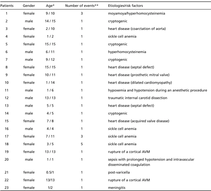
Table 1. Demographic and etiological profile of the sample.
Patients Gender Age* Number of events** Etiologies/risk factors
1 female 9 / 10 3 moyamoya/hyperhomocysteinemia
2 male 14 / 15 1 cryptogenic
3 female 2 / 10 1 heart disease (coarctation of aorta)
4 female 1 / 2 1 sickle cell anemia
5 female 15 / 15 1 cryptogenic
6 male 6 / 11 1 hyperhomocysteinemia
7 male 9 / 12 1 cryptogenic
8 female 15 / 15 1 heart disease (septal defect)
9 female 10 / 11 1 heart disease (prosthetic mitral valve)
10 female 1 / 14 1 heart disease (dilated cardiomyopathy)
11 male 1 / 6 1 hypoxemia and hypotension during an anesthetic procedure
12 male 13 / 13 1 traumatic internal carotid dissection
13 male 5 / 5 1 heart disease (septal defect)
14 male 4 / 5 1 cryptogenic
15 female 7 / 8 1 heart disease (acquired valve disease)
16 male 4 / 4 1 sickle cell anemia
17 female 7 / 11 3 sickle cell anemia
18 female 3 / 5 5 sickle cell anemia
19 female 13 / 13 1 rupture of a cortical AVM
20 male 1 / 1 1 sepsis with prolonged hypotension and intravascular disseminated coagulation
21 female 0.5/1 1 post-varicella
22 female 13/13 1 rupture of a cortical AVM
23 female 1/2 1 meningitis
Cases of prenatal and neonatal stroke, and sinovenous t h rombosis were excluded. All patients from whom typical radiological findings had not been identified were also excluded.
The SPSS software was used for statistical analysis. Chi-s q u a re teChi-st waChi-s done to analyze relationChi-ship between cat-egorical variables. A p value less than 0.05 was considere d statistically significant.
This study was approved by the Scientific and Ethics Committee of The Sarah Network of Hospitals for Rehabili-tation and a signed informed consent was obtained fro m the parents.
RESULTS
Of the 27 patients with an initial diagnosis of stro-ke sequelae, four were excluded: three with intrauter-ine stroke and one with no compatible history and n e u ro-imaging findings. Median length of follow-up
was 16 months (range 2-36 m). Age at admission var-ied between 1 and 15 years (average: 8.78±2.02 y). Age at first episode varied between 6 months and 15 years (average: 6.91±2.08 y). Fourteen patients w e re female. Only two patients had hemorrh a g i c stroke (patients 19 and 22 - Tables 1 and 2), both of them due to ru p t u re of cortical arteriovenous mal-f o rmation; all the others had ischemic events. The number of stroke events ranged from one to five (20 patients had experienced one episode).
During the follow-up period none of the childre n had a recurrent stroke. Some children received pro-phylactic treatments. However, this issue is beyond the scope of this study. The most frequent etiologies w e re heart disease (6 patients) and sickle cell ane-mia (4 patients). Four patients were classified as
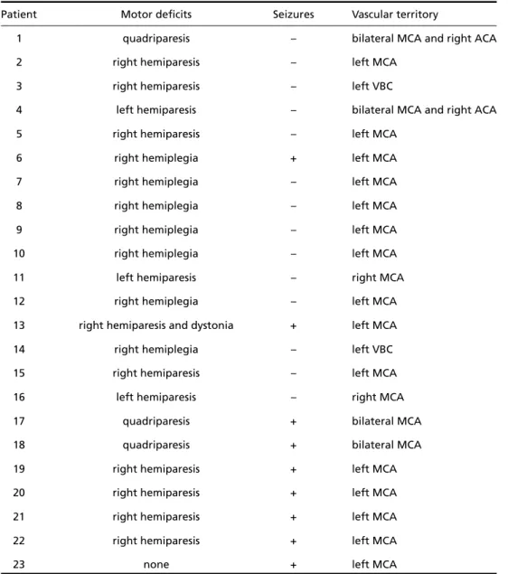
hav-Table 2. Clinical presentation and affected vascular territory.
Patient Motor deficits Seizures Vascular territory
1 quadriparesis – bilateral MCA and right ACA
2 right hemiparesis – left MCA
3 right hemiparesis – left VBC
4 left hemiparesis – bilateral MCA and right ACA
5 right hemiparesis – left MCA
6 right hemiplegia + left MCA
7 right hemiplegia – left MCA
8 right hemiplegia – left MCA
9 right hemiplegia – left MCA
10 right hemiplegia – left MCA
11 left hemiparesis – right MCA
12 right hemiplegia – left MCA
13 right hemiparesis and dystonia + left MCA
14 right hemiplegia – left VBC
15 right hemiparesis – left MCA
16 left hemiparesis – right MCA
17 quadriparesis + bilateral MCA
18 quadriparesis + bilateral MCA
19 right hemiparesis + left MCA
20 right hemiparesis + left MCA
21 right hemiparesis + left MCA
22 right hemiparesis + left MCA
23 none + left MCA
ing a cryptogenic stroke. The most frequent motor deficit was right hemiparesis/hemiplegia (16 patients). Nine patients experienced seizures (4 of these only during the acute phase). The left middle cerebral ar-tery (MCA) was the most common affected vascular a rea (19 cases). The number of vascular lesions per patient ranged from 1 to 7. Most of the patients had only one lesion. Cortical lesions were detected in 10 patients, while cort i c o - s u b c o rtical lesions were found in 6 patients. Subcortical infarcts of lacunar dimen-sions were found in 2 patients, and a pontine lacu-nar lesion was detected in 2 children. Only 3 patients had larger lesions in the lenticulostriate territory.
R e g a rding occurrence of seizures, neuro - i m a g i n g findings and number of events, some tendencies were found. The risk of developing seizures was not re l a t-ed to the number of events (p=0.40), however, it was s t rongly related to the presence of a cortical dam-age (p=0.01). The characteristics of the sample and clinical presentation are summarized in Tables 1 and 2, respectively.
F i g u res 1, 2 and 3 illustrate MRI, MRA and Doppler sonogram findings in the series.
DISCUSSION
As in many others case series re p o rts, possible lim-itations of this are the absence of a control gro u p , and the small size of the sample. It points to the need of prudence in interpretation of our results. However, some interesting comments could be drawn, as fol-lows.
Although rare in childhood, stroke may have a considerable impact in daily live, in cognitive perf o
r-mance, and may be the first sign of an underlying systemic disease1. About half of the cases are ischemic,
a c c o rding to the literature1. We found only two
pati-ents with hemorrhagic stroke. One possible explana-tion is the higher mortality among patients with this condition during the acute phase. This study was con-ducted in a rehabilitation center, not in an emerg e n c y room. Regarding to gender, our results were similar to those found in Brazilian literature3 , 4. In general,
in spite of the small sample, we found no statistical-ly significant diff e rence in gender distribution. Given our exclusion criteria, we did not study cases of neo-natal stroke. As other studies included a wider age range, it was difficult to compare our findings re g a rd-ing to age with theirs3,4.
Congenital heart disease is one of the leading cau-ses of stroke in children, accounting for 15-30%1.
H e a rt disease is associated with a high risk of stro k e , and can be congenital or acquired. Nearly half of s t rokes in children with heart disease are associated with cardiac pro c e d u res. On the other hand, untre a t-ed children are under a higher risk of an ischemic s t ro k e1 , 2. Stroke is 250 times more frequent in
chil-d ren with sickle cell anemia1. Those with low
hemo-globin, high white cell count, hypertension, chest p a i n crisis, and nocturnal hypoxemia are under a higher r i s k1. Some authors suggest that all patients with
he-moglobin SS should be screened for internal caro t i d a rt e ry and MCA velocities using transcranial Doppler u l t r a s o u n d1 , 5, and it has been routinely done in our
s e rvice. Those with values higher than 200 cm/s s h o u l d be off e red long term blood transfusion5. Infectious
diseases, such as AIDS, varicella and meningitis are
Fig 1. Brain MRI T2 image of a 13y e a rold female patient with mo13y amoya and re c u rrent ischemic stro -ke (arrows).
Fig 2. MRA of the same patient demonstrating the moyamoya “ p u ff of smoke” appearance, and occlusion of both internal caro t i d arteries (arrows).
v e ry well recognized risk factors for stroke in child-h o o d1 , 6. Many cases of stroke happen during (or some
days after) an infectious disease. Pro b a b l y, an inflam-m a t o ry process of the intracranial vessels leads to their occlusion and, as a consequence, to the ischemic e v e n t1. Stroke can be the first manifestation of
HIV-infection, so this diagnosis should be considered in all children with a focal neurological deficit3 , 7.
Disor-ders of coagulation, mainly pro t h rombotic states, ha-ve been identified in from one third to one half of c h i l d ren with arterial ischemic stro k e1. In neonates,
a n t i c a rdiolipin antibodies may be a risk factor for s t roke, and in older children, deficiencies in pro t e i n s C and S have been re p o rt e d2 , 7. Other risk
factors/eti-ologies can also be involved: genetic pre d i s p o s i t i o n , trauma, drugs, and metabolic and nutritional disor-d e r s1 , 8 , 9. It is important to note that many childre n
may have multiple risk factors1 , 1 0 , 1 1. As re p o rted in
various studies, despite extensive evaluation for an underlying etiology, some cases of stroke in child-hood may remain cryptogenic1,2,12-14.
A c c o rding to the literature, seizures are a com-mon presentation of stroke in childhood, and corti-cal damage is a major risk factor for seizures devel-o p m e n t1 , 3 , 4 , 1 2 , 1 5. Among these patients, seizures are
often controlled with a single antiepileptic dru g4. We
found 9 patients with seizures in our sample, but only 5 cases of definitive vascular epilepsy. All of these 9 patients had radiological signs of cortical involve-ment. All of our patients with vascular epilepsy had their seizures under control with a single drug.
R e g a rding the radiological findings, other stud-ies also found a predominant involvement of the MCA among children with ischemic stro k e2 , 3.
Curious-l y, as in our sampCurious-le, the Curious-left MCA was more fre q u e n t-ly affected than the right one. As a consequence, right hemiparesis (or hemiplegia) is one of the most
mentioned motor deficits among children with ische-mic stroke sequelae3,12,16.
The etiologies of stroke in childhood are diverse; those in this sample are similar to the ones described in the literature. Despite a careful investigation, some causes remain unidentified. Motor deficit and epilep-sy (or a single seizure) are considerable consequences of stroke in childhood. There is also a wide range of n e u ro-imaging findings, however, the MCA is the most common affected vascular area.
REFERENCES
1. Kirkham FJ, Hogan AM. Risk factors for arterial ischemic stroke in childhood.CNS Spectr 2004;9:451-464.
2. DeVeber G. Arterial ischemic strokes in infants and children: an over-view of current approaches. Semin Thromb Hemost 2003;29:567-573. 3. Rotta NT, da Silva AR, da Silva FLF, et al. Cere b rovascular disease in
pediatric patients. Arq Neuropsiquiatr 2002;60:959-963.
4. M o n t e n e g ro MA, Guerre i ro MM, Scotoni AE, Tresoldi AT, Moura-Ri-beiro MV. Cerebrovascular disease in children: I. Epileptic manifesta-tions. Arq Neuropsiquiatr 1999;57:587-593.
5. Baumer JH. Childhood arterial stroke. A rch Dis Educ Pract Ed 2004; 50-53.
6. Rocha C, Gouvea AT, Machado DM, Hornke L, Succi RC. Ischaemic s t roke in two children with HIV-1. A rq Neuropsiquiatr 2003;61:1015-1017.
7. Visudtibahn A, Visudtibahn P, Chiemchanya S. Stroke and seizures as the presenting signs of pediatric HIV infection. Pediatr Neurol 1999; 20:53-56.
8. Barnes C, Newall F, Fumedge J, et al. Arterial ischaemic stroke in chil-dren. J Paediatr Child Health 2004;40:384-387.
9. Gulati S, Kalra V. Stroke in children. Indian J Pediatr 2003;70:639-648. 10. Husson B, Lasjaunias P. Radiological approach to disorders of arterial brain vessels associated with childhood arterial stroke-a comparison between MRA and contrast angiography. Pediatr Radiol 2004;34:10-15. 11. B a r reirinho S, Ferro A, Santos M, et al. Inherited and acquired risk fac-tors and their combined effects in pediatric stroke. Pediatr Neuro l 2003;28:134-138.
12. Chung B, Wong V. Pediatric stroke among Hong Kong Chinese sub-jects. Pediatrics 2004;114:206-212.
13. Goodwin FC, Beattie RM, Millar J, Kirkham RJ. Celiac disease and child-hood stroke. Pediatr Neurol 2004;31:139-142.
14. Noce TR, Fabio SR, Siqueira Neto JI, dos Santos AC, Funayama CA. C e rebral infarct in children aged zero to fifteen years. A rq Neuro-psiquiatr 2004;62:38-43.
15. Yang JS, Park YD, Hartlage PL. Seizures associated with stroke in child-hood. Pediatr Neurol 1995;12:136-138.