Arq Neuropsiquiatr 2006;64(3-A):592-595
Division of Neurosurgery - University of São Paulo Medical School - São Paulo SP, Brazil: 1MD;2MD, PhD.
Received 25 October 2005, received in final form 27 January 2006. Accepted 6 April 2006.
Dr. Wagner Malagó Tavares - Rua Alves Guimarães 642 / 125 - 05410-001 São Paulo SP - Brasil. E-mail: [email protected]
CSF MARKERS FOR DIAGNOSIS OF BACTERIAL
MENINGITIS IN NEUROSURGICAL
POSTOPERATIVE PATIENTS
Wagner Malagó Tavares
1, Andre Guelman Machado
2,
Hamilton Matushita
2, Jose Pindaro P. Plese
2ABSTRACT -Objective:To evaluate the diagnostic usefulness of cerebral spinal fluid (CSF) cellularity, pro-tein, neutrophils, glucose and lactate for detection of postoperative bacterial meningitis. Method:This p rospective study was conducted in 28 postoperative neuro s u rgical patients from 2002 to 2005 at University of São Paulo. The CSF markers were plotted in a receiver operating characteristic (ROC) curve to evaluate their accuracy. Results:Based on the area under ROC curve CSF glucose, cellularity, and lactate were con-s i d e red good tecon-stcon-s. Polymorphonuclear and protein did not achieve enough accuracy to be ucon-sed clinical-l y. Conclusion:The CSF glucose, lactate, and cellularity can be used for the diagnosis of bacterial menin-gitis. Moreover, it can be helpful to differentiate bacterial from aseptic meninmenin-gitis.
KEY WORDS: CSF, bacterial meningitis, neurosurgery, ROC analysis, postoperative infection.
M a rc a d o res liquóricos para o diagnóstico de meningite bacteriana em pós-operatório neuro-cirúrgico
RESUMO -Objetivo:Para avaliar a utilidade diagnóstica dos marc a d o res liquóricos de celularidade, con-centração de proteína, neutrofilia, concon-centração de glicose e lactato para a detecção da meningitie bac-teriana no pós-operatório neuro c i r ú rg i c o . Método:Esse estudo foi conduzido de maneira pro s p e c t i v a na Universidade de São Paulo no período de 2002 a 2005 em 28 pacientes no pós-operatório neuro c i r ú r-gico. Os marc a d o res liquóricos foram colocados em uma curva ROC (receiver operating characteristic) para avalição da sua acurácia. Resultados:Baseadas na área sob a curva ROC, glicorraquia, celularidade e con-centração de lactato foram considerados bons testes. A contagem de polimorf o n u c l e a res e a pro t e í n o r-raquia não atingiram acurácia suficiente para serem utilizadas clinicamente. Conclusão:A glicorraquia, a concentração de lactato e a celularidade podem ser utilizadas clinicamente para o diagnóstico da menin-gite bacteriana. Esses marc a d o res também podem ser úteis na diferenciação entre meninmenin-gite bacteriana e asséptica.
PALAVRAS-CHAVE: meningite bacteriana, LCR, neurocirurgia, curva ROC, infecção pós-operatória.
Postoperative meningitis is an uncommon com-plication (0.3 to 1.5%)1following intradural pro c
e-d u re, ane-d is potentially fatal with mortality of 20 to 5 0 %2. The diagnosis of meningitis in postsurgical
pa-tients is often difficult especially if the patient has been subjected to a ventricular or posterior fossa pro-cedure. The clinical signs of fever, meningismus and mental status alteration are non-specific and insuf-ficient for the diagnosis of postoperative meningi-tis3.
Analysis of cerebral spinal fluid (CSF) can be help-ful in many cases. However, postoperative CSF mark-ers including lactate, glucose, protein and cellulari-ty can also differ because of brain manipulation,
un-derlying brain pathology or bleeding into CSF follow-ing intradural pro c e d u re4 - 6. The desirability of
avoid-ing unnecessary administration of antibiotics to non-infected patients, the potentially devastating conse-quences associated with administration of cort i c o s-t e roids s-to pas-tiens-ts wis-th aseps-tic meningis-tis, and s-the need to establish diagnostic usefulness of CSF mark-ers have prompted this study.
METHOD
Patients –Lumbar or external ventricular CSF samples
purpos-Arq Neuropsiquiatr 2006;64(3-A) 593
es of this study. Patients had at least one clinical manifes-tation of bacterial meningitis (BM) including fever, menin-gismus and alteration of mental status. The group com-prised 9 females and 19 males; mean age was 41 years (ran-ge 2 to 69 years old). Samples were collected between days 1 to 21 after initial intradural procedure.
Sample collection and analysis –We first rinsed the
e x t e rnal ventricular derivation (EVD) for 10 minutes with iodide povidine. CSF was collected into sterile polystyre n e tubes and immediately submitted for analysis. Assessment of CSF markers CSF glucose and lactate were determ i n e d using Roche™ kit with Hitachi equipment. Total CSF pro-tein was determined in Hitachi equipment using the ben-zethonium chloride precipitation technique standard i z e d to the biuret method. Cell counts were determined using a calibrated Fuchs-Rosenthal chamber after staining with toluidine blue. CSF analysis to distinguish polymorphonu-clear leukocytes (PMNs) and lymphocytes was done using Shandon cytocentrifuge and Pappenheim stain.
Statistical analysis –Statistical analyses were perf o rm e d with SPSS™ for Windows. The CSF markers were analyzed to determine specificity and sensitivity of each marker and combinations between them. We used the Receiver Ope-rating Characteri stic (ROC) curve t o evaluate clinical use-fulness of each marker. The ROC curve re p resents the pro ability of true results in a disease as a function of the pro
b-ability of false positive results of a test. The area under the c u rve re p resents the validity of a test with 1.00 being the highest and 0 the lowest. A classification for accuracy of a diagnostic test considers .90 to 1.00=excellent; .80 to .89= good; .70 to .79=fair; .60 to .69=poor; .50 to.59=failure.
RESULTS
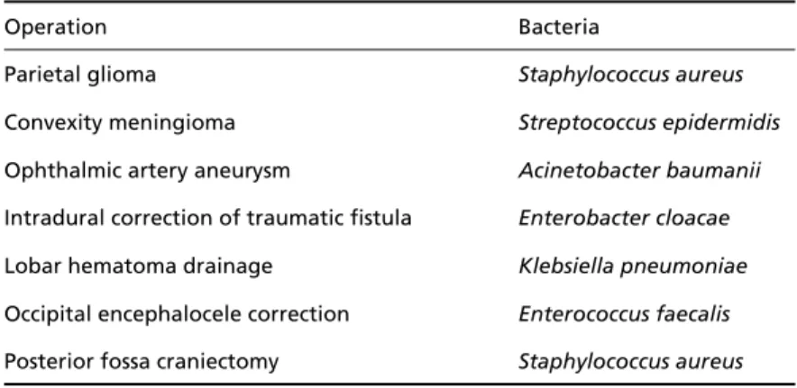
Of 28 patients, 7 had positive Gram stain and cul-t u re for baccul-teria. Table 1 presencul-ts cul-the cul-types of oper-ations and bacteria involved in the infection. We sum-marized the CSF markers according to highest ROC under the curve, sensitivity, specificity and pre d i c t i v e value (Table 2). The aforementioned values and the c u t o ff values were based on the point of the ROC curve which presented highest sensitivity and speci-f i c i t y. Markers were classispeci-fied according to area under the curve as excellent, good, fair, poor and failure as defined under methods. Based on these criteria, glu-cose, lactate, and cellularity were classified as good tests. Number of PMNs was classified as fair and and p rotein as poor. No tests achieved the excellent clas-sification.
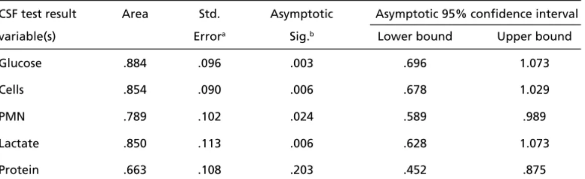
Table 3 describes the ROC analysis of CSF markers with their respective confidence intervals. All of the tests presented an excellent specificity (90.5%) but
Table 1. Types of procedure and the nature of infection.
Operation Bacteria
Parietal glioma Staphylococcus aureus
Convexity meningioma Streptococcus epidermidis Ophthalmic artery aneurysm Acinetobacter baumanii Intradural correction of traumatic fistula Enterobacter cloacae
Lobar hematoma drainage Klebsiella pneumoniae
Occipital encephalocele correction Enterococcus faecalis Posterior fossa craniectomy Staphylococcus aureus
Table 2. CSF markers diagnostic power.
CSF markers Cutoff* Area under Sensitivity Specificity Positive Negative
ROC curve predictive predictive
value value
Glucose 30 mg/dL .884 86% 90.5% 75% 95%
Cellularity 768/mL .854 71.4% 90.5% 83% 91%
Lactate 49 mg/dL .850 86% 90.5% 75% 95%
Polymorphonuclear 575/mL .789 42.9% 90.5% 50% 67%
Protein 370.5 mg/dL .663 28.6% 90.5% 25% 79%
a poor sensitivity with the exception of lactate and glucose (86%). In order to improve sensitivity we changed the cutoff values for all markers but this change decreased specificity to unacceptable levels (<70%). We found high predictive levels for glucose and lactate (positive predictive value 75%, negative p redictive value 95%) and also cellularity (positive p redictive value 83%, negative predictive value 91%). All the marker values were statistically significant with 95% confidence.
DISCUSSION
The postoperative period after neuro s u rg e ry may be complicated by nofrequent but life-thre a t e n-ing BM7 - 9. Neuro s u rgical patients frequently have
ventricular drains, bladder and intravenous catheters that may be present for a long time. All of these fac-tors together with type and duration of the opera-tion can increase the chance of infecopera-tion after neu-ro s u rg e ry. The onset of BM may be insidious and dif-ficult to diff e rentiate from the onset of other infec-tions that cause similar symptoms. More o v e r, postope-rative changes caused by opening of the meninges and breakdown of blood products may contribute to the clinical picture of fever, confusion and menin-gismus sometimes observed after neuro s u rg e ry. Clini-cal features are not reliable to diff e rentiate between BM and aseptic meningitis3. Therefore, diagnosis of
BM can be made solely by detection of bacteria on Gram stain or isolation in culture, but CSF culture s remain negative in 70% of clinically suspected cas-e s7. The sensitivity and specificity of bacterial
isola-tion from CSF can be raised if gene amplificaisola-tion is p e rf o rmed on the specimen1 0. This technique re q u i re s
trained personnel and equipment not available in many clinical laboratories perf o rming studies on CSF.
BM necessitates prompt and early diagnosis be-cause of its devastating consequences8. Use of bro a d
s p e c t rum antibiotics is to be avoided because of high
cost and risk to unaffected patients2 , 3. M o re o v e r, the
use of intravenous antibiotics and corticosteroids in suspected cases of postoperative BM impairs the diag-nostic value of CSF markers2. Our present study
ana-lyzes postoperative CSF to establish cutoff values for c e l l u l a r i t y, lactate, glucose, protein, and PMNs for the diagnosis of BM.
C o rtes-Lopez et al.1 1found a sensitivity of 51.5%
and specificity of 95% using CSF leukocyte count of
1700/mL. Using this cutoff we had a similar re s u l t of 57.1% and 100%, re s p e c t i v e l y, but this cutoff did not represent the spot on the ROC curve with high-est sensitivity and specificity. We lowered the cutoff value to 768 cells/mL and found a sensitivity o 71.4% and specificity of 90.5%. Cortes-Lopez et al.1 1a rg u e d
that the CSF number of PMNs, protein concentration and glucose were not diff e rent enough to discrimi-nate. We agree in part, since we found a high are a under the ROC curve, as well as high sensitivity and specificity for lactate and glucose. The number of PMNs and protein concentration were not classified as good tests for the diagnosis of BM. Lactate analy-sis in this study has inferior sensitivity and specifici-ty compared to previous work (88 and 98% re s p e c-t i v e l y )2. Results of this study were also diff e rent fro m
those published by Ross et al.3. Reported diff e re n c e s
in the diagnostic power of CSF markers could be ex-plained by: 1) the type of study design ( re t ro s p e c-tive studies are more susceptible to bias related to data and population selection); 2) use of antibiotics and steroid which may decrease inflammation and CSF culture growth thus lowering the diagnostic val-ue of CSF analysis; 3) prevalence of BM in the study g roup which correlates directly with diagnostic pow-er of the tests undpow-er evaluation. In this study the pre-valance of BM was 25%, close to the value in other studies2,3,10.
One may argue that in cases of BM the number of neutrophils could raise the concentration of
lac-594 Arq Neuropsiquiatr 2006;64(3-A)
Table 3. Area under the curve and their respective confidence intervals.
CSF test result Area Std. Asymptotic Asymptotic 95% confidence interval
variable(s) Errora Sig.b Lower bound Upper bound
Glucose .884 .096 .003 .696 1.073
Cells .854 .090 .006 .678 1.029
PMN .789 .102 .024 .589 .989
Lactate .850 .113 .006 .628 1.073
Protein .663 .108 .203 .452 .875
Arq Neuropsiquiatr 2006;64(3-A) 595
tate in CSF. However, several authors in clinical, in v i t ro and animal studies did not find a significant p roduction of lactate by neutro p h i l s1 2 - 1 4. More o v e r,
CSF lactate level is not affected by the presence of red blood cells and it can be used in postoperative neurosurgical patients2,15,16.
All patients in our study received antibiotics and s t e roids as soon as BM was suspected. As alre a d y mentioned, this use can lower the diagnostic power of CSF markers. As shown by Salord et al.10,
sensitiv-ity and specificsensitiv-ity of CSF markers and hence the pre-valence of BM can be increased if PCR and gene am-plification techniques are applied to CSF samples. Use of D(-) lactate, tumor necrosis factor, interleukin-1, interleukin-6 and interleukin-8 can also incre a s e sensitivity and specificity1,11but these assays are not
readily available in clinical laboratories receiving CSF for diagnostic testing. Assays for the CSF markers evaluated in this study are relatively inexpensive, rap-id and established in most laboratories.
Our current results show that for postoperative n e u ro s u rgical patients, CSF cellularity, glucose and lactate can be more reliable for the diagnosis of BM than clinical parameters alone. These CSF markers are available routinely in clinical laboratories, how-e v how-e r, if thhow-erhow-e is still doubt about thhow-e diagnosis, how- empir-ical treatment should be started17.
Acknowledgements– To Jean H. Priest (re t a i red pro-fessor of the Emory University, Atlanta USA) for helping the translation of this paper.
REFERENCES
1. S a l o rd F, Boussaid O, Eynard N, Perret C, Grando J, Chacornac R. Intere t du dosage du D(-) lactate pour le diagnostic rapide de meningite après
craniotomie: étude pre l i m i n a i re. Ann Fr Anesth Reanim 1994;13: 647- 653.
2. Leib SL, Boscacci R, Gratzl O, Zimmerli W. Predictive value of cere-brospinal fluid (CSF) lactate level versus CSF/Blood glucose ratio for the diagnosis of bacterial meningitis following neurosurgery. Clin Inf Dis 1999;29:69-74.
3. Ross D, Rosegay H, Pons V. Diff e rentiation of aseptic and bacterial meningitis in postoperative neuro s u rgical patients. J Neuro s u rg 1988; 69:669-674.
4. Carmel PW, Fraser RAR, Stein BM. Aseptic meningitis following pos-terior fossa surgery in children. J Neurosurg 1974;41:44-49. 5. Kaufman BA, Tunkel, AR, Pryor JC, et al. Meningitis in the
neurosur-gical patient. Infect Dis Clin N Am 1990;4:677-701.
6. Kim YS, Pons VG. Infectious in the neuro s u rgical intensive care unit. Neurosurg Clin N Am 1994;5:741-754.
7. Blomstedt GC. Infections in neuro s u rgery: a re s t rospective study of 1143 patients and 1517 operations. Acta Neurochir (Wien) 1985;78: 81-90.
8. Buckwold FJ, Hand R, Hansebout RR. Hospital-acquired bacterial meningitis in neurosurgical patients. J Neurosurg 1977;46:494-500. 9. Mollman HD, Haines SJ. Risk factors for postoperative neurosurgical
wound infection: a case-control study. J Neurosurg 1986;64:902-906. 10. S a l o rdF, Druel B, Grando J, et al. Aseptic meningitis: demonstration
of bacterial DNA in cerebrospinal fluid by gene amplification. Ann Fr Anesth Reanim 1995;14:320-325.
11. Lopez-Cortes LF, Marquez-Arbizu R, Jimenez-Jimenez LM, et al. C e re b rospinal fluid tumor necrosis factor-, interleukin-1, interleukin-6, and interleukin-8 as diagnostic markers of cere b rospinal fluid infec-tion in neurosurgical patients. Crit Care Med 2000;28:215-219. 12. Bland, RD, Lister RC, Ries JP. Cere b rospinal fluid lactic acid level and
pH in meningitis: aids in diff e rential diagnosis. Am J Dis Child 1974; 128:151-156.
13. G u e r r a - R o m e ro L, Tauber MG, Fournier MA, Tu reen JH. Lactate and glucose concentrations in brain interstitial fluid, cere b rospinal fluid, and serum during experimental pneumococcal meningitis. J Infect Dis 1992;166:546-550.
14. Tauber MG, Borschberg U, Sande MA. Influence of granulocytes on brain edema, intracranial pre s s u re, and cere b rospinal fluid concentra-tions of lactate and protein in experimental meningitis. J Infect Dis 1998; 157:456-464.
15. Begovac J, Bace A, Soldo I, Lehpamer B. Lactate and glucose in cere-b rospinal fluid heavily contaminated with cere-blood. Acta Med Cro a t i c a 1991;45:341-345.
16. C a m e ron PD, Boyce JM, Ansari BM. Cere b rospinal fluid lactate in meningitis and meningococcaemia. J Infectol 1993;26:245-252. 17. Martins R, Ciquini O Jr, Matushita H, Cabral ND, Plese JPP. Infections