Arq Neuropsiquiatr 2006;64(3-A):650-653
1Adjuvant teacher of Medical School, Minas Gerais Federal University, MG, Brazil. Neuro s u rgeon of Felício Rocho Hospital, Belo Horizonte, MG, Brazil (FRH);2Adjuvant Teacher of Biological Sciences Institute, Minas Gerais Federal University, MG, Brazil. Neurosurgeon of FRH; 3Neurosurgery Resident FRH.
Received 17 November 2005, received in final form 30 March 2006. Accepted 9 May 2006.
Dr. Rodrigo Moreira Faleiro - Rua Caraça 518 / 301 - 20220-260 Belo Horizonte MG - Brasil. E-mail: [email protected]
ENDOSCOPIC ASSISTED MICRONEUROSURGERY
FOR GASSERIAN PORTION OF TRIGEMINAL
NEUROMA
Two cases
Luiz Carlos Mendes Faleiro
1, Rodrigo Moreira Faleiro
2,
Luiz Fernando Vilar Barroso
3, Daniel Andrade Gripp
3ABSTRACT - We re p o rt two cases of trigeminal neuroma that were operated on by the neuro s u rg e ry team at Felício Rocho Hospital, Belo Horizonte, Minas Gerais State, Brazil. Endoscopic assisted micro s u rg e ry was the technique used to approach the gasserian region tumor with good results.
KEY WORDS: trigeminal schwannoma, posterior fossa tumors, endoscopic brain surg e ry, micro n e u ro s u rg e ry.
M i c ro n e u ro c i ru rgia assistida por endoscópio para abordagem da porção gasseriana do neuri-noma do trigêmeo: relato de dois casos
RESUMO - D e s c revemos dois casos de neurinoma do trigêmeo operados no Serviço de Neuro c i ru rgia do Hospital Felício Rocho, Belo Horizonte, Estado de Minas Gerais, Brasil. Utilizamos com sucesso a micro c i ru r-gia assistida por endoscopia para abordagem de tumores da porção gasseriana .
PA L AV R A S - C H AVE: schwanoma trigeminal, tumores de fossa posterior, neuro c i ru rgia endoscópica, micro n e u-rocirurgia.
Trigeminal neuroma (TN) was described in 1849
by Smith and it was first surgically treated by Frazier1.
It is also known as schwannoma or neurilemona. TN is a rare tumor, corresponding to 0.1-0.4% of intracra-nial tumors and 1-8% of neuromas. After vestibular n e u roma, it is the most common intracranial neuro-ma. Prevalence is higher in the forth and fifth deca-des. Most are histologically benign (schwannoma or
n e u ro f i b ro m a )2 , 3. Few malignant cases have been
described in the literature3. It may present in
associ-ation with other cranial nerves neuromas like in neu-ro f i b neu-romatosis. TN may be located in the subdural space (cere b e l l a r-pontine angle), interdural (lateral wall of cavernous sinus), epidural or extracranial space
(orbit and pterigopalatyne fossa)2. Most common
clinical presentation is sensory deficit in corre s p o n-ding side of the face, for months to years. Lancinating pain or paresthesias may also occur. Other re l a t e d
symptoms are headache, hemifacial spasm, hearing deficits, focal seizures, hemiparesis, intracranial hyper-tension, otalgya, cerebellar signs and gait disturban-c e1 , 4. Oculomotor, trochlear and abducens nerves may
also be affected4.
Many other entities at the cere b e l l a r-pontine an-gle and Meckel’s cave region are to be differentiat-ed: metastases, lymphomas, bony tumors (chord o m a and chondro s a rcoma) and meningiomas. Meningio-ma is the Meningio-main diff e rential diagnosis when at the
M e c k e l ’s cave location3. Imaging methods are useful
in diff e rentiating between lesions at the cere b e l l a r-pontine angle and Meckel’s cave region. On magnet-ic resonance imaging (MRI), neurinoma presents as hypointense (low weight) on T1 and hyperintense ( h a rd weight) on T2. Contrast enhancement is not common. Hyperostosis and intratumoral calcifications
Arq Neuropsiquiatr 2006;64(3-A) 651
TN is classified according to its location: (1) at the posterior fossa (P), (2) at the middle fossa including the Gasser ganglion (M), (3) supra infratentorial (SIT)
(dumbbell-shape) and (4) at the periphery (E)2 , 4.
Supra-infratentorial TN (SIT) is equally distributed among posterior and middle cranial fossa. Combined a p p roaches are usually re q u i red to accomplish its total resection with opening of the lateral wall of
the cavernous sinus (associated morbidity)5. On those
patients with SIT-TN whose extra ocular movements and hearing capacity are pre s e rved, a less invasive approach is needed.
Two cases of SIT-TN in which endoscopic assisted m i c ro s u rg e ry allowed good resection of the lesion without further deficits are presented.
CASES
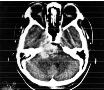
Case 1 –Woman aged 48 presented at our hospital in 1999, with right frontal region paresthesias, which later p ro g ressed to right hemiface. She than began to notice headache, disequilibrium and diplopia. She had been pre-viously diagnosed with systemic arterial hypertension. At examination, she was alert, oriented and had a norm a l speech. Fundoscopic examination revealed redded optic discs with moderately shaded borders. Extra ocular move-ments were normal; a right jaw shift, abolished right cor-nean reflex, normal index-nose test, and an ataxic gait with a wide open base was noted. Computer tomography (CT) and MRI revealed a supra-infratentorial lesion at the right trigeminal nerve location (Fig 1). On July 1999, Dolenc’s extradural approach with incomplete removal of intra-gasserian portion of the neuroma was perf o rmed. On August 1999, a transpetrosal approach to remove posteri-or fossa lesion was added (Fi g 3). A diagnosis of TN with sub-total removal was made. On post-operative period, she p resented with hypoesthesia an the right side of the face. A CT scan on 2000 revealed a s mall residual lesion at the Gasser ganglion location. On 2003, patient re t u rns pre s e n t-ing pain on the right side of the face and a new MRI scan revealed a lesion in Meckel’s cave and posterior fossa (Fig 2). Endoscopic assisted micro s u rg e ry (EAM) was pro p o s e d to remove tumor re c u rrence. After resection under micro-scopic vision at the posterior fossa, the endoscope was applied to remove the gasserian portion, with the help of m i c ro - c u rettes and micro - f o rceps, under direct endoscop-ic vision. The use of the mendoscop-icroscope alone would not allow direct visualization of this part of the tumor.
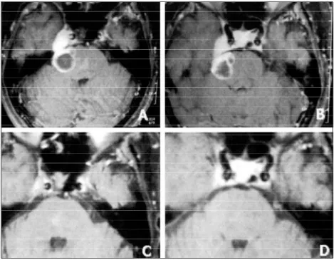
Case 2 – On 2002, a 66-year-old woman presented to us with pain on the right side of the face for the last few months. Neurological examination was considered norm a l . A MRI revealed a partially cystic, partially solid lesion at the right cere b e l l a r-pontine angle and gasserian region (Fig 4A and 4B). A trigeminal neurinoma was suspected; patient began to take carbamazepine and surgery was proposed. On January 2003, a re t rosigm oid approach and posterior fossa exploration was undertaken, with partial removal of
Fig 1. Cerebral tomography with right trigeminal nerve lesion.
Fig 2. New MRI with a lesion in Meckel’s cave and posterior fossa.
Fig 3. Endoscopic and microneurosurgical instruments.
attempt-ed. As with in case 1, endoscope was important in the com-plete removal of the lesion. On post-operative period, pa-tient presented with hyperesthesia on the right trigeminal n e rve terr i t o ry. On Febru a ry 2003, the patient re t u rn e d with MRI showing complete removal of the TN (Fig 4C and 4D). She still suff e red from hyperesthesia at the face, despite using carbamazepine 200 mg a day. On December 2003, a new MRI was considered normal. Nowadays there is still mild numbness on the face.
DISCUSSION
Yoshida et al.2, have classified 27 cases of TN
ac-c o rding to its loac-cation (Table 1). The M type inac-cludes tumors of the middle fossa originating on the Gasser ganglion or its branches on to the lateral wall of the cavernous sinus. P type tumors include those on the posterior fossa with origin on the root of the TN; type E tumors are those with origin on the extracranial p o rtion of the TN. Type MP includes those dumbbell-shaped tumors located equally both in the middle and posterior cranial fossa. Type ME is dumbbell-shap-ed locatdumbbell-shap-ed in the middle fossa and extracranial com-p a rtment; tycom-pe MPE is tumors in the com-posterior and middle fossa and also in the extracranial compart-ment2,3.
Yoshida et al.2reviewed 251 cases of TN from 1985
to 1999 and the post-operative complication most commonly observed was facial hypoesthesia (70%). C e rebellar disturbances, abducens nerve palsy, intra-tumoral hemorrhage or subarachnoid hemorrh a g e were less frequent.
Suboccipital re t rosigmoid approach to the
poste-rior fossa is one of the most used approaches in ro s u rg e ry and allows a good exposure of the neu-rovascular stru c t u res, micro s c o p i c a l l y. When a lesion is present inside the cavernous sinus, a combined ap-p roach is necessary. Aap-pap-proach to the ap-posterior fossa has its associated morbidity: cerebellar edema and infraction, hearing loss, facial palsy, cere b ro - s p i n a l fluid leak5.
Endoscopic assisted micro s u rg e ry (EAM) may sim-plify surgical approach to those lesions, and tumor removal, allowing low morbidity. EAM has been
giv-en a lob of attgiv-ention recgiv-ently6-10. Its indications and
limits are still not yet well defined. It allows less inva-sive surg e ry and lower associated morbidity, goals of any surgical treatment. Advances in imaging meth-ods have contributed to improve diagnosis and
sur-gical planning10.
652 Arq Neuropsiquiatr 2006;64(3-A)
Table 1. Classification of TN for localization2.
Type Cases %
M 4 14.8
P 5 18.5
E 1 3.7
MP 10 37
ME 5 18.5
MPE 2 7.4
Total 27 100
Arq Neuropsiquiatr 2006;64(3-A) 653
The utilization of the endoscope to assist in iden-tification of stru c t u res in the posterior fossa is not a recent pro c e d u re, although with few re p o rts on the l i t e r a t u re5 - 9. O’Donoghue and O’Flynn8d e s c r i b e d
endoscopic anatomy of the cere b e l l a r-pontine angle in 1993. In 1999, Magnan and Sanna published an atlas with detailed surgical anatomy of the cranial
n e rves of the base of the skull9. The endoscope has
also been used in neurovascular decompressions at the posterior fossa, minimizing retraction of stru
c-tures and widening lateral angle of vision5,6.
Endoscopic pro c e d u res have been classified ac-c o rding to its role on the neuro s u rgiac-cal pro ac-c e d u re1 0.
They are: pure endoscopic neuro s u rg e ry (NE), endo-scopic assisted micro neuro s u rg e ry (EAM), endoscop-ic controled mendoscop-icro n e u ro s u rg e ry(ECM), and endosco-pic inspection (EI). NE uses the endoscope exclusive-ly and surgical instruments inside it, having a single cranial hole as entry site. EAM is a micro n e u ro s u rg e ry p ro c e d u re visually assisted by the endoscope. ECM uses micro n e u ro s u rg e ryinstruments but not the mi-c rosmi-cope itself and vision is provided by the endosmi-co- endosco-pe. EI may be used in any surgical procedure to
ins-pection only1 0. EAM improves light and definition
besides allowing better lateral view of areas next to the main stru c t u res. Endoscope allows a less traumat-ic pro c e d u re, better light and better view of places that the microscope would not be to re a c h5 , 6. Also it
makes possible inspection of bony orifices without
tissue removal or retraction5,11-13.
E A M ’s characteristic is to combine the advantages of micro n e u ro s u rg e ry and endoscopic surg e ry1 0.
Com-plex maneuvering is made through the microscope, and the endoscope allows identification of
inacces-sible regions5,6, improving surgical outcomes.
The main disadvantages of the endoscope are re-lated to the excessive amount of heading in the sur-gical field disturbing vision and lack of thre e - d i m e
n-sional view and cranial nerve injury risk6. King et al.6
re p o rted 10 patients with trigeminal neuralgia aged 16 through 67 (16 male and 3 female) in the period of 1997 through 2000 in which EAM had been used. All patients presented with total improvement or sat-i s f a c t o ry sat-improvement of symptomatology wsat-ithout
associated morbidity. Jarrahy et al.7used EAM to
t reat 21 patients suffering from trigeminal neural-gia. The endoscope was used to study the neuro v a s-cular complex, with 52 neurovass-cular conflicts being identified. In 14 patients (27%), that would not have been possible without the assistance of the
endo-scope. Perneczky et al.1 0followed 36 patients thro u g h
a period of 44 months who were operated on by EAM in the treatment of intracranial cysts (arachnoid and intraventricular cysts). As a result, 20% rates of success, 25% inalterated and 5% worst were achiev-ed. There was a diminished cerebellar retraction and intracranial neuropathies with the risk of CSF leak
l o w e red from 9% to 0%. Al-Mefty et al.3re v i e w e d
25 cases of TN between 1989 and 2000, with 76% of the cases being of the dumbbell-shapped tumor. They re p o rt difficulties in dealing with those tumors be-cause of the lack of good visualization of the caver-nous sinus.
There were no reports of EAM being used in the t reatment of the Gasser portion of the trigeminal neuroma in the literature reviewed.
REFERENCE
1. B o rdi L, Compton J, Symon L. Trigeminal neuroma. Surg Neurol 1989; 31:272-276.
2. Yoshida K, Kawase T. Trigeminal neurinomas extending into multiple fossae: surgical methods and review of the literature. J Neurosurgery 1999;91:202-211.
3. Al-Mefty O, Ayoubi S, Gaber E. Trigeminal schwannomas: removal of dumbbell-shaped tumors through the expanded Meckel cave and out-comes of cranial nerve function. J Neurosurg 2002;96:453-463.
4. Winn H. Youmans Neurological Surg e r y, 5t hed. Philadelphia: Sanders,
2003:1343-1350.
5. Fries G, Perneczky A. Endoscope-assisted brain surg e r y. Part 2: analy-sis of 380 procedures. Neurosurgery 1998;42:226-230.
6. King W A, Wackym P A, Sen C, Meyer G, et al. Adjunctive use of endos-copy during posterior fossa surgery to treat cranial neuro p a t h i e s . Neurosurgery 2001;49:108-115.
7. Jarrahy R, Berci G. Shahinian H K. Endoscope-assisted microvascular d e c o m p ression of the trigeminal nerve. Otolaringol Head Neck Surg 2000;123:218-223.
8. O’Donoghue GM, O’Flynn P. Endoscopic anatomy of the cere b e l o p o n-tine angle. Am J Otol 1993;14:122-125.
9. Magnan J, Sanna M. Endoscopy in neuro - o t o l o g y. New York: Thieme, 1999:7-23.
10. Perneczky A. Hopf N J. Endoscopic neuro s u rgery and endoscope-assist-ed micro n e u ro s u rgery for the treatment of intracranial cysts . Neurosurgery 1998;43:1130-1136.
11. Perneczky A, Fries G. Endoscope-assisted brain surg e r y. Part 1: evolu-tion, basic concept and current technique. Neuro s u rgery 1998;42: 219-224.
12. Mobbs R, Teo C. Endoscopic assisted posterior fossa descompre s s i o n . J Clin Neurosci 2001;8:343-344.
13. Göksu N, Bayazıt Y, Kemaloglu Y. Endoscopy of the posterior fossa