D
ESCRIPTION
OF THE CLINICAL PICTURE OF DENGUE
HEMORRHAGIC FEVER/DENGUE SHOCK
SYNDROME (DHF/‘DSS) IN ADULTS
A. L&z, 2 G. Ko~ti,~ M. G. Gu~m~n,~ L. Lobuinu, 5 J. Bwvo, G A. R&z, 7 A. R.amo~,~ and R. Marthex
I
NTRODUCTION
Prior epidemic dengue out- breaks in the United States (1922), South Africa (1927), and Greece (1928) afflicted adult patients who developed hemorrhagic manifestations and shock (I). But since 1954, when dengue hem- orrhagic fever/ dengue shock syndrome (DHF/DSS) was reported as a new phe- nomenon in the Philippines (2), it has affected mainly children, and there have only been a few isolated descriptions of the severe picture in adults (3).
For over 30 years, up to 1977, there was no evidence of dengue virus circulation on Cuba; but in that and the following year a classic dengue epidemic occurred that affected a large part of the
Cuban population (4). Then, in the summer of 1981, a major epidemic of DHF/DSS occurred for the fast time in Cuba and the Americas. That epidemic struck all age groups, both whites and blacks, and both sexes (r).
The present article describes the clinical manifestations of DHFlDSS in adults on the basis of observations made during the 1981 epidemic. Such clinical description has previously been provided only by isolated reports within our area. It is therefore expected that the description provided here will prove use- ful to other countries in interpreting the clinical-epidemiologic situation-a situ- ation that, with regard to DHF/DSS, is becoming progressively more complex and dangerous.
’ The work reported here was carried out by the Pedro Kouil Institute of Tropical Medicine in Havana, Cuba, with financial support from the International Develop- ment Research Center in Ottawa, Canada. This article will also be published in Spanish in the Boletin de la Of&a Sanirasia Panamericana, vol. 104, 1988. -. 2 Internist, Pedro Kouri Institute of Tropical Medicine,
Apartado 601, Zona Postal Marianao 13, Havana, Cuba.
3 Director and Virologist, Pedro Kouri Institute. 4 Chief, Arbovirus Department, Pedro Kouri Institute. 5 Virologist, Pedro Kouri Institute.
‘ Researcher, Pedro Kouri Institute. ’ Internist, Pedro Koud Institute. a Internist, Pedro Kouri Institute.
M
ATERIALS AND
METHODS
We examined the clinical his- tories of patients whose diagnosis upon hospital admission was “hemorrhagic dengue” and who had entered the Ca- lixto Garcia, Salvador Allende , and Enri- que Cabrera hospitals in the city of Ha- vana during the DHFlDSS epidemic of 1981.. From among those whose histories showed a disease picture appropriate for the study, 200 were selected at random. Efforts were then made to retrospectively confirm that the patients involved had experienced dengue-2 infections and to determine whether they had been previ- ously exposed to dengue- 1.
Specifically, sera from these 200 subjects were tested for the presence of antibodies to dengue viruses 1 and 2
by the plaque reduction neutralization method (G) . Test sera were deemed posi- tive for dengue-1 or dengue-2 antibodies if they reduced the number of plaques of the respective virus by 50% or more. It was considered that all persons with neu- tralizing dengue-2 antibodies had been infected during the 1981 epidemic, and that all those with neutralizing antibod- ies to both viruses had experienced sec- ondary infections. All subjects in whom dengue-2 infection could not be serolog- ically confirmed were excluded from the study.
The remaining 104 subjects,
in whom dengue-2 infection was con- firmed, came to constitute Group 1. All of these subjects were told about the study’s aims and significance, and all agreed voluntarily to be included.
A second study group (Group 2) was composed of 26 adults who had died (out of a total of 57 adult fatalities recorded during the epidemic), these be- ing subjects for whom complete autopsy reports were available and whose clinical histories were considered appropriate for the study. In this group the diagnosis of DHF/DSS was clinical and anatomo- pathologic. (It should be noted that no other hemorrhagic fever exists in Cuba, and that all the cases involved were re- ported during the height of the epi- demic.)
Because the criteria for defin- ing cases as DHF/DSS have previously been developed only for children, while the aim of the study reported here was to offer the elements needed to diagnose and define clinical cases in adults, we de- cided to include those subjects with a hospital discharge diagnosis of hemor- rhagic dengue who were confirmed sero- logically to have neutralizing antibodies for dengue-2. Regarding the fatalities, those subjects were included who had a clinical diagnosis of DHF I DSS with ana- tomopathologic lesions compatible with hemorrhagic dengue and who had died during the epidemic. Naturally, the di- agnostic criteria developed by the WHO
expert group (7) served as a basis for se- lection of these latter cases.
R
ESULTS
Of the 104 Group 1 subjects selected for the study, 102 (98%) were found to have had confirmed secondary dengue infections (dengue-1 plus den- gue-2).
Age, Sex, and Race
The age distribution of the 104 Group 1 subjects and 26 Group 2 subjects generally conformed to that of Cuba’s adult population, there being no evident clustering by age among these adult subjects.
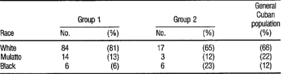
Regarding race (Table l),
81% of the Group 1 subjects were white, while only 65% of the fatally afflicted Group 2 subjects were white, this latter figure being similar to the percentage of whites among the Cuban population ac- cording to the last (1981) census. (De- spite the relatively high percentage of whites in the Group 1 study sample, the disease also affected mulattoes and blacks. )
Women accounted for 65 % of all the Group 1 subjects (Table 2), a pre- dominance that is statistically signifi- cant (p < 0.05). Most (62%) of the 26 Group 2 subjects were also women; but, partly because of the group’s smaller size, this latter predominance was not statistically significant (p > 0.05).
TABLE 2. Pmptins af men and women in the two groups studied.
Gmup 1 Group 2 No. (%I No. (“/PI Women 68 (65) ;; (62)
Men 36 (35) (38)
Total 104 (100) 26 uw
Clinical Picture
From the clinical standpoint, four syndromes were identified-these being general (constitutional manifes- tations), digestive, hemorrhagic, and shock syndromes. Table 3 shows the per- centages of Group 1 and Group 2 sub- jects with constitutional manifestations. As may be seen, fever, headache, asthe- nia, and arthralgia predominated in both the nonfatal (Group 1) and fatal (Group 2) cases. Rash was found infre- quently in both groups, being especially uncommon in Group 2.
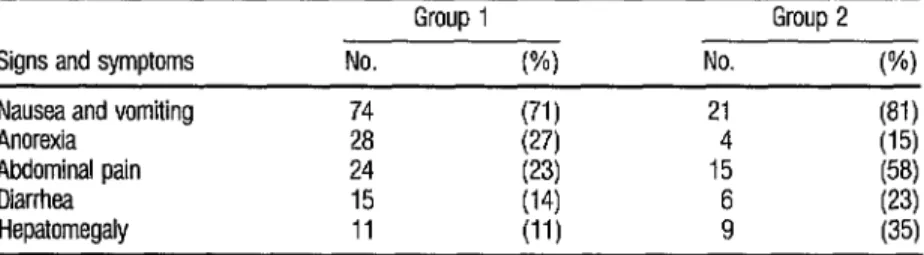
In both groups, symptomatic involvement of the digestive system was characterized by nausea and vomiting (Table 4). Hepatomegaly occurred three
TABLE 1. Racial composition of the two gmups studied alnd the general Cuban population (1981 census).
Race
General ‘i . Group 1 Group 2 population Cuban < No. (%I No. (%I (%I ,Q White 84 631) 17 4
Mulatto 14 3
TABLE 3. General (classical) dengue signs and symptoms exhibited by members of the two groups studied.
Signs and symptoms Fever Headache
Asthenia Arthralgia Myalgia Retroorbiial pain Rash
Group 1 Group 2 No. (%I No. (%I 101 80 1;:; 23 14 I::;
60 (58) 17
56 (54) IO ~~~; 46 20 Iti{ 11 7 I::; 13 (13) 2 (8)
TABLE 4. Digestive system signs and symptoms found among members of the two groups studied.
Group 1 Group 2 Signs and symptoms No. W) No. (“/I Nausea and vomiting
Anorexia Abdominal pain Diarrhea Hepatomegaly
21 4
24 15 (23) (14) 15 6 I;:; 11 (11) 9 (35)
times as frequently among the fatal (Group 2) cases as it did among the non- fatal (Group 1) cases. Similarly, abdomi- nal pain apparently afflicted 58% of the Group 2 subjects but only 23% of the Group 1 subjects.
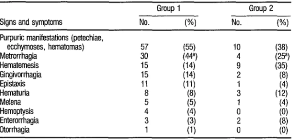
Despite the fact that all the subjects were diagnosed as having hem- orrhagic dengue, manifestations of the hemorrhagic syndrome were confirmed in only 90% of the Group 1 patients and 65 % of the Group 2 patients (Table 5). The predominant hemorrhagic symp- toms in both groups were purpuric mani- festations ( petechiae, ecchymoses, he- matomas). Hematemesis was found in a higher percentage of the subjects dying (Group 2) than of those surviving (Group 1).
Shock syndrome, which was present in all the fatal cases, appeared on the average around the ftith day of the
disease. Appearing at the earliest on the third day and at the latest on the eighth, it was frequently irreversible. Clinically, the shock syndrome was characterized by an increased respiration rate, reduced di- uresis, a rapid pulse exhibiting narrow pulse pressure until the pulse disap- peared, a decline in arterial blood pres- sure, distal cyanosis, and lack of any re- sponse to symptomatic medication.
TABLE 5. Signs and symptoms of dengue hemorrhagic fever (DHF) found in members of the two groups studied.
Signs and symptoms
Purpuric manifestations (petechiae, ecchymoses, hematomas) Metrorrhagia
Hematemesis Gingivorrhagia Epistaxis Hematuria Melena Hemoptysis Enterorrhagia Utorrhagia
a Percentage oliemles only.
Group 1 Group 2 No. (%I No. WI z; (55) 10 (38)
[T’ 4
15 9 I:; 15 2
11 Ii] 1 It; 8 (8) 3 (12) 5 1
4 1:; 0 1:; 3 (3) 2
1 (1) 0
Shock preceded death in all of In the fatal cases (Group 2) the fatal (Group 2) cases; but it was only
classified as posthemorrhagic in half of these cases, since the remainder did not show major accompanying hemorrhagic manifestations. No instances of con- firmed shock were found among the Group 1 subjects, whose cases evolved in a satisfactory manner.
the initial clinical picture was similar, in- volving various general and digestive symptoms compatible with classical den- gue. Abdominal pain began occurring on the third day, and hemorrhagic mani- festations on the fifth, these latter coin- ciding with the onset of shock and a worsening of the clinical picture that led to death within a few hours.
Clinical Course of the Disease
Among the Group 1 patients the disease lasted an average of seven days, while the Group 2 patients died, on the average, during the sixth day.
Clinical Laboratory Results
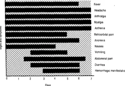
At the outset, the clinical pic- ture among the Group 1 subjects tended
to be characterized by fever, headache, asthenia, myalgia, arthralgia, and re- troorbital pain (Figure 1). Symptoms in- volving the digestive system began around the second day and lasted three to four days. The first hemorrhagic manifestations began to appear on the third day.
Platelet counts and hemo- grams were obtained from all the sub- jects in a systematic fashion upon their admission to the hospital. If some change in a person’s condition was noted, these analyses were repeated every
12 or 24 hours. Hematocrits were not ob- tained from all of the subjects.
Thrombocytopenia was found in 80.3% of the Group 1 cases and 7 1.8 % of the Group 2 cases. In contrast,
FIGURE 1. Clinical course of the disease during days one through seven among the study subjects who recovered (Group 1).
0 1 2 3 4 5 6 Days
tration we also used clinical criteria, find- ing hemoconcentration in all subjects who presented clinical, radiologic, or au- topsy evidence of a shift of fluid into their body cavities.)
Leukocytosis (2 10,000 leu- kocytes per cubic millimeter), with a pre- dominance of neutrophils and lympho- penia, was found more commonly in Group 2 subjects (44 % ) than in Group 1
subjects (9.3 %).
Antecedent Pathologies
Relatively high percentages of the Group 2 subjects were found to have had histories of bronchial asthma (11.5%) and sickle cell anemia (15.3%). In contrast, the prevalences of these dis- eases among the adult Cuban population were 7 % for bronchial asthma and
0.08% for sickle cell anemia. These dif-
ferences are statistically significant (p < 0.05). Diabetes mellitus was also found
lfestatlons 7
relatively frequently among the Group 2 subjects\ but the difference between this
prevalence and that found in the general Cuban population was not statistically significant.
D ISCUSSION
AND
CONCLUSIONS
Our results show that 98% of the subjects whose cases evolved favor- ably (those in Group 1) had been in- fected by both the dengue-1 and den- gue-2 viruses. This fact, together with the results of other research carried out by our group that yielded similar results
(8, 91, suggests that such secondary infec- tion is a necessary condition (though not the only condition) required for develop- ment of hemorrhagic dengue. It also supports what Halstead has postulated regarding the etiopathology of this dis- ease (10).
Among adults, the 1981
the age distribution of the affected adults being similar to that of the Cuban population as a whole (11). In Southeast Asia and the islands of the Western Pa- cific, the disease is reported to occur pri- marily among children (people under age 14). This circumstance could relate to the broad, permanent, and sometimes simultaneous circulation of the four den- gue virus serotypes in these regions (l)-
a circumstance implying that practically the entire population, on reaching adult- hood, may already be immune to den- gue viruses.
Since Cuba experienced in- tense circulation of dengue-1 in l977- 1978 (4) and dengue-2 in 1981 (51, it was possible for the disease to arise in chil- dren and adults whose only two experi- ences with these viruses occurred within a four-year period. (As previously noted, there is no evidence of dengue virus cir- culation in Cuba between 1945, when a limited outbreak occurred in the city of Havana (12) and 1977, when dengue-1 was introduced.)
Recently, Fang et al. (13) re- ported on the epidemic of DHFlDSS that occurred in Malaysia in 1982, when some 56% of the persons affected were adults. The authors state that the reason for the observed age displacement to- ward those over 15 “is not clear, but may be due to the immune status of the com- munity in which the virus circulated.”
In Cuba, although DHF/DSS affected whites, mulattoes, and blacks, the highest frequency of cases was found in whites. Bravo et al. (9), in a study of three groups of Cuban patients with DHF/DSS, reports that the disease oc- curred at a significantly greater rate among whites. White fatalities were not found to predominate in that study, but this could conceivably be explained by the presence in the study group of two black patients with sickle-cell anemia (a genetic condition associated with
blacks), this latter being a risk factor for onset of the severe disease (9). If these two patients were removed from the sample, the percentage of fatalities among whites would exceed the percent- age of fatalities among members of other races.
Overall, the work reported here underscores the fact that hemor- rhagic dengue is not limited to people of Asian origin, and that whites, mulattoes, and blacks are all susceptible. (In this vein, it is worth noting that initial re- search results obtained by our group have shown that the phenomenon of immunologic amplification was not found to occur in macrophages from the vast majority of a group of blacks studied-14).
Regarding sex, other authors
(1, 3, 15) have reported a relatively high incidence of hemorrhagic dengue among young girls over four years old. In our study women were more frequently af- fected. These findings can be related to the apparently greater immunocompe- tence of females (3).
From the clinical standpoint, it should be noted that hepatomegaly, abdominal pain, and hematemesis were relevant signs in fatal cases and could be taken to suggest a poor prognosis. Other
authors have made similar reports on af- 2
flitted children (IG-19). 3
The hemorrhagic manifesta- 3 tion most frequently observed was pur-
pura, a fmding also reported by other au- 5 thors studying childhood cases (8, 1~5, 19).
2
Hematemesis was observed
9
twice as often among the Group 2 fatali- LF ties as it was among the Group 1 patients .
< P % I!$ Q
who evolved favorably. It also appeared before or on the same day that shock emerged, a finding that differs from what has been reported for children (both on Cuba and in Southeast Asia), who have been found to experience bleeding as a terminal picture with some frequency after irreversible shock has been established (7).
Among the Group 1 adults
whose cases evolved favorably, shock was not observed. (This finding contrasts sharply with observations made of Cu- ban children-20, 21.) In general, it can
be said that shock was observed less fre- quently among adults than among chil- dren, but that when it appeared in an adult it was more serious and generally led to death. In contrast, shock estab- lished in a child often proved reversible, so that relatively few fatalities occurred. This circumstance suggests there may be important differences between the pathogenesis of shock in children and adults; and it may help explain a pattern observed in our region-one involving an increased rate of adult fatalities that is not accompanied by a corresponding in- crease in severe adult cases (22, 23).
On the basis of our experi- ence, we are able to offer data that ap- pear useful for clinical diagnosis of DHF/ DSS in adults. Specifically, a diagnosis of DHF/DSS can be made when the epide- miologic conditions necessary for it are
St
present, and when an individual of any Y age, race, or sex shows appropriate gen-
.
s eral manifestations (fever, headache, as- 2 thenia, myoarthralgia), gastrointestinal .,g manifestations (nausea, vomiting), mi-
u nor or major hemorrhagic manifesta- 3 tions, or laboratory evidence of hemo- 2 concentration (> 20% hematocrit in-
2
crease) and thrombocytopenia (< 100,000 platelets per cubic millimeter).
For practical purposes, we consider that the epidemiologic criteria
140 must always be met, Regarding the clini-
cal criteria, we feel at least two general manifestations and one hemorrhagic symptom (or thrombocytopenia) must be present in order to justify the diag- nosis .
It should be mentioned that the clinical diagnosis is always presump- tive, and that whenever possible sero- logic or virologic confirmation should be sought. Anatomopathologic data can add confirmatory elements to the diag- nosis by verifying the existence of lesions associated with the disease.
Among the most interesting clinical laboratory findings of this inves- tigation were confirmation of thrombo- cytopenia in a high percentage of the pa- tients, both among those dying and those recovering, and detection of hemo- concentration in only 34% of those re- covering but in 92 % of those dying. Ob-
viously, the presence of these two disease indicators was not uniform among the study subjects, possibly because of the times when blood samples were taken or because of treatments the patients were receiving. In any case, both severe and fatal cases were seen in which neither thrombocytopenia nor hemoconcentra- tion could be confirmed. However, these findings should not be regarded as re- ducing the value of these two indicators for DHF/DSS diagnosis, prognostica- tion, and monitoring. Indeed, they are the most important laboratory indicators to keep in mind, especially since throm- bocytopenia can substitute for the clini- cal hemorrhagic manifestation required for diagnosis.
In our experience, the appear- The fatal picture of the dis- ance of thrombocytopenia in a patient
who otherwise had only general symp- toms determined hospital admission. If hematemesis, abdominal pain, or hepa- tomegaly appear, the case should be con- sidered grave and should be watched closely. Ultimately, the severity of the clinical picture will depend on the mag- nitude of the hemorrhagic syndrome and appearance of the shock syndrome.
As previously reported (9, 24), bronchial asthma, diabetes mellitus, and sickle-cell anemia have been identi- fied as discrete risk factors in the groups of adults studied. The mechanisms by which these chronic diseases have a nega- tive impact on the disease’s evolution are currently being investigated. At present it appears that these mechanisms may somehow be related to certain character- istics favoring the infection of macro- phages in dengue patients.
ease that we observed was somewhat more prolonged than that described by Nisalak et al. (26) in a study of fatal childhood cases. This latter study found death to occur, on the average, during the fourth or f&h day following the on- set of symptoms. Our study found a somewhat different situation, with the clinical picture deteriorating between the third and seventh day, the disease lasting an average of six days among the patients who died and seven days among those who recovered.
In cases taking a favorable clinical course the leukogram appears normal, with a differential count show- ing a predominance of lymphocytes (1).
In contrast, the fatal cases that we stud- ied exhibited moderate leukocytosis with neutrophilia and lymphopenia. This ob- servation coincides with what has been reported by other authors (7, 19-Z>),
who state that the leukogram may show variable values, that mild to moderate leukocytosis may be observed, and that this leukocytosis may increase, especially before hemorrhage occurs.
Keeping in mind that the cri- teria adopted by WHO for clinical diag- nosis and for classification according to degrees of severity have served as a practi- cal guide in many parts of the world, we consider that these criteria are valid for the adult patient and that their use should be continued. The data provided by this study indicate some evolutionary and clinical peculiarities in the adult pa- tient that should be valid for prospective studies conducted at the time of another DHP/DSS epidemic involving a substan- tial number of adults. The results pre- sented here are in no way regarded as conclusive, however, because our studies have been retrospective and have been based upon data obtained during the ep- idemic that were analyzed several years z later. Nevertheless, we consider them 3 highly useful, especially in providing in-
formation suited to making an effective
2
contribution to future studies.
5
2 Q
A
CKNOWLEDGMENT
g
.
The authors wish to thank Professor S. B. Halstead for his assistance and advice in planning this study.
S
UMMARY
Previous descriptions of den- gue hemorrhagic fever/ dengue shock syndrome (DHF/DSS) in adults have been limited. This article assesses the dis- ease cases of 130 Cuban adults who be-
came ill during the 1981 dengue epi- demic in that country and who were diagnosed as having hemorrhagic den- gue. One hundred and four of these sub- jects (comprising Group 1) were admit- ted to one of three major Havana hospitals and recovered. The other 26 (comprising Group 2) died. The infor- mation cited was obtained from the two groups’ clinical histories, serologic test- ing of the Group 1 subjects, and autopsy reports on the Group 2 subjects.
Age did not appear to be a risk factor among these adult subjects; but race and sex appeared to play a role in the disease, a disproportionate share of both groups being white and female. Classical dengue symptoms (fever, head- ache, asthenia, myalgia, arthralgia, ret- roorbital pain, and occasionally rash) predominated in both groups. Digestive symptoms (nausea and vomiting, ano- rexia, abdominal pain, diarrhea, and hepatomegaly) were also common. Hemorrhagic symptoms were confirmed in 90% and 65 % of the Group 1 and Group 2 subjects, respectively, and shock occurred in all the fatal cases.
The study subjects typically developed classical dengue symptoms at the outset, followed by digestive and hemorrhagic symptoms. The Group 1
subjects recovered after an average of seven days of illness. The Group 2 sub- jects began to show shock symptoms on the fifth or sixth day and a worsening of their condition that led to death in a few hours.
Hemoconcentration and throm- bocytopenia (especially the latter) were
found in both groups. Leukocytosis was found in both groups but was more com- mon in Group 2. Antibodies to both dengue-1 and dengue-2 viruses were found in the sera of nearly all (98%) of the Group 1 subjects.
Overall, these and other results (8, 9) suggest that such secondary infection is a necessary condition (though not the only condition) required for development of hemorrhagic den- gue. They also suggest that numerous adults were afflicted in 1981 because a large adult population existed that had previously been infected with dengue- 1
in 1977-1978 but had never been ex-
posed to dengue-2.
Also, the evidence from this study indicates that shock rarely occurred among the adults who recovered, but that it invariably occurred and led fre- quently to death in those who died. This constitutes a noteworthy departure from the typical course of DHF/DSS in chil- dren, who commonly experience and re- cover from shock symptoms.
R
EFERENCES
Schlesinger, R. W. Dengue Virmes. Virology Monographs, No. 16. Springer-Verlag; Wien, New York, 1977.4 Pan American Health Organization. Dengue in the Caribbean, 1977. PAHO Scientific Pub- lication 375. Washington, D.C., 1979.
5
6
7
8
9
10
11
12
13
Kouri, G., F! Mk, M. G. Guzmkn, M. Soler, A. Goyenechea, and L. Morier. Dengue hem- orrhagic fever in Cuba, 1981: Rapid diagnosis of the etiologic agent. BuL Pan Am Hea& Organ 17:126-132, 1983.
Morens, D. M., S. B. Halstead,. and L. K. Lar- sen. Comparison of dengue vnus plaque-re- duction neutralization by macro and “semi- macro” methods in LLC-MK2 cells. Microbial
~mmlcno~29:1197-1205, 1985.
World Health Organization. G&de for Diag- no.& Eeatment and Con& of Denpue Hemorrhagic Fever: Technical Adv~sosory Czm- mittee on Dengue Hemorrhagic Fever for the South-East Arian and Western Pacif;c Regions (Second ea.). Geneva, 1980.
Guzm;in, M. G., G. Kouri, J. Bravo, M. Soler, M. Santos, R. Villaescusa, P. Basanta, G. In- dan, and J. M. Ballester. Dengue hemorrhagic fever in Cuba: II. Clinical investigations. Trans R Sot Trap MedHyg 78:239-241, 1984.
Bravo, J.. M. G. Guzmti, and G. Kouri. Why dengue hemorrhagic fever in Cuba? I. Individ- ual risk factors for dengue hemorrhagic fever/ dengue shock syndrome (DHF/DSS). Tram K Sot Tmp Med Hyg (in press).
Halstead, S. B. The Alexander D. Langmuir Lecture: The pathogenesis of dengue: Molecu- lar e idemiology in infectious disease. Am J EpdmioI 114632-648, 1981.
Cuba. National Population and Housing Cen- sus of Cuba. Havana, 1981.
Pittaluga, G. Sobre un brote de “dengue” en la Habana. Rev&a de Medicina Tropical’ y ParasitoLogta, Bacteriolo&a, Cknica y Lubora-
tori0 ll:l-3, 1945.
Fang, R., L. Lo, and T W. Lim. The 1982 den- gue epidemic in Malaysia: Epidemiological, serological, and virological aspects. Southeast Asian Journal of Tropical Mea’icine and Pubhc Health 15:51-58, 1984.
14
15
16
17
18
19
20
21
22
23
Morier, L., G. Kouri, M. G. Guzm%n, and M. Soler. Race as a factor in dengue hemorrhagic feverldengue shock syndrome (DHFIDSS) in Cuba. Luncet (in press).
Nimmanitya, S., S. B. Halstead, S. N. Cohen, and M. R. Margotta. Dengue and chikun- gunya virus infection in man in Thailand, 1962-1964: I. Observations on hospitalized patients with hemorrhagic fever. Am J %op MedHyg 18:954-971, 1969.
Kuberski? T, L. Rosen, D. Reed, and J. Ma- taika. Chnical and laboratory observations on patients with primary and secondary dengue type 1 infections with hemorrhagic manifesta- tions in Fiji. Am J sop MedHyg 26~775-783, 1977.
Gubler, D. J., W. Suharyono, L. Lubis, S. Eram, and J. Sulianti Saroso. Epidemic den- gue hemorrhagic fever in rural Indonesia: I. Virological and epidemiological studies. Am J Fop MedHyg 28:701-710, 1979.
Erarn, S., Y. Setyabudi, T. I. Sadono, D. S. Sutrisno, D. J. Gubler, and J. Sulianti Saroso. Epidemic dengue hemorrhagic fever in rural Indonesia: II. Clinical studies. Am J T7op Mea’ Hyg 28: 711-716, 1979.
George, R., and C. Duraisarny. Bleeding man- ifestations of dengue hemorrhagic fever in Ma- laysia. Acta ??op 38~71-78, 1981.
Rojo, M., M. Carriles, C. Coto, L. Lahoz. C. Bosch, B. Acosta, M. Calderhn, A. Saavedra, R. Marrero, and M. Rodriguez. Dengue he- morragico: Estudio clinic0 de 202 pacientes pediltricos. Rev Cubana Pediatn’a 54:519-
538, 1982.
Guzm&n, M. G., G. Kouri, E. Martinez, J. Bravo, R. River&, M. Soler, S. Vh uez, and L. Morier. Clinical and serologic stu 1 y of Cu- ban children with dengue hemorrhagic fever/ dengue shock syndrome (DHF/DSS). BuU Pan Am Hedth Organ 21(3):271-279, 1987.
Kouri, G., M. G. Guzman, J. R. Bravo, M. Soler, S. Vbquez, M. Valdes, J. Arguello, and L. ValdCs. Epidemic Dengue in Nicaragua, 1985. Dengue Surveillance Summarv. No. 32. San Juan gboratories,. Dengue Bra&h, D&i- sion of Vector-Borne Vrral Diseases, CDC; San Juan, Puerto Rico; April 1986, p. 2.
24 Kouri, G.. M. G. GuzmBn, and J1 Bravo. Dengue hemorragico en Cuba: cronica de una epidemia. Bol Of Sanit Panam 100:322-329, 1986.
25 Benson, M. D. Fiebre hemorragica por virus
26 Nisalak, A., S. B. Halstead, I! Singharaj, S. Udomsakdi, S. W. Nye, and K. Vinijchaikul. Observations related to pathogenesis of den- gue hemorrhagic fever: II. Virological studies
dengue. Tratado de Medicina Intema (Six- teenth ed., vol. I), Interamericana, Mexico City, 1978, pp. 264-286.
of fatal disease. Yale J Bio/ Med 42:293-310, 1970.
XIII World Conference
on
Health
Education
To Be Held
Health experts from around the world will dis- cuss strategies for combatting such critical global health problems as AIDS, hunger, cancer, and drug abuse at the XIII World Conference on Health Education, to be held 28 August-2 Sep- tember 1988 in Houston, Texas, USA. The theme of the conference, “Participation for All in Health,” will be highlighted and developed through discussion of four subthemes-“ln- volving People and Community” “Supporting Community Access,” “Involving the Total Heafth System,” and “Gaining Intersectoral Support.” Almost 4,000 health professionals from more than 75 countries are expected to attend. Dr. C. Everett Koop, Surgeon General of the United States, will present the opening address. The International Union for Health Education convenes the conference every three years and is a co-organizer this year with the U.S. Centers for Disease Control, the National Center for Health Education, and the United States Host Committee. The World Health Organization, the Pan American Health Organization, and the United Nations Children’s Fund (UNICEF) are cosponsors of the event.