Quality control of gene expression in the
mammalian cell nucleus
Noélia Maria Fernandes Custódio
Doutoramento em Ciências Biomédicas
Especialidade de Ciências Morfológicas
A impressão desta dissertação foi aprovada pela Comissão Coordenadora
do Conselho Científico da Faculdade de Medicina de Lisboa
Quality control of gene expression in the
mammalian cell nucleus
Noélia Maria Fernandes Custódio
Tese orientada pela Professora Doutora Maria Carmo-Fonseca
Doutoramento em Ciências Biomédicas
Especialidade de Ciências Morfológicas
Controlo de qualidade da expressão génica
no núcleo de células de mamífero
Dissertação de Candidatura ao Grau de Doutor em Ciências Biomédicas,
especialidade de Ciências Morfológicas, apresentada à Faculdade de
Este trabalho foi parcialmente financiado pela Fundação para a Ciência e a
Tecnologia, no âmbito do Programa PRAXIS XXI, através da Bolsa de
TABLE OF CONTENTS
PREFÁCIO...iii RESUMO………....…...vii PREFACE……….…....xv ABSTRACT………..……..xvii KEYWORDS....………...xxi ABBREVIATIONS……….………..xxiiiCHAPTER I
INTRODUCTION
11. The cell nucleus 3
2. Gene expression as a multistep process 3
2.1 Transcription by RNA polymerase II 5
2.2 Pre-mRNA processing 8
2.3 Nuclear export and translation 18
3. Coupling between steps of gene expression 22
3.1 Connecting transcription with pre-mRNA processing 23
3.2 Interaction between processing steps 33
3.3 Coupling transcription and processing with mRNA export 35
4. Integrating gene expression with nuclear architecture 43
4.1 The concept of sub-nuclear compartments 43
4.2 Organisation of RNA polymerase II transcription sites in the nucleus 45
4.3 Pre-mRNA processing within the nucleus 45
4.4 Dynamics of mRNA within the nuclear environment 47
5. Quality control mechanisms of gene expression in the nucleus 48
5.1 A nuclear turnover pathway for mRNA 49
5.2 A polyadenylation-dependent checkpoint at the transcription site 50
5.3 A final checkpoint at the nuclear periphery 52
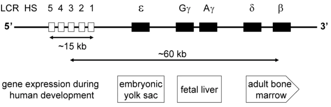
6. The human β–globin as a model to study quality control of gene expression 53
6.1 The human β-globin gene cluster and the LCR 54
6.2 The LCR/MEL expression system 57
6.3 Using the LCR/MEL expression system to study quality control of gene
expression in the nucleus 58
CHAPTER II
RESULTS
611. Inefficient processing impairs release of RNA from the site of transcription 63 2. In vivo recruitment of exon junction complex proteins to transcription sites in
…mammalian cell nuclei 77
3. Splicing- and cleavage-independent requirement of RNA polymerase II CTD
....for mRNA release from the transcription site 91 4. Abundance of the largest subunit of RNA polymerase II in the nucleus is
…regulated by nucleo-cytoplasmic shuttling 103
CHAPTER III
CONCLUDING REMARKS AND FUTURE PERSPECTIVES
117PREFÁCIO
Nesta dissertação apresentam-se os resultados do trabalho de investigação desenvolvido entre os anos de 1997 e 2007 na Faculdade de Medicina da Universidade de Lisboa, sob orientação da Professora Doutora Maria Carmo-Fonseca. O trabalho foi iniciado no Instituto de Histologia e Embriologia da Faculdade e continuado, desde 2003, na Unidade de Biologia Celular do Instituto de Medicina Molecular, Faculdade de Medicina da Universidade de Lisboa.
Este trabalho teve como principal objectivo elucidar o mecanismo de controlo de qualidade que leva à retenção nuclear dos transcritos do gene da β-globina humana com mutações que afectam o processamento do pré-mRNA.
Como previsto no Artigo 40º do Regulamento de Estudos Pós-graduados da Universidade de Lisboa (Deliberação nº 961/2003), a presente dissertação encontra-se redigida em língua inglesa, contendo um resumo alargado (mais de 1200 palavras) em língua portuguesa.
A dissertação encontra-se dividida em três capítulos: no primeiro capítulo -
Introduction, é feita uma revisão geral sobre as vária etapas da expressão génica com
principal incidência nas etapas que ocorrem ao nível do núcleo. Inicialmente é feita uma descrição de cada uma das etapas e da maquinaria nela interveniente. É dada especial relevância às inter-relações que hoje se conhecem entre as várias etapas da expressão génica e é feita uma revisão sobre o seu controlo de qualidade ao nível do núcleo. Finalmente apresenta-se o modelo utilizado e os principais objectivos deste estudo.
No segundo capítulo - Results, são apresentados os resultados originais obtidos neste estudo sob a forma de artigos publicados.
Finalmente, no terceiro capítulo - Concluding Remarks and Future Pespectives, são realçadas as principais conclusões do trabalho e expostas as perspectivas que se abrem para futuros estudos.
Como previsto no Decreto de Lei 388/70, art. 8º, parágrafo 2, parte integral dos resultados encontra-se publicada nos seguintes artigos:
Custódio, N., Carmo-Fonseca, M., Geraghtly, F., Pereira. S.H., Grosveld, F., Antoniou, M. (1999) Inefficient processing impairs release of RNA from the site of transcription. EMBO J. 18: 2855-2866.
Custódio, N., Carvalho, C. Condado, I., Antoniou, M., Blencowe, B., Carmo-Fonseca, M. (2004) In vivo recruitment of exon junction complex proteins to transcription sites in mammalian cell nuclei. RNA 10: 622-633.
Custódio N, Antoniou M, Carmo-Fonseca M. (2006) Abundance of the largest subunit of RNA polymerase II in the nucleus is regulated by nucleo-cytoplasmic shuttling. Exp Cell
Res. 312: 2557-2567.
Custódio, N., Vivo, M., Antoniou, M. and Carmo-Fonseca, M. (2007) Splicing- and cleavage-independent requirement of RNA polymerase II CTD for mRNA release from the transcription site. J. Cell Biol. 179: 199-207.
A realização deste trabalho não teria sido possível sem a colaboração de pessoas e instituições a quem desejo deixar expressos os meus agradecimentos.
Em primeiro lugar, gostaria de endereçar um agradecimento especial à Professora Doutora Maria Carmo-Fonseca, pelo privilégio que me concedeu ao admitir-me como membro da sua equipa de investigação e mais tarde como membro da equipa de docentes de Biologia Molecular da Célula. Agradeço-lhe ainda pelas excelentes condições de trabalho que me proporcionou e pelo empenho com que sempre orientou o meu trabalho e a minha formação científica.
Ao Professor Doutor J.F. David-Ferreira agradeço o facto de me ter recebido enquanto Director do Instituto de Histologia e Embriologia da Faculdade de Medicina da Universidade de Lisboa, para a realização do meu estágio de licenciatura.
Ao Doutor Michael Antoniou agradeço a sua colaboração no trabalho apresentado nesta dissertação e o facto de me ter recebido no seu laboratório no Department of
Experimental Pathology, Guy’s Hospital Campus, Londres, onde efectuei um estágio de
alguns meses em 1998 e 99. Os seus conselhos, assim como os dos seus colaboradores Selina Raguz e Desmond Chow, foram imprescindíveis à realização de uma parte do trabalho aqui apresentado.
Os trabalhos publicados apresentados nesta dissertação contaram ainda com a participação dos seguintes co-autores: Helena Sofia Pereira, Inês Condado, Célia Carvalho, Maria Vivo (Instituto de Histologia e Embriologia e Instituto de Medicina Molecular), Finola Geraghty, Michael Antoniou (King’s College London School of Medicine, London, UK), Frank Grosveld (Erasmus University, The Netherlands), Benjamin J. Blencowe (University of Toronto, Canada) a quem agradeço a valiosa contribuição.
Agradeço à Fundação para a Ciência e a Tecnologia o apoio financeiro que me concedeu no âmbito de uma Bolsa de Doutoramento (Bolsa PRAXIS XXI/BD/9439/96) durante a fase inicial do trabalho aqui apresentado.
A todos os meus colegas de trabalho do Instituto de Histologia e Embriologia e mais tarde do Instituto de Medicina Molecular, quero agradecer a amizade constante, os conselhos sempre úteis e a troca de ideias e conhecimentos que ajudou a ultrapassar muitos obstáculos ao longo dos anos. Não posso deixar de expressar aqui uma palavra especial para todos aqueles que conheci como colegas e que hoje considero também amigos: Ana Garcia, Ângelo Calado, Anita Gomes, Fátima Almeida, Hélia Neves, Isabel Alcobia, Maria Vivo, Marta Agostinho, Patrícia Calado, Peter Jordan, Sandra Caldeira, Sandra Martins, Sofia Pereira, Teresa Raquel, Zé Braga e Zé Rino. Por fim, gostaria de agradecer aos meus colegas com quem partilhei durante muitos anos a docência de Biologia Molecular da Célula e com quem muito aprendi: Célia Carvalho, Joana Desterro, João Ferreira, Margarida Gama-Carvalho e Teresa Carvalho.
Desejo também deixar expresso um agradecimento muito especial à minha família, pelo seu apoio e confiança.
RESUMO
Nos eucariontes, a transcrição dos genes pela RNA Polimerase II (RNA Pol II) origina moléculas de RNA mensageiro precursoras (pré-mRNA), sendo necessárias várias etapas de processamento até à formação do mRNA funcional que é transportado para o citoplasma, onde serve de molde para a síntese proteica. Essas etapas de processamento consistem em três processos distintos: na adição de uma trifosfoguanosina metilada na extremidade 5' trifosfato, estrutura conhecida como cap; na remoção de sequências não codificantes presentes no pré-mRNA (intrões) e junção das sequências codificantes flanqueadores (exões), num processo designado por splicing; e finalmente na formação da extremidade 3’ do mRNA que consiste na clivagem e na adição à extremidade 3’OH resultante, de múltiplos nucleótidos de adenina, num processo designado por poliadenilação. Cada uma destas etapas de processamento é levada a cabo por uma maquinaria própria. No caso da adição do cap são necessárias três enzimas: uma RNA 5’ trifosfatase, uma guanililtransferase e uma metiltransferase. O mecanismo de splicing envolve a ligação sequencial ao pré-mRNA de várias partículas ribonucleoproteicas nucleares pequenas (snRNP) que, com o auxílio de vários factores proteicos, formam uma estrutura complexa denominada spliceossoma. A maquinaria de poliadenilação é constituída por vários complexos proteicos, pela polimerase de poly(A) (PAP) e pela proteína nuclear de ligação ao poli(A) (PABPN1, anteriormente designada PABP2) (revisto por Proudfoot et al., 2002).
O correcto processamento das moléculas de pré-mRNA é um requisito fundamental para o transporte do mRNA para o citoplasma. Existem evidências de que moléculas de pré-mRNA com mutações nas regiões de consenso 5' e 3' dos seus intrões, que permitem a formação do spliceossoma mas que impedem a remoção dos intrões, são retidas no núcleo, enquanto que moléculas de pré-mRNA com mutações que não permitem a formação do spliceossoma são rapidamente exportadas para o citoplasma (Chang and Sharp, 1989; Hamm and Mattaj, 1990; Legrain and Rosbash, 1989). Do mesmo modo, mutações que interferem com a correcta poliadenilação do pré-mRNA também impedem a sua exportação para o citoplasma (Antoniou et al., 1998). Apesar destes resultados apontarem para a existência, no núcleo, de um mecanismo de controlo da qualidade do mRNA exportado, sabe-se muito pouco sobre o modo como esse controlo é efectuado. Com o objectivo de compreender o mecanismo de controlo de qualidade do mRNA exportado em células de mamífero iniciou-se
o estudo da organização intranuclear do RNA da β-globina humana. O gene da β-globina humana foi escolhido para este estudo devido ao grande número de mutações pontuais que afectam o processamento do pré-mRNA e que estão na base de uma doença genética designada por β-talassémia (Antonioiu, 1995). A expressão deste gene está limitada a células da linhagem eritróide, durante o processo de diferenciação (Tang et al., 2002). Assim, como modelo de estudo, utilizaram-se células de eritroleucemia murina (MEL, Murine ErythroLeukemia), que podem ser induzidas a diferenciar-se em cultura e expressar os genes específicos das células eritróides, onde se incluem os genes das globinas. Estas células foram transfectadas de forma estável com o gene da β-globina humana (βWT) e com mutantes do referido gene, nomeadamente com mutações que impedem o splicing do segundo intrão (βSM) e uma mutação que impede a poliadenilação (βIVSI). Quando este estudo foi iniciado sabia-se que os genes mutados eram transcritos e que o respectivo RNA não se acumulavam no citoplasma das células MEL, ao contrário do que acontecia com o mRNA da β-globina humana normal (Antoniou et al., 1998). O primeiro objectivo deste estudo foi então determinar a localização nuclear dos transcritos mutados. Para alcançar este objectivo foi optimizada a técnica de hibridação in situ para detecção dos transcritos do gene da β-globina humana nas células MEL. Esta experiência mostrou que os transcritos do gene normal eram detectados no núcleo, junto ao local de transcrição e acumulavam-se no citoplasma, enquanto que os transcritos dos genes mutados eram detectados apenas junto ao local de transcrição. Para avaliar a cinética de libertação do RNA do local de transcrição foram efectuadas experiências com o inibidor de transcrição actinomicina D. Este inibidor actua muito rapidamente após a sua adição ao meio de cultura (Darnell et al., 1971) e exerce o seu efeito através do bloqueio da elongação, uma vez que se intercala nas moléculas de DNA (Reich and Goldberg, 1964). Após 5 minutos de tratamento com actinomicina D os transcritos do gene da β-globina humana normal deixaram de ser detectados no núcleo, o que sugeriu que o RNA previamente transcrito já tinha sido libertado do local de transcrição. Por outro lado, os transcritos que têm uma mutação na região de consenso 5' do segundo intrão (βSM), que permite a formação do spliceossoma, mas inibe o splicing (Lamond et al., 1987) permaneceram junto ao local de transcrição após o tratamento com actinomicina D. Os transcritos que não possuem o segundo intrão (βIVSI) e por isso não são poliadenilados (Collis et al., 1990) ficaram também retidos no núcleo, junto ao local de transcrição, após o tratamento com actinomicina D. Estes resultados mostraram que os transcritos do gene da β-globina humana são rapidamente libertados do local de transcrição, enquanto que os
transcritos que possuem mutações que impedem o splicing e a poliadenilação ficam retidos próximo do gene, sugerindo que o mecanismo de controlo de qualidade do mRNA exportado actua ao nível do local de transcrição (Custódio et al., 1999).
O objectivo seguinte foi determinar qual o mecanismo molecular responsável pelo controlo de qualidade do mRNA ao nível do local de transcrição. Estudos efectuados na última década têm revelado a existência de inúmeras interacções entre a maquinaria de transcrição, de processamento do pré-mRNA e de exportação do mRNA para o citoplasma (ver Bentley, 2005). Uma das hipóteses colocadas para explicar a retenção dos transcritos com mutações junto ao local de transcrição foi a de que neste caso poderia não haver recrutamento de proteínas essenciais para a libertação e/ou transporte do mRNA a partir do respectivo gene. A outra hipótese colocada foi a de que a retenção poderia ocorrer por intermédio de proteínas que estão ligadas aos transcritos nascentes durante o processamento e que se libertam quando este se completa, mas que no caso dos mutantes ficam bloqueadas devido ao facto de o processamento do pré-mRNA não se completar.
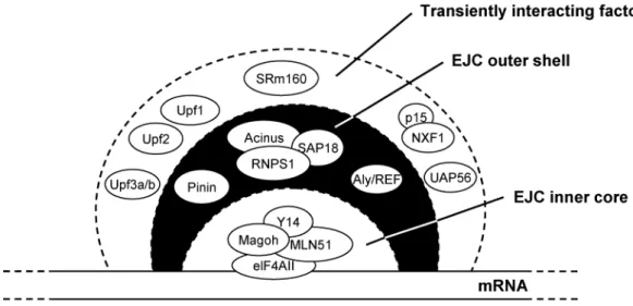
Para testar a primeira hipótese decidimos estudar o recrutamento, para o local de transcrição, de proteínas que se ligam ao mRNA. Assim testamos o recrutamento de proteínas que se ligam ao mRNA após o splicing, designadas de proteínas do complexo junção de
splicing (EJC, exon junction complex), e de factores de exportação do mRNA. Para tal,
conjugou-se a técnica de hibridação in situ para detecção dos transcritos com a localização das proteínas em questão (por imunomarcação com anticorpos específicos ou transfecção para expressão de proteína com marcador fluorescente). Assim, efectuou-se detecção de proteínas do EJC (REF/Aly, Y14, SRm160, UAP56, RNPS1 e Magoh), de componentes do spliceossoma (proteínas Sm e proteína B’’ do snRNP U2) e dos factores de exportação do mRNA (NXF1/TAP e p15) simultaneamente com a detecção dos transcritos da β-globina humana. Os resultados obtidos indicaram que as proteínas EJC e os componentes do spliceossoma são recrutados para os locais de transcrição do gene normal, no entanto, não se detectou acumulação dos factores de exportação junto ao local de transcrição. Estes resultados sugerem que as proteínas do EJC se ligam estavelmente ao pré-mRNA co-transcricionalmete, mas que os factores de exportação se ligam imediatamente antes da libertação dos transcritos do gene ou após estes se terem libertado. Quando se efectuou a mesma análise num mutante de processamento não se detectou recrutamento para o local de transcrição, nem das proteínas do EJC, nem dos componentes do spliceossoma. Estes resultados sugerem que o splicing dos transcritos da β-globina é essencial para a acumulação das proteínas do EJC no local de
transcrição e possivelmente para o posterior direccionamento para a via de exportação do mRNA para o citoplasma (Custódio et al., 2004). Embora os resultados obtidos não descartem a hipótese de que o recrutamento eficiente de proteínas do EJC e/ou de factores de exportação do mRNA possa contribuir para a libertação do mRNA do local de transcrição, são ainda necessários mais estudos para que se possa concluir se estas proteínas desempenham um papel essencial neste mecanismo.
Sabe-se que a associação entre a maquinaria de transcrição e a maquinaria de processamento do pré-mRNA é mediada pela extremidade carboxílica da subunidade grande da RNA polimerase II (RNA Pol II LS), normalmente designada de CTD (Carboxyl-Terminal Domain) (revisto por Bentley, 1999). O CTD da RNA Pol II apresenta uma constituição invulgar, sendo formado por repetições em tandem de um heptapéptido com o consenso Tirosina-Serina-Prolina-Treonina-Serina-Prolina-Serina, seguido de um motivo C-terminal constituído por 10 aminoácidos (Corden et al., 1985). O número de heptapéptidos presentes varia entre os eucariontes, apresentando os mamíferos 52 unidades repetitivas (Corden et al., 1985) e as leveduras apenas 26 (Allison et al., 1985). As primeiras evidências para uma ligação entre o CTD e o processamento do pré-mRNA vieram de estudos que apontavam para a existência de uma interacção entre determinados factores de splicing (Chabot et al., 1995; Kim et al., 1997; Mortillaro et al., 1996), de poliadenilação (McCracken et al., 1997b) e
capping (McCracken et al., 1997a) com o CTD da RNA Pol II LS. Essas evidências foram
fortalecidas quando se mostrou que uma forma da RNA Pol II LS com apenas 5 unidades repetitivas no CTD não era capaz de assegurar nem o splicing nem o processamento da extremidade 3’ do pré-mRNA (McCracken et al., 1997b). Estudos mais recentes mostraram que as unidades repetitivas 1-15 e 1-25 são capazes de assegurar o capping, mas não o
splicing ou a formação da extremidade 3’ do pré-mRNA, enquanto que as unidades 27-52 em
conjunto com os 10 aminoácidos do domínio C-terminal asseguram o capping, o splicing e o processamento da extremidade 3’ (Fong and Bentley, 2001). Foi também mostrado que a alteração do motivo C-terminal reduz a eficiência do splicing e da clivagem da extremidade 3’ do pré-mRNA (Fong et al., 2003) e impede a libertação do RNA do local de transcrição (Bird et al., 2005).
Tendo em conta estes dados, a hipótese de que a retenção junto ao local de transcrição pode ocorrer por intermédio de proteínas que se ligam aos transcritos nascentes e não se libertam devido ao bloqueio no processamento do pré-mRNA ganhou um candidato preferencial, o CTD da RNA Pol II. Assim, a retenção poderá ocorrer por intermédio da
maquinaria de processamento ineficaz ou inactiva que permanece associada ao CTD da RNA Pol II, pressupondo-se que a maquinaria de processamento se associa ao CTD durante a reacção e se desliga do CTD no final da mesma, permitindo a libertação do RNA para posterior transporte para o citoplasma. No caso dos mutantes, em que ocorre associação da maquinaria de processamento mas a reacção não se completa devido à mutação, o pré-mRNA poderá permanecer ligado ao CTD via essa mesma maquinaria, acabando por ser degradado. Para testar esta hipótese a abordagem experimental escolhida consistiu na construção de um modelo celular onde a transcrição dos genes é efectuada por uma RNA Pol II com delecção parcial do CTD. Esta abordagem é facilitada pela existência de formas da RNA Pol II LS resistentes ao inibidor de transcrição α-amanitina, o que permite anular a RNA Pol II endógena. Foram estabelecidas linhas celulares estavelmente transfectadas com o gene da RNA Pol II LS com 5 repetições no CTD (Δ5), com 31 repetições (Δ31) e com 52 repetições (controlo, wild-type). Foi efectuada uma primeira triagem a estas linhas para avaliar a sua capacidade de transcrever na presença de α-amanitina. Nenhuma das linhas Δ5 testadas apresentou capacidade de transcrever o gene da globina murina β-major na presença de α-amanitina. Este resultado foi inesperado, uma vez que este mutante já tinha sido utilizado em estudos anteriores, onde foi mostrado que apresentava capacidade de transcrever o gene da β-globina humana sob controlo do promotor SV40 (McCracken et al., 1997b). No entanto, estudos posteriores onde se analisou a capacidade transcricional da RNA Pol II Δ5 por run-on em mais de 500 genes, indicaram que a este mutante não é funcionalmente activo para a transcrição de genes endógenos (Meininghaus et al., 2000). Estes resultados impossibilitaram a utilização dos clones Δ5 nos estudos posteriores. Das linhas Δ31 e wild-type que apresentaram capacidade de transcrever na presença de α-amanitina foi seleccionada uma para transfecção com o gene da β-globina humana. Cada uma das linhas foi transfectada quer com a versão normal do gene (βWT), quer com uma versão que possui uma mutação de splicing (βSM) para a qual se mostrou anteriormente que há retenção do respectivo RNA junto ao local de transcrição. Os transcritos da β-globina humana foram detectados no núcleo das células transfectadas como um foco que corresponde ao RNA junto do local de transcrição. Quando se efectuou o tratamento com o inibidor de transcrição actinomicina D os resultados indicaram que uma grande proporção dos transcritos βWT feitos pela RNA Pol II Δ31 ficavam retidos no local de transcrição. Apesar deste resultado, observou-se recrutamento de proteínas do EJC, incluindo Aly/REF, Y14 e SRm160 para os locais de transcrição. Uma análise bioquímica dos transcritos βWT feitos pela RNA Pol II Δ31 indicou que sofriam
splicing e processamento na extremidade 3’ correctamente. Estes resultados indicam que a
RNA Pol II LS com apenas 31 unidades repetitivas é capaz de transcrever mas não permite a libertação eficiente do mRNA do local de transcrição. Isto sugere que a estrutura do CTD é importante para a libertação do mRNA do local de transcrição, possivelmente porque assegura o recrutamento de proteínas importantes para uma maturação final do mRNA após o splicing e o processamento da extremidade 3’, que o transforma numa partícula ribonucleoproteíca competente para ser exportada. Propomos assim que as unidades repetitivas em falta no mutante Δ31 da RNA Pol II poderão ser essenciais para o recrutamento destas proteínas (Custódio et al., 2007).
A RNA Pol II eucariota é uma enzima complexa, composta por 12 sub-unidades distintas, que se encontra presente na célula em níveis relativamente baixos (revisto por Shilatifard et al., 2003). A transcrição do pré-mRNA pela RNA Pol II envolve ciclos de fosforilação e desfosforilação do CTD da sua maior subunidade. O CTD encontra-se hipofosforilado quando a RNA Pol II se liga ao complexo de pré-iniciação e assume uma forma hiperfosforilada quando o transcrito tem cerca de 25 bases (O'Brien et al., 1994). A primeira fosforilação ocorre na Serina que se encontra na posição 5 do heptapéptido e durante a elongação a Serina na posição 2 é fosforilada (Komarnitsky et al., 2000; Lu et al., 1991). Pelas razões já mencionadas, estabelecemos linhas celulares murinas que expressam uma forma da RNA Pol II LS resistente ao inibidor de transcrição α-amanitina. Foi efectuada uma primeira triagem a essas linhas celulares para seleccionar aquelas que apresentavam a capacidade de sobreviver e transcrever na presença da α-amanitina, assegurando que nessas linhas a RNA Pol II LS exógena é funcional. Com o objectivo de estudar a localização sub-celular da RNA Pol II LS seleccionaram-se duas linhas com níveis diferentes de expressão da subunidade exógena. Resultados de imunomarcação e western-blotting indicaram que a RNA Pol II LS sobre-expressa se encontra predominantemente hipofosforilada e se acumula sobretudo no citoplasma. A forma activa (fosforilada na Serina 2 do heptapéptido), pelo contrário, encontra-se exclusivamente no núcleo, em níveis constantes, independentemente do grau de expressão da proteína. A presença, no citoplasma, da RNA Pol II LS sobre-expressa foi descrita num outro estudo onde se procedeu à sobre-expressão de uma forma conjugada à proteína fluorescente EGFP (Sugaya et al., 2000). Este resultado pode ser explicado por uma importação ineficiente para o núcleo do excesso de proteína produzida no citoplasma, ou por o excesso de proteína que é importado para o núcleo ser posteriormente exportado para o citoplasma. Para investigar a possibilidade da RNA Pol II LS poder
deslocar-se do núcleo para o citoplasma comparámos a distribuição sub-celular das formas endógena e exógena sobre-expressa, na presença de leptomicina B (LMB), um inibidor de exportação nuclear dependente do transportador CRM1 (Fornerod et al., 1997; Kudo et al., 1999). Os resultados indicaram que o LMB diminui a acumulação citoplasmática da proteína sobre-expressa e aumenta os níveis nucleares da proteína endógena. Estes resultados indicaram, pela primeira vez, que a RNA Pol II LS tem a capacidade de efectuar um mecanismo de vai-e-vem entre o núcleo e o citoplasma. Propomos que este mecanismo poderá ser um meio a que a célula recorre para regular o número do moléculas desta unidade presentes no núcleo, de modo a manter uma relação equilibrada das várias sub-unidades e assim assegurar a formação de complexos de transcrição funcionalmente activos (Custódio et al., 2006).
Em conclusão, o trabalho apresentado nesta dissertação foi pioneiro na identificação de um mecanismo de controlo de qualidade do mRNA, que actua ao nível do local de transcrição, no núcleo de células de mamífero (Custódio et al., 1999). Este mecanismo evita que moléculas de pré-mRNA com mutações que impedem o seu correcto processamento cheguem ao citoplasma, onde poderiam original proteínas defeituosas com consequências negativas quer para a célula quer para o organismo. O trabalho apresentado foi também o primeiro a mostrar a importância da libertação do mRNA do local de transcrição, actualmente considerada uma etapa importante da síntese do mRNA (ver Vasudevan and Peltz, 2003). Estudos efectuados em S. Cerevisiae vieram mostrar que um mecanismo de controlo de qualidade semelhante também funciona em levedura, onde transcritos com defeitos na poliadenilação ficam retidos no núcleo, ao nível do local de transcrição (Hilleren et al., 2001; Jensen et al., 2001b; Libri et al., 2002; Thomsen et al., 2003). O trabalho aqui apresentado contribuiu ainda para a compreensão do mecanismo responsável pela retenção dos transcritos com defeitos no processamento junto ao local de transcrição. Mostrámos que estes transcritos não são capazes de recrutar eficientemente proteínas do complexo junção de splicing (Custódio et al., 2004) e que o CTD da RNA Pol II desempenha um papel importante na etapa de libertação do mRNA do local de transcrição (Custódio et al., 2007).
PREFACE
This thesis presents the results of research that was carried out between 1997 and 2007 at the Faculty of Medicine, University of Lisbon, under the supervision of Professor Maria Carmo-Fonseca. The work was started at the Institute of Histology and Embryology of the Faculty and continued, since 2003, in the Cell Biology Unit of the Institute of Molecular Biology, Faculty of Medicine, University of Lisbon.
The main goal of this work was to elucidate the quality control mechanism responsible for the retention within the nucleus of human β-globin transcripts with mutations that impaired their processing.
This thesis is divided in three chapters: the first chapter - Introduction - presents a general overview of the steps of gene expression with a special focus on those that occur within the nucleus. A special focus is given to what is currently known on the coupling between the steps of gene expression and to the quality control mechanisms of gene expression that operate in the nucleus. In the end of this chapter, the model system used in these work and the general goals of the study are presented. In the second chapter - Results - the original results of this work are presented as published journal articles. Finally, the last chapter - Concluding Remarks and Future Perspectives - summarizes the most important conclusions and future perspectives that have emerged from this dissertation.
The work presented in this dissertation was possible due to the contribution of people and institutions to whom I would like to express my gratitude.
I am deeply grateful to Professor Maria Carmo-Fonseca for giving me the opportunity to be a member of her research group. I acknowledge her for having provided me excellent working conditions and for her supervision and encouragement throughout the work presented in this dissertation.
To Professor J.F. David-Ferreira I thank the opportunity to start my reseach work, still as an undergraduate student, at the Institute of Histology and Embryology, Faculty of Medicine.
I am grateful to Dr. Michael Antoniou, for his collaboration in the work presented here and for the generosity with which he received me in his laboratory in the Department of Experimental Pathology, Guy’s Hospital Campus, London.
Special thanks to all the co-authors of the work presented in this dissertation for their valuable contribution: Helena Sofia Pereira, Inês Condado, Célia Carvalho, Maria Vivo (Instituto de Histologia e Embriologia and Instituto de Medicina Molecular), Finola Geraghty, Michael Antoniou (King’s College London School of Medicine, London, UK), Frank Grosveld (Erasmus University, The Netherlands), Benjamin J. Blencowe (University of Toronto, Canada).
I wish also to acknowledge Fundação para a Ciência e a Tecnologia, for supporting me with the Fellowship PRAXIS XXI/BD/9439/96.
I would like also to express my gratitude to all my colleagues over the years for their permanent help, support and friendship. A special note of gratitude to those I initially met as colleagues that today I consider my friends: Ana Garcia, Ângelo Calado, Anita Gomes, Fátima Almeida, Hélia Neves, Isabel Alcobia, Maria Vivo, Marta Agostinho, Patrícia Calado, Peter Jordan, Sandra Caldeira, Sandra Martins, Sofia Pereira, Teresa Raquel, Zé Braga e Zé Rino. Finally, a special word to my colleagues with whom I shared for many years the experience of teaching and with whom I learned so much: Célia Carvalho, Joana Desterro, João Ferreira, Margarida Gama-Carvalho e Teresa Carvalho.
ABSTRACT
Protein-encoding genes are transcribed in the nucleus by RNA polymerase II as precursor RNAs that undergo extensive processing before being translocated to the cytoplasm for translation by the ribosomes. This spatial and temporal separation between RNA and protein synthesis offers an immense opportunity for regulation and quality control.
When this study was initiated it was known from biochemical studies that human β-globin (HBB) transcripts with mutations that impaired splicing or 3’ end formation were unable to be exported to the cytoplasm, a phenotype identical to that seen in β-thalassemia patients harbouring similar mutations (Antoniou et al., 1998). This result was consistent with the retention in the nucleus of incorrectly processed transcripts. Based on this initial observation we hypothesised the existence of a quality control mechanism to retain the incorrectly processed transcripts in the nucleus and proposed to elucidate the mechanism responsible for the observed retention. The first goal of this work was to determine the intranuclear localisation of the retained transcripts. To address this we used as a model system murine erythroleukemia (MEL) cells stably transfected with either wild-type or processing mutant HBB genes. The experimental approach was based on the direct visualisation of both normal and defective HBB transcripts using fluorescence in situ hybridisation and confocal microscopy. Nuclear transcripts of both wild-type and mutant HBB genes were detected only as intranuclear foci co-localising with the template gene locus. To determine the kinetics of transcript release from the site of transcription the cells were treated with the transcriptional inhibitors actinomycin D, α-amanitin and DRB. These drugs induced the rapid disappearance of nuclear foci corresponding to wild-type HBB RNA. In contrast, pre-mRNA mutants defective in either splicing or 3’ end formation and which fail to be transported to the cytoplasm were retained at the site of transcription. These results indicated that the quality control mechanism responsible for the nuclear retention of incorrectly processed transcripts operates at the site of transcription and suggest that splicing and 3’ end processing are rate limiting for release of mRNA from the transcription site.
The next goal of this work was to determine the molecular players involved in the quality control mechanism that operates at the transcription site. Studies over the past years have indicated that there is extensive coupling between transcription, pre-mRNA processing and nuclear export of mRNA (reviewed by Bentley, 2005). One hypothesis to explain the
retention of the mutant transcripts could be the absence of recruitment of proteins essential for the release and/or transport of the transcripts from the gene locus. Alternatively, the retention could be mediated by proteins that are bound to the nascent transcripts and become stalled due to incomplete processing.
To test the first hypothesis we decided to study the recruitment of exon junction complex (EJC) proteins and mRNA export factors to the retention sites in the nucleus. To achieve this we visualised the distribution of EJC proteins and RNA export factors relative to the sites HBB transcription in the nucleus. Using in situ hybridisation and confocal microscopy, we observed accumulation of EJC proteins (REF/Aly, Y14, SRm160, UAP56, RNPS1 and Magoh) and core spliceosome components (U snRNPs) at sites of wild-type HBB transcription. This suggests that EJC proteins bind stably to pre-mRNA co-transcriptionally. No concentration of the export factors NXF1/TAP or p15 was detected on nascent transcripts, arguing that in mammalian cells these proteins bind the mRNA shortly before or after release from the sites of transcription. Contrasting with the results obtained in MEL cells expressing wild-type HBB transcripts, mutant pre-mRNA defective in splicing and 3’ end processing do not co-localise with SRm160, REF, UAP56 or Sm proteins. These results suggest that the accumulation of EJC proteins at transcription sites requires efficient processing of the nascent pre-mRNA. Although the results obtained do not discard the hypothesis that efficient recruitment of EJC proteins and/or export factors may contribute to the release of the transcripts from the transcription site, further studies are required to conclusively show or discard the involvement of these proteins in transcription site release.
In the second hypothesis the best candidate to mediate the retention is the carboxyl-terminal domain (CTD) of the largest subunit of RNA polymerase II (RNA Pol II LS). The mammalian CTD comprises 52 heptad repeats followed by a terminal 10 amino acid motif (Corden et al., 1985). Several reports in the last decade showed that both splicing and 3’ end processing factors can associate with the CTD of RNA Pol II (reviewed by Bentley, 2005). Since the processing mutants analysed are able to assemble some processing machinery, the release of the transcripts could be blocked by the stalled or abnormal processing machinery associated with the CTD. In order to test this hypothesis we generated cell lines that express either the wild-type HBB gene or a mutant version defective in splicing and an α-amanitin resistant form of RNA Pol II LS with either 52 (wt), 31 (Δ31) or 5 (Δ5) repeats of the CTD. When these cells were treated with actinomycin D to stop transcription the results showed that a large proportion of the wild-type HBB transcripts made by RNA Pol II with only 31 CTD
repearts were retained at the site of transcription. Despite this result we observed recruitment of several EJC proteins, includind Aly/REF, Y14 and SRm160 to the transcription sites and RNase protection assays showed that the HBB transcripts produced by RNA Pol II LS Δ31 are correctly spliced and 3’ end cleaved. These results show that the form of RNA Pol II LS with only 31 repeats of the CTD is competent in transcription and processing but fails to allow the efficient release of the transcripts from the transcription site. These results provide evidence that mRNA release from the transcription site requires the heptad repeat structure of the CTD and we propose that the missing heptads in the truncated CTD mutant are required for binding of proteins implicated in a final co-transcriptional maturation of spliced and 3’ end cleaved mRNAs into export-competent ribonucleoprotein particles.
Eukaryotic RNA polymerase II is a complex enzyme composed of 12 distinct subunits that is present in cells in low abundance (reviewed by Shilatifard et al., 2003). Transcription of mRNA by RNA polymerase II involves a phosphorylation/ dephosphorylation cycle of the CTD of the enzyme’s largest subunit. The hypophosphorylated form assembles into pre-initiation complexes and the CTD becomes first phosphorylated on Serine at position 5 of the heptad repeat and during elongation on Serine at position 2 (Komarnitsky et al., 2000; Lu et al., 1991). As mentioned before we have generated stable murine cell lines expressing an α-amanitin resistant form of RNA Pol II LS. The cell lines generated were screened for survival and transcription in the presence of α-amanitin ensuring the functionality of the exogenous subunit. To characterise these cell lines we studied the localisation of the exogenous subunit and observed that over-expressed RNA Pol II LS was predominantly hypophosphorylated, soluble and accumulated in the cytoplasm in a CRM1-dependent manner. Our results further showed that the transcriptionally active form of RNA Pol II LS containing phosphoserine in position 2 of the CTD repeats was restricted to the nucleus and its levels remained remarkably constant. These results suggest that the nuclear-cytoplasmic distribution of RNA Pol II LS may be regulated by shuttling and we propose that this may provide a mechanism to control the pool of RNA polymerase subunits that is accessible for assembly of a functional enzyme in the nucleus.
KEYWORDS
Transcription;
RNA polymerase II;
CTD;
Pre-mRNA processing;
mRNA export;
ABBREVIATIONS
A - adenosine
ADAR - adenosine deaminase that act on
RNA
Br-UTP - bromouridine triphosphate C - cytidine
CBC - cap binding complex
CDAR - cytidine deaminase acting on
RNA
cDNA - complementary DNA Ceg1 - RNA guanylyltransferase Cet1 - RNA triphosphatase CF - cleavage factors
CFIA - cleavage and polyadenylation
factor IA
CFIB - cleavage and polyadenylation
factor IB
ChIP - chromatin immunoprecipitation CoTC - co-transcriptional cleavage
CPF - cleavage and polyadenylation factor CPSF -
cleavage-polyadenylation-specificity factor
CstF - cleavage stimulatory factor CTD - carboxyl-terminal domain CTE - constitutive transport element DRB -
5,6-dichloro-1-beta-D-ribofuranosylbenzimidazole
DSIF - DRB sensitivity-inducing factor dsRNA - double-stranded RNA
EJC - exon junction complex ESE - exonic splicing enhancer GFP - green fluorescent protein GMP - guanosine-5'-monophosphate GTFs - general transcription factors GTP - guanosine-5'-triphosphate
HBB - human β-globin gene
hnRNP - heterogeneous nuclear
ribonucleoprotein
HS - DNase I hypersensitive site I - inosine
IGC - interchromatin granule clusters IIa - hypophosphorylated form of CTD IIo - hyperphosphorylated form of CTD LCR - locus control region
MEL - murine erythroleukemia
MEX - mRNA export Mlp - myosin-like protein
mRNP - mensenger ribonucleoprotein
particle
MTR - mRNA transport
NELF - negative elongation factor NMD - nonsense-mediated mRNA decay NPC - nuclear pore complexes
Nups - nucleoporins
NXF - nuclear export factor proteins Pab1p - yeast poly(A) tail-binding protein PABP - poly(A) tail-binding protein PABPC - cytoplasmic poly(A)-binding
protein
PABPN1 - nuclear poly(A)-binding
protein
PAN - poly(A)-specific nuclease PAP - poly(A) polymerase PML - promyelocytic leukaemia Poly(A)+ - polyadenylated
Pre-mRNA - precursor messenger RNA P-TEFb - positivetranscription elongation factor b
Py - polypyrimidine
RAT - ribonucleic acid trafficking
REF - RNA export factor binding protein RNA Pol II - RNA polymerase II
RNA Pol II LS - largest subunit of RNA
polymerase II
RNAi - RNA interference rRNA - ribosomal RNA
RRP6 - ribosomal RNA processing 6
SFC - splicing factor compartments snoRNA - small nucleolar RNAs snRNA - small nuclear RNA
snRNP - small nuclear ribonucleoprotein
particle
SR - serine/arginine TF - transcription factor
Tpr - translocated promoter region protein TREX - transcription and export complex U - uridine
U2AF - U2 snRNP auxiliary factor βLCR - HBB micro-locus control region
CHAPTER I
INTRODUCTION
1. The cell nucleus
The nucleus is the hallmark of all eukaryotic cells. It houses the genome of the cell serving as the repository of genetic information and functioning as the control center of the cell. The nuclear envelope, a double membrane that can be regarded as a specialized extension of the endoplasmic reticulum (ER), separates the contents of the nucleus from the cytoplasm and provides the structural framework of the nucleus (see Cooper and Hausman, 2007). Many essential processes take place in the cell nucleus, including DNA replication, repair and recombination as well as the initial steps of gene expression (transcription and RNA processing). Only the final stage of gene expression (translation) takes place in the cytoplasm (Cooper and Hausman, 2007). Prokaryotes do not have a nucleus but carry out many of the processes that take place inside the nucleus in eukaryotic cells. So what functional advantages can the nucleus bring to a cell? By separating the genome from the cytoplasm, the nuclear envelope provided an opportunity for the evolution of new regulatory possibilities for gene expression that are unique to eukaryotes. For example, whereas prokaryotic mRNAs are translated while their transcription is still in progress, eukaryotic mRNAs undergo extensive posttranscriptional processing before being transported to the cytoplasm for translation (Cooper and Hausman, 2007). This spatial and temporal separation between transcription and protein synthesis offers new opportunities for regulation and opens a new window for the control of the quality of gene expression.
2. Gene expression as a multistep process
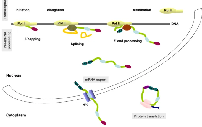
Gene expression can be defined as the process by which the genetic instructions of the DNA are transformed into functional proteins in a cell. Eukaryotic gene expression is a complex multistep process that requires several complex cellular machines. Each machine is responsible for a specific step in this process, which includes transcription, several precursor messenger RNA (pre-mRNA) processing steps (5’ capping, splicing, 3’ end processing and editing), export of the mature mRNA to the cytoplasm and translation into protein sequence (Figure 1). Identifying the protein components of each of this cellular machines and understanding how they work has been a major goal of molecular biologists for the last 25 years. For that purpose, each of the major steps outlined above has been carried out
independently in vitro. However, in the last decade numerous studies have provided evidence that they are not independent reactions in vivo. In contrast to a simple linear assembly line in which the transcript is made and passes from one processing reaction to the next, it is now clear that the processing reactions are co-transcriptional and interlinked in such a way that they influence one another’s specifically and efficiently. The emerging picture now is that a complex and extensively coupled network exists to coordinate the activities of the several gene expression machines (reviewed in Hirose and Manley, 2000; Maniatis and Reed, 2002).
The goal of this section is to introduce the major steps in gene expression as well as the major players in each one of the steps. The connections between them will be the focus of section 3.
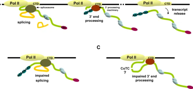
Figure 1 - The multiple steps of gene expression. The different steps in the pathway from
gene to protein include transcription (initiation, elongation and termination), several precursor messenger RNA (pre-mRNA) processing steps (5’ capping, splicing and 3’ end processing), export of the mature mRNA to the cytoplasm and translation into protein sequence. In the figure the mRNA is represented by a green line and the introns by a yellow line. The cap binding complex (CBC) added to the 5’ end of the mRNA is represented by a purple oval, the splicing machinery by a green oval and the 3’ end processing machinery by a red oval. The blue ovals on the mRNA represent the several proteins that bind to it forming a messenger ribonucleoprotein particle (mRNP). The ribosome is represented by two pink ovals and the nascent peptide by a blue line (see text for more details).
2.1 Transcription by RNA polymerase II
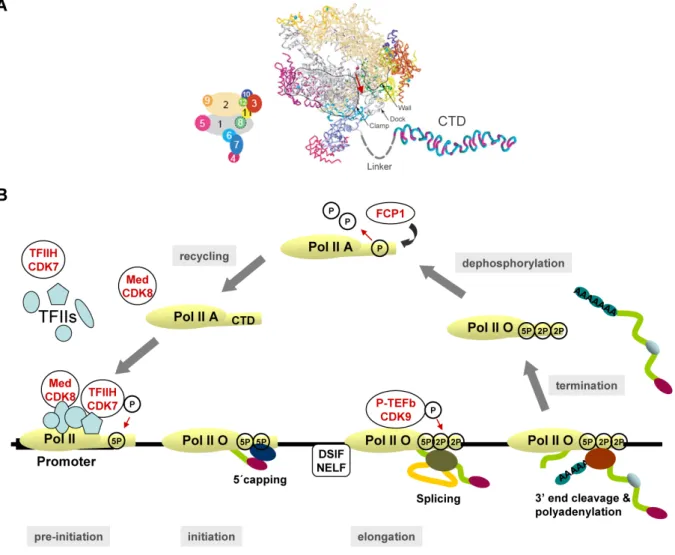
Transcription, the DNA-directed synthesis of RNA, is the first step in the sequence of events that lead to gene expression. The many thousands of genes coding for proteins in eukaryotes are transcribed by the RNA polymerase II (RNA Pol II) transcription machinery. In a simplistic view, this machinery can be divided in three major components: the 12-subunit polymerase (Figure 2A), capable of synthesizing RNA and proofreading the nascent transcript; a set of five general transcription factors (GTFs), denoted TFIIB, -D, -E, -F and -H, which are responsible for promoter recognition and for unwinding the promoter DNA; and the Mediator complex, composed of 20 subunits, which transduces regulatory information from activator and repressor proteins to the RNA polymerase and is unique to eukaryotes (reviewed in Boeger et al., 2005; Woychik and Hampsey, 2002). The RNA polymerase can be regarded as the core of the machinery because it is the platform upon which all the components are assembled. The transcription machinery is composed of a total of nearly 60 subunits and a mass of approximately 3 million Daltons (reviewed in Boeger et al., 2005).
The transcription cycle can be divided mechanistically in three basic stages: initiation, elongation and termination (Figure 2B). During each of these stages, RNA Pol II associates transiently with many different factors. Initiation involves the binding of transcription factors and RNA Pol II to DNA sites adjacent to the start site of transcription (the promoter regions) and the onset of RNA synthesis. After productive initiation, transcription proceeds into elongation mode and this is accompanied by a partial disassembly of the initiation complex and the association with elongation factors, which enable efficient production of long transcripts. In this stage the polymerase moves 5’ to 3’ along the DNA and makes an RNA copy of the gene. Finally, the termination stage involves release of the transcripts from RNA polymerase and release of the polymerase from the DNA template, allowing recycling of the polymerase and preventing it from perturbing promoters of genes located downstream from the transcription unit (reviewed by Howe, 2002; Orphanides and Reinberg, 2002).
The largest of the 12 subunits of RNA Pol II possesses a unique domain, not related to regions of any known protein, at its carboxyl terminus, termed the carboxyl-terminal domain (CTD) (Figure 2A). The CTD consists of heptapeptide repeats with the consensus Tyr1-Ser2 -Pro3-Thr4-Ser5-Pro6-Ser7 (Corden et al., 1985), which has been conserved through evolution (Allison et al., 1988; Barron-Casella and Corden, 1992). The number of repeats varies among species, ranging from 26-27 in yeast (Allison et al., 1985) to 52 in mammals (Corden et al.,
1985). The CTD is rich in potential phosphoacceptor amino acid residues and undergoes a cycle of phosphorylation and dephosphorylation during the transcription cycle (Figure 2B) (for reviews see Howe, 2002; Kobor and Greenblatt, 2002).
Figure 2 - RNA Polymerase II and the transcription cycle. (A) Model of the complete
12-subunit RNA Polymerase II (Armache et al., 2003; Bushnell and Kornberg, 2003). The view is from the top. Cyan spheres and a pink sphere depict eight zinc ions and an active site magnesium ion, respectively. The red arrow indicates the direction of RNA exit. A key to subunit colour is shown on the left with subunits numbered 1-12. The carboxyl-terminal domain (CTD) of the largest subunit of RNA polymerase II (RNA Pol II LS, subunit 1) is shown in a β-spiral model (Meinhart and Cramer, 2004). The CTD is connected to the structured core of RNA Pol II with the mobile linker of approximalely 90 amino acid residues in length. Adapted from (Cramer, 2004; Meinhart and Cramer, 2004). (B) CTD phosphorylation and the transcription cycle. After recycling the “free” RNA Pol II core enzyme is not phosphorylated on the CTD. It may assemble with coactivators such as the mediator complex (Med) thus forming a holoenzyme. Premature CTD phosphorylation by CDK8 prevents the assembly of RNA Pol II on the promotor. For pre-initiation the unphosphorylated RNA Pol II core or holoenzyme assembles onto the promoter sequences with general transcription factors (TFIIs) thus forming a pre-inititiation complex of
transcription. The CTD is phosphorylated on serines at position 5 (5P) by the CDK7 subunit of TFIIH. Initiation follows and transcription begins. The phosphorylated CTD recruits the capping enzymes (dark blue oval) and the nascent transcript (geen line) is capped at its 5’ end (purple oval). During elongation the CTD is phosphorylated on serines at position 2 (2P) by the CDK9 subunit of the positive transcription elongation factor (P-TEFb) which is required to remove the block opposed by the DSIF/NELF factors and elongate transcription. The phosphorylated CTD recruits the splicing machinery (green oval) to remove introns (yellow
line) and finally recruits the cleavage and polyadenylation factors (red oval) that cleave the
transcript and add a poly(A) tail at its 3’ end. This step signals transcription to terminate and RNA Pol II falls off its DNA template. The resulting mRNA is exported to the cytoplasm. The blue ovals on the mRNA molecules represent proteins that are bound to the mRNA forming a messenger ribonucleoprotein particle (mRNP). To be recycled for another transcription round, RNA Pol II is dephosphorylated by the FCP1 CTD phosphatase. Adapted from (Palancade and Bensaude, 2003).
RNA Pol II with a hypophosphorylated CTD is recruited to promoters (Lu et al., 1991). Shortly after transcription begins, the CTD becomes phosphorylated on Ser5. This modification signals the polymerase to clear the promoter and shift into an elongation mode. During the elongation stage of the transcription cycle, phosphorylation of Ser2 predominates (Komarnitsky et al., 2000). During or after transcription termination, the CTD is dephosphorylated by CTD phosphatases, the best studied of which is FCP1, resulting in recycling of the largest subunit of RNA Pol II (Figure 2B) (Cho et al., 1999; Kobor et al., 1999).
The high-resolution crystal structure of RNA Pol II has provided detailed insight into how a catalytically active polymerase is structured and has revealed important aspects of its function (reviewed in Boeger et al., 2005; Woychik and Hampsey, 2002). It is now known that the RNA leaves the polymerase active center cleft via the exit tunnel and then disengages from the enzyme surface. When the RNA reaches lengths if 26 and 29 nt, its 5’ end associates with RNA Pol II at the base of the dock domain (Andrecka et al., 2008). Interestingly, the CTD was shown to be placed near the exit tunnel (Cramer et al., 2001; Douziech et al., 1999) and the RNA was shown to extend toward the linker connecting to the CTD (Andrecka et al., 2008). These structural observations are consistent with the coupling of transcription with the processing events and to the role of the CTD as the major key player in this coupling, as will be discussed in detail in section 3.
2.2 Pre-mRNA processing
In eukaryotes, before a gene transcript is ready to be transported out of the nucleus, it has to undergo three major processing events to produce the fully translatable mRNA. These comprise the acquisition of a cap structure at the 5’ terminus, the splicing out of non-coding intervening sequences (introns) from the pre-mRNA, and the generation of a 3’ end, usually modified by the addition of a poly(A) tail. Some transcripts are also subject to an additional processing event termed RNA editing.
Capping
The 5’ end of the pre-mRNA is modified soon after its synthesis, when the transcript is about 25–30 nucleotides in length, by the addition of 7-methyl guanosine, usually called the cap structure (Coppola et al., 1983; Moteki and Price, 2002; Rasmussen and Lis, 1993). The addition of the cap structure is carried out by three enzymatic activities in a process known as capping (for a review see Gu and Lima, 2005). First, RNA 5’-triphosphatase hydrolyses the triphosphate of the first nucleotide to a diphosphate. Then, RNA guanylyltransferase catalyses the fusion of a GMP moiety from GTP to the diphosphate end of the pre-mRNA. To finalize the cap structure, the RNA-(guanine-7) methyltransferase adds a methyl group to the N7 position of the transferred GMP to form the m7G(5’)ppp(5’)N cap. In metazoans, both the RNA 5’-triphosphatase and the RNA guanylyltransferase activities are part of the same polypeptide at the N-terminus and C-terminus, respectively, called the capping enzyme. In
Saccharomyces cerevisiae (S. cerevisiae), the capping enzyme consists of two polypeptides,
RNA triphosphatase (Cet1) and RNA guanylyltransferase (Ceg1), which form a heterodimer (reviewed by Shuman, 2001).
The initial cap structure is recognised by the cap binding complex (CBC), which contains two proteins, CBP20 and CBP80. The cap structure bound to CBC is believed to play a major role in the stabilization of the mRNA, since it provides an obstacle for 5’ to 3’ exonucleases. Upon export through the nuclear pore complex, the nuclear cap binding proteins are replaced by the cytoplasmic translation initiation factor, eIF-4E. In the cytoplasm, cap bound to eIF-4E and other translation initiation factors enhances translation by promoting the engagement of the ribosomal subunits with the mRNA (see Mitchell and Tollervey, 2001).
Splicing
In the majority of mammalian genes the coding sequences are interrupted by non-coding intervening sequences (introns) that must be removed from the primary transcript (pre-mRNA) in order to generate a mature functional mRNA for translation (for a review see Sharp, 2005). In humans, a typical pre-mRNA contains seven or eight introns (Lander et al., 2001). The precise removal of the introns and joining of the coding sequences (exons), by a process known as splicing, is a critical step in gene expression. The splicing reaction is carried out by a large macromolecular complex termed the spliceosome. This is a highly dynamic and complex molecular machine whose composition and structure undergoes several rearrangements during each cycle of splicing. To date, two spliceosomes of unique composition have been characterised. The first one to be identified, now called U2-dependent or major spliceosome, is found in all eukaryotes and splices the most commonly encountered class of introns (the U2-type introns). It is composed of five small nuclear ribonucleoprotein particles (snRNPs; U1, U2, U4, U5 and U6), each of them containing a single uridine-rich small nuclear RNA (snRNA) that is associated to a common core of Sm proteins and other proteins specific of each particular snRNP. This spliceosome contains also a large number of non-snRNP proteins, known as splicing factors that exert auxiliary functions in the splicing reaction (reviewed by Jurica and Moore, 2003). The major class of non-snRNP proteins are the serine-arginine (SR) proteins, characterised by the presence of one or two RNA-binding domains and a carboxyl-terminal SR-rich domain (RS-domain) (Graveley, 2000). A less abundant spliceosome was discovered more recently in a subset of eukaryotes and is termed U12-dependent or minor spliceosome. This consists of the U11, U12, U5 and U4atac/U6atac snRNPs and an unknown number of non-snRNP proteins. Members of the SR protein family have been shown to function also in the U12-dependent splicing. Both the major and the minor spliceosomes coexist in eukaryotic cells and pre-mRNAs containing both types of introns serve as substrates for both splicing machineries. Although U12-type introns represent less than 1% of all introns present in human cells, they are found in genes carrying out essential cellular functions (for a review see Will and Luhrmann, 2005). It was recently reported that the two splicing pathways (minor and major) are spatially separated in the cell.
In situ hybridisation on tissue sections from adult zebrafish and in mammalian cultured cells
revealed a perinuclear and cytoplasmic staining for the U12 and U6atac, compared to the well-established nuclear distribution of the U2 snRNA of the major spliceosome. Functional analysis revealed that the cytoplasmic minor spliceosome is active and pointed to a role in cell
proliferation (Konig et al., 2007). U2-dependent splicing was also reported in the cytoplasm of platelets and dendritic cells but the full functional and physiological relevance of cytoplasmic splicing is not entirely clear yet (Denis et al., 2005; Glanzer et al., 2005).
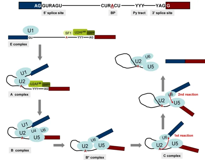
The spliceosome acts through RNA-RNA, RNA-protein and protein-protein interactions to recognise the exon-intron junctions and catalyse removal of the introns and the joining of the exons in the correct order (see Faustino and Cooper, 2003). There are four short sequences that define an intron: the exon-intron junction at the 5’ and 3’ ends of the introns (5’ and 3’ splice sites), the branch point sequence, which includes an adenosine residue and the polypyrimidine tract (Py tract) that precedes the 3’ splice site (see Ast, 2004). Different splice sites and branch point sequences are found in U2- and U12- type introns. U2- type introns of higher eukaryotes have the sequence AG/GURAGU at the 5’ splice site, CURACU at the branch point sequence and YAG/G at the 3’ splice site (where / denotes an exon-intron boundary, A is the branchpoint adenosine, R is a purine and Y a pyrimidine). The vast majority of U2-type introns have GU and AG dinucleotides at their 5’ and 3’ ends, respectively (Figure 3A) (Will and Luhrmann, 2005). By comparison, consensus sequences delineating the U12-type 5’ splice site –A(or G)UAUCCUUU– and branch point sequence – UCCUUAACU– are longer and more tightly constrained. The 3’ splice sites of U12-type introns are typically denoted by YAC/ or YAG/, but various other dinucleotides can serve as U12- type 3’ splice sites. Similarly to U2-type introns present in yeast, U12-type introns lack a polypyrimidine tract. Because the first set of U12-type introns identified contained the dinucleotides AT and AC at their 5’ and 3’ ends, respectively, they were originally referred to as ATAC introns. Further studies, however, revealed that some U2-type introns also end in AT-AC and a large number of U12-type introns also end in GT-AG (see Will and Luhrmann, 2005). The splicing consensus sequences are short and weakly conserved, thus the splice site recognition requires additional RNA sequences that can be present in both introns or exons and function as splicing enhancers or repressors modulating the ability of the spliceosome to recognise nearby splice sites (reviewed by Matlin et al., 2005). In most organisms, exons or introns are recognised as a unit in “exon definition” for short exons or “intron definition” for short introns (Robberson et al., 1990).
In chemical terms, the splicing reaction involves two trans-esterification reactions and the U2-type and U12-type introns appear to be removed by an identical mechanism. First the 2’OH group of the branch point adenosine acts as a nucleophile to attack the phosphate at the 5’ splice site, and trans-esterification results in a free 5’ exon and a lariat-shaped molecule
consisting of the intron sequences and the 3’ exon. In the second reaction, the 3’OH group of the free 5’ exon attacks the phosphate at the 3’ end of the intron. The subsequent trans-esterification results in the fusion of the two exon sequences and the release of the lariat-shaped intron (see Proudfoot et al., 2002; Will and Luhrmann, 2005).
Figure 3 - Splicing of U2-type introns. (A) Schematic representation of a U2-type intron
with its splicing consensus sequences; 5’ and 3’ splice sites, branch point (BP) and polypyrimidine (Py) tract. The intron is flanked by two exons (blue and red), the branching adenosine is underlined and highlighted in red, R is a purine and Y a pyrimidine. (B) Simplified view of the spliceosome cycle for U2-type introns. Only the snRNPs U1, U2, U4, U5 and U6 and the splicing factors SF1, U2AF65 and U2AF35 are shown. Different spliceosome conformations can be found at specific time points and purified as stable (E, A, B and C) complexes. Splicing proceeds through two trans-esterification reactions (red arrows) within the active spliceosome complex. After the second splicing reaction the mRNA is released, the post-spliceosomal complex containing the excised intron and the U2, U5 and U6 snRNPs disassembles and the snRNPs are recycled for new rounds of splicing (see text for more details) (Ast, 2004; Jurica and Moore, 2003; Will and Luhrmann, 2005).
The dissection of the splicing reaction through in vitro studies gave rise to the knowledge we have today about the spliceosome cycle for U2-type introns. In these experiments, splicing is uncoupled from other pre-mRNA processing events and the spliceosome assembly proceeds through a stepwise series of assembly events giving rise to short-lived intermediate stages, named E, A, B and C complexes (Figure 3B).
The initial events of spliceosome assembly require interaction of the U1 snRNA with the 5’ splice site. In higher eukaryotes, the 3’ splice site and adjacent Py tract are identified through interactions with U2 snRNP auxiliary factor (U2AF) composed of a large subunit of 65 kDa (U2AF65) and a small subunit of 35 kDa (U2AF35). U2AF65 is an essential splicing
factor that binds to the polypyrimidine tract and contacts also the branch point (Guth et al., 2001; Kent et al., 2003), while U2AF35 binds to the conserved AG dinucleotide at the 3’ splice site and is dispensable for splicing of some introns that contain “strong” polypyrimidine tracts (Pacheco et al., 2006; Pacheco et al., 2004). The branch point is also specifically recognised by the splicing factor 1 / mammalian branch point binding protein (SF1/mBBP), in a cooperative binding with U2AF65 (Selenko et al., 2003). These ATP independent events lead to the formation of an E (early) complex that commits the pre-mRNA to the splicing pathway. These interactions help the recruitment of the U2 snRNP to the branch point in an ATP dependent manner (Sanford and Caceres, 2004). U2 snRNA base pairs with the branch point sequences generating the pre-spliceosome or A complex. Several proteins of the SR family are thought to mediate interactions between adjacent 5’ and 3’ splice sites, both across the intron and over the exon helping to determine the correct splice site selection and to stabilize the A complex (Hertel and Graveley, 2005; Shen and Green, 2004). This complex recruits the pre-formed U4/U6.U5 tri-snRNP to form the mature spliceosome or B complex. The U4 snRNA does not directly interact with the pre-mRNA but plays an essential role in bringing U5 and U6 into the spliceosome. At this step, the U1 base pairing interaction with the 5’ splice site is replaced by a similar interaction involving the U6 snRNA, while U5 binds to sequences in the 3’ exon, thus bringing the two exons closer together. Extensive conformational changes promote the dissociation of U1 and U4 from the complex and the spliceosome is activated (B* complex) and capable of catalyzing the first splicing reaction whereby the branch point adenosine is connected to the 5’ end of the intron, which is cleaved from the upstream exon. The intron is now in a lariat configuration and the C complex is formed. More conformational changes are required for the second trans-esterification reaction to occur, in which the 3’ end of the upstream exon is joined with the 5’
end of the downstream exon, cleaving the intron from the 3’ end splice site. After this final step, the spliced mRNA is released from the post-spliceosomal complex formed by the lariat intron and the snRNPs, which must disassemble and recycle for another round of splicing (Figure 3B) (reviewed by Hastings and Krainer, 2001; Jurica and Moore, 2003). Assembly of the U12-dependent spliceosome is similar to that of the U2-dependent, with the U11, U12 and U4atac/U6atac being functionally similar to the U1, U2 and U4/U6. The major difference occurs at the earliest assembly step in which the U11 and U12 snRNPs form a highly stable di-snRNP that binds cooperatively to the 5’ splice site and branch point, which is equivalent to the A complex of the U2-dependent spliceosome (reviewed by Will and Luhrmann, 2005).
While many introns are constitutively excised during splicing, other introns and exons can either be included or excluded from the mature mRNA, in different combinations in a process called alternative pre-mRNA splicing (for reviews see Blencowe, 2006; Matlin et al., 2005). By this process a single pre-mRNA yields different mRNAs leading to the production of different proteins creating proteome diversity. It has been estimated that more than 50% of all human genes are, at some stage, expressed by alternative splicing (see Modrek and Lee, 2002).
3’ end processing
The 3’ end processing of the transcripts consists in a two-step reaction involving site-specific endonucleolitic cleavage of the pre-mRNA and synthesis, at the newly generated 3’ product, of a poly(A) tail of 70–90 adenosine residues in yeast (S. cerevisiae) and 200–250 adenosine residues in higher eukaryotes. The two steps of the reaction (cleavage and polyadenylation) are considered to be tightly coupled, as cleaved 3’ ends that were not polyadenylated have not been observed in vivo. With the exception of replication-dependent histone transcripts (in higher eukaryotes), that possess a stem-loop structure at their 3’ end, all protein encoding mRNAs contain a poly(A) tail (see Proudfoot et al., 2002; Zhao et al., 1999). The poly(A) tail is an important determinant of mRNA metabolism and function, largely through the action of cellular poly(A) tail-binding proteins (PABPs) (for a review see Mangus et al., 2003). PABPs have a minimal binding site of 11–12 adenosines and, as a consequence, a single poly(A) tail is bound by multiple PABP molecules (Mangus et al., 2003). While higher eukaryotes have multiple PABPs, including several cytoplasmic forms (PABPC) and one nuclear (PABPN1), yeast cells have a predominantly cytoplasmic poly(A)-binding protein (Pab1p) although cell fractionation indicates that some Pab1p is also nuclear (Zhao et