UNIVERSIDADE TÉCNICA DE LISBOA
Faculdade de Medicina Veterinária
THE SYLVATIC AND SYNANTHROPIC CYCLES OF ECHINOCOCCUS SPP., TAENIA SPP. AND TOXOCARA SPP. IN PORTUGAL: COPROLOGIC AND MOLECULAR DIAGNOSIS IN
CANIDS
DIOGO RIBEIRO ALMEIDA GUERRA
CONSTITUIÇÃO DO JÚRI ORIENTADOR
Doutor Virgílio da Silva Almeida Doutor Peter Deplazes Doutor Luís Manuel Madeira de Carvalho
Doutor José Augusto Farraia e Silva Meireles CO-ORIENTADOR
Doutor Peter Deplazes Doutor Luís Manuel Madeira de Carvalho
2012 LISBOA
UNIVERSIDADE TÉCNICA DE LISBOA
Faculdade de Medicina Veterinária
THE SYLVATIC AND SYNANTHROPIC CYCLES OF ECHINOCOCCUS SPP., TAENIA SPP. AND TOXOCARA SPP. IN PORTUGAL: COPROLOGIC AND MOLECULAR DIAGNOSIS IN
CANIDS
DIOGO RIBEIRO ALMEIDA GUERRA
DISSERTAÇÃO DE MESTRADO EM MEDICINA VETERINÁRIA
CONSTITUIÇÃO DO JÚRI ORIENTADOR
Doutor Virgílio da Silva Almeida Doutor Peter Deplazes Doutor Luís Manuel Madeira de Carvalho
Doutor José Augusto Farraia e Silva Meireles CO-ORIENTADOR
Doutor Peter Deplazes Doutor Luís Manuel Madeira de Carvalho
2012 LISBOA
i
iii
Acknowledgements
Firstly, to Prof. Dr. Peter Deplazes, my supervisor, for accepting me at the Institute of Parasitology, University of Zurich (IPZ-UZH) and allowing me to use all the infrastructures to develop my project. I am truly grateful for all the guidance, all the knowledge transmitted and for setting such a good example of professionalism and leadership. It was an honor.
Secondly, to Prof. Dr. Luís Manuel Madeira de Carvalho, my co-supervisor, and, more than that, a genuine friend. I thank you for believing in me and for creating the enthusiasm I now have for Parasitology and Wildlife Medicine. Thank you for all the support, guidance and endless good mood. Again, it was an honor.
To Dr. Teresa Armua, postdoc at the IPZ-UZH, with whom I learnt so much and whose help was more than essential to develop the present work. I thank you for all support and friendship.
To Prof. Dr. Alexander Mathis, Dr. Felix Grimm and Dr. Daniel Hegglin from the IPZ-UZH, for the availability and all the questions answered. Also, to Jeannine Hauri, Katja Huggel and Dr. Iskender Ziadinov for teaching me all the techniques.
To the remaining staff of the IPZ-UZH for welcoming me so well and confirming that Switzerland is indeed an amazing country to work, and, more thant that, to live.
To António Valverde, Dr. Marta Silva, Dr. Inês Bravo, Dr. Miguel Minas and Dr. Nuno Santos, for collecting the samples and letting me use them. Without their work nothing of this would have been possible.
To Dr. Lídia Nabais and all the students at the Parasitic Diseases Routine Laboratory, Faculty of Veterinary Medicine, Technical University of Lisbon, for all the help and support during my training period.
To all of you in Lisbon, you know who you are, for accompanying me during all these years. Thank you for all the laughs and amazing support. You made everyting so much easier.
Again to my parents, to whom this work I dedicate, for believing in me and allowing me to follow my will.
Finally, to my grandparents, with whom I learnt what was like to be on a farm, surrounded by animals.
v
Abstract
THE SYLVATIC AND SYNANTHROPIC CYCLES OF ECHINOCOCCUS SPP., TAENIA SPP. AND TOXOCARA SPP. IN PORTUGAL: COPROLOGIC AND MOLECULAR DIAGNOSIS IN
CANIDS
Echinococcus spp., Taenia spp. and Toxocara spp. are important parasites of domestic and wild
canids and neglected zoonotic helminths. Despite their relevance in Public Health, little is known about their prevalence in Portugal. An epidemiological study was conducted to clarify the role of canids in the sylvatic and synanthropic cycles of these pathogens in our country.
Fecal samples from dog (n = 51), red fox (n = 62) and Iberian wolf (n = 68) were collected from two regions. Toxocara spp. and taeniid eggs were isolated through a Sieving-flotation technique. Species identification in taeniids was made using Multiplex-PCR followed by sequencing of amplified material; in Toxocara spp. by measuring the eggs.
Taenia hydatigena, T. serialis, T. pisiformis, T. polyacantha, Echinococcus canadensis (G7) and Toxocara canis were detected in wolves. This was the first time taeniid species were studied in
Portuguese Iberian wolf populations, with the first records of T. polyacantha and E. canadensis in Iberian wolves. T. hydatigena and Toxocara canis were found in both dogs and foxes and
T. polyacantha in foxes.
Dogs were considered the most important link between domestic and synanthropic cycles but wolves and foxes can be regarded as the most relevant hosts in maintaining the sylvatic and synanthropic cycles of taeniids and Toxocara spp., respectively. Control programs should consider these species as part of their measures, and dogs, since they are more easily reached, should be dewormed more frequently.
vii
Resumo
OS CICLOS SILVÁTICOS E SINANTRÓPICOS DE ECHINOCOCCUS SPP., TAENIA SPP. E
TOXOCARA SPP. EM PORTUGAL: DIAGNÓSTICO COPROLÓGICO E MOLECULAR EM
CANÍDEOS
Echinococcus spp., Taenia spp. e Toxocara spp. são parasitas importantes de canídeos
domésticos e silvestres e agentes de zoonoses negligenciadas. Apesar da sua relevância em Saúde Pública, pouca informação existe acerca da prevalência em Portugal. Foi realizado um estudo epidemiológico para compreender o papel que espécies de canídeos poderiam desempenhar nos ciclos silvático e sinantrópico destes parasitas no nosso país.
Foram recolhidas amostras fecais de cão (n =51), raposa (n = 62) e lobo ibérico (n = 68) de duas regiões. Os ovos de Toxocara spp. e de tenídeos foram isolados por uma técnica de Filtração-Flutuação. As espécies de tenídeos foram identificadas por Multiplex-PCR e sequenciação do material amplificado; para Toxocara spp. foram medidos os ovos.
Em lobos foram detectadas as espécies Taenia hydatigena, T. serialis, T. pisiformis,
T. polyacantha, Echinococcus canadensis (G7) e Toxocara canis, sendo este o primeiro estudo
das espécies de tenídeos na população portuguesa de lobo ibérico. T. polyacantha e E.
canadensis (G7) foram detectados pela primeira vez em lobo ibérico. T. hydatigena e Toxocara canis foram encontrados em raposas e cães e T. polyacantha apenas nas raposas.
Considerou-se que os cães serão o principal elo de ligação entre os ciclos doméstico e sinantrópico, enquanto os lobos e as raposas o serão para os ciclos silvático e sinantrópico de tenideos e Toxocara spp., respectivamente. As medidas dos programas de controlo deverão, por isso, focar-se também nestas espécies, e os cães, por serem mais facilmente manipulados, devem ser desparasitados mais frequentemente.
ix
Table of Contents
1. Introduction ... 1
1.1. Training Period Activities ... 1
2. Literature Review ... 3
2.1. Echinococcus spp. ... 3
2.1.1. Taxonomy ... 3
2.1.2. Morphology ... 5
2.1.3. Life Cycle ... 6
2.1.4. Pathology and Immunity ... 7
2.1.5. Diagnosis ... 9
2.1.5.1. Diagnosis in Definitive hosts ... 9
2.1.5.2. Diagnosis in Intermediate hosts ... 11
2.1.6. Epidemiology ... 12 2.1.6.1. Definitive Hosts ... 12 2.1.6.2. Intermediate Hosts ... 13 2.1.6.3. Genetic Characterization ... 14 2.1.6.4. Environmental Contamination ... 15 2.1.6.5. Hydatidosis in Humans ... 15
2.1.7. Treatment and Control ... 17
2.2. Taenia spp. ... 20
2.2.1. Taxonomy ... 20
2.2.2. Morphology ... 22
2.2.3. Life Cycle ... 24
2.2.4. Pathology and Immunity ... 24
2.2.5. Diagnosis ... 26
2.2.6. Epidemiology ... 26
2.2.7. Treatment and Control ... 30
2.3. Toxocara spp. ... 31
2.3.1. Taxonomy ... 31
2.3.2. Morphology ... 31
2.3.3. Life Cycle ... 33
2.3.4. Immunity and Pathology ... 34
2.3.5. Diagnosis ... 36
2.3.6. Epidemiology ... 37
2.3.7. Treatment and Control ... 42
3. Main Objectives of the Study ... 43
x
4.1. Sample Population and Study Areas ... 44
4.2. Decision Tree for the samples analysis ... 46
4.3. Note concerning safety procedures ... 47
4.4. Taeniid and Toxocara spp. eggs isolation through sieving and flotation procedure ... 47
4.5. Measurement of Toxocara spp. eggs ... 48
4.6. Multiplex-PCR for identification of taeniid eggs in feces from carnivores ... 49
4.7. Taenia spp. and Echinococcus granulosus amplicons sequencing ... 50
4.8. Statistical Analysis ... 51
5. Results ... 52
5.1. Taenia spp. and Echinococcus spp. ... 52
5.2. Toxocara spp. ... 55
5.3 Other parasites ... 59
6. Discussion ... 60
6.1. Taenia spp. and Echinococcus spp. ... 60
6.2. Toxocara spp. ... 64
6.3. Other Parasites ... 66
7. Conclusions and Further Studies ... 68
8. References ... 70
Annex ... 92
xi
List of Figures
Figure 1 – Morphology of different stages of Echinococcus granulosus. (Original illustrations) ... 6
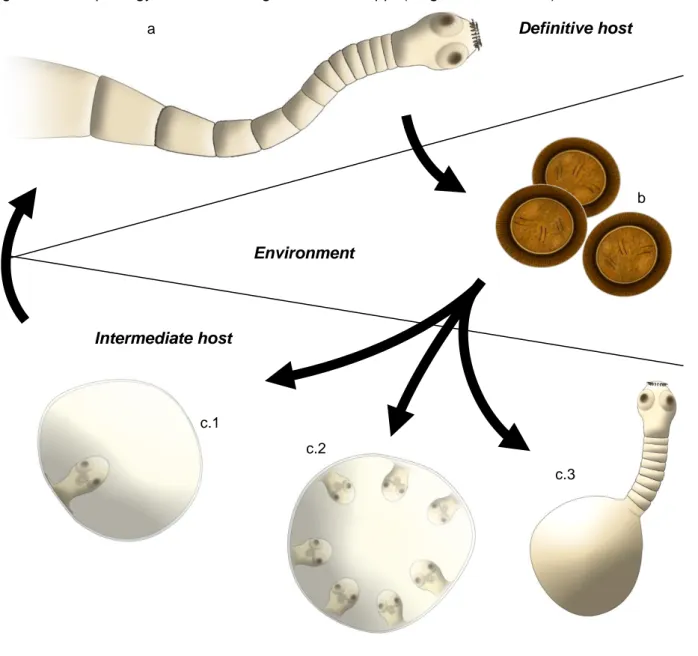
Figure 2 – Morphology of different stages of Taenia spp. (Original illustrations) ... 23
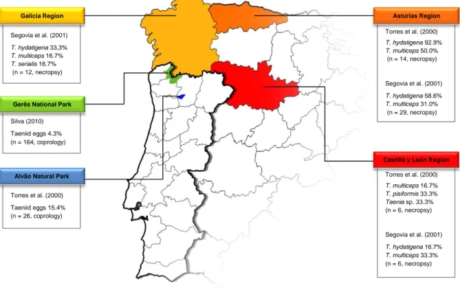
Figure 3 – Taenia species infecting Iberian wolf in Northwestern Iberian Peninsula (Original) ... 27
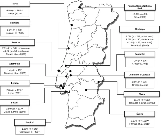
Figure 4 – Prevalence of Taeniid eggs in dog’s fecal samples collected from public places. (Original map) ... 29
Figure 5 – Morphology of different stages of T. canis and T. cati. (Original illustrations) ... 32
Figure 6 – Prevalence of Toxocara sp. eggs in dog’s fecal samples collected from public places. (Original map) ... 37
Figure 7 – Areas of study and geographical distribution of fecal samples. (Original map) ... 44
Figure 8 – Decision Tree for samples analysis ... 47
Figure 9 – A 21-μm sieve for egg isolation. ... 48
Figure 10 – Taeniid eggs in a wolf fecal sample positive for T. hydatigena. (Original pictures)... 52
Figure 11 – Example of Multiplex-PCR results of two samples (Original picture)... 52
Figure 12 – Approximate geographic location of samples positive for taeniid species. ... 53
Figure 13 – (a) unembryonated (b) embryonated and (c) larvated Toxocara spp. eggs recovered from fecal samples (Original pictures). ... 56
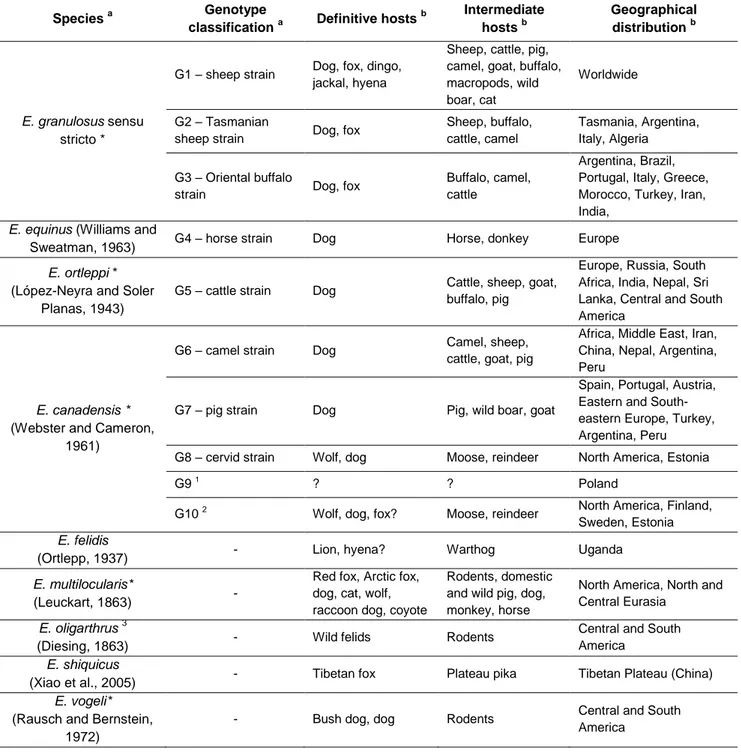
List of Tables Table 1 – Taxonomy of genus Echinococcus and main characteristics of the different species. ... 4
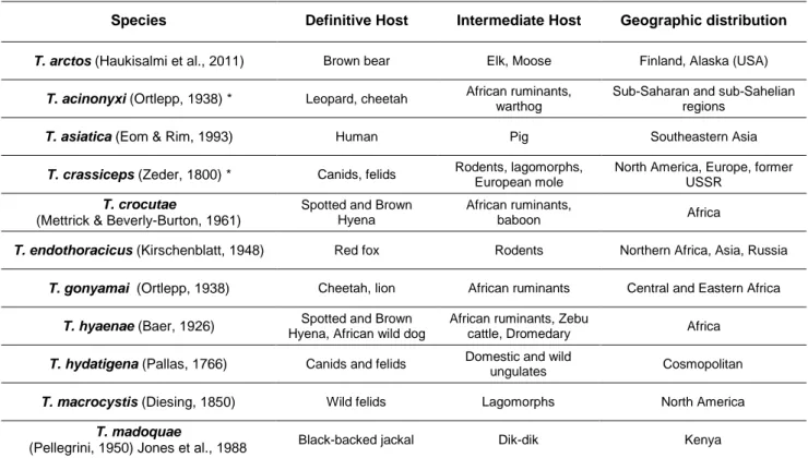
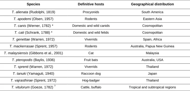
Table 2 – Species within the genus Taenia. ... 20
Table 3 – Main features of metacestodes from Taenia spp. ... 23
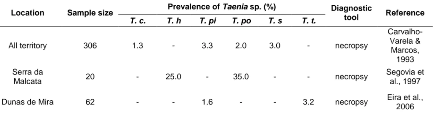
Table 4 – Prevalence of different Taenia species in Red fox populations from Portugal. ... 28
Table 5 – Some species within the genus Toxocara. ... 31
Table 6 – Prevalences of Toxcara egg and T. canis in wild hosts from Portugal and Spain. ... 38
Table 7 – Prevalences of T. cati in cats from Portugal and Spain. ... 39
Table 8 – Overall number of species collected by host species and area of study. ... 44
Table 9 – Distribution of samples, according to each wolf pack territory in the Northern area of study. ... 45
Table 10 – Distribution of samples, according to each hunting association area in the Municipality of Elvas. ... 46
Table 11 – Primers targets and sequences amplified for Multiplex-PCR (adapted from Trachsel et al.; 2007). ... 50
Table 12 – Positive fecal samples for Taeniid eggs and results of Multiplex PCR and Sequencing. ... 53
Table 13 – Prevalence of Taeniid species per subpopulation of Iberian wolf. ... 54
Table 14 – Epg of Taeniid samples per host species. ... 54
Table 15 – Frequency Table comparing two techniques to isolate taeniid eggs from fecal samples of the Northern Area of study. ... 55
Table 16 – Positive fecal samples for Toxocara sp. eggs per area of study. ... 55
Table 17 – EPG, egg classification and maximum and minimum length of Toxocara spp. eggs. ... 56
Table 18 – EPG and comparison of positive results of Sieving-flotationg technique with the ones obtained previously. ... 57
Table 19 – Frequency Table comparing two techniques to isolate Toxocara spp. eggs from fecal samples. ... 58
xii
List of Abbreviations and Symbols
bp – base pairs bw – body weight
DNA – Deoxyribonucleic Acid e.g. – exempli gratia
ELISA – Enzyme-linked Immunosorbent Assay EPG – Eggs per gram
g – gram i.e. – id est kg – kilogram mg – milligram ml – milliliter N – normal n – sample size No. – number
OIE – World Organization for Animal Heath PCR – Polymerase Chain Reaction
pi – post infection
WHO – World Health Organization x g – gravity
1
1. Introduction
In September 2005, a meeting of the World Health Organization (WHO) in Geneva, Switzerland, developed the concept of Neglected Zoonotic Diseases to address the increasing concern towards a group of pathogens with specific characteristics. All these were ancient diseases, largely known by the human populations, with severe clinical signs and deep economic impact. Most importantly, they presented an endemicity that was strongly supported by the interaction between humans, domestic animals and wildlife. Underdiagnosis led to underestimation of their real importance which resulted in few control measures being taken nowadays (World Health Organization [WHO], 2010).
Since its creation, the list of neglected zoonotic diseases increased every year. Two diseases included in this list are Cystic Echinococcosis/Hydatidosis and Cysticercosis/Taeniasis, both present in Portugal. Although not a part of this list, Toxocarosis still fulfills the majority of the criteria and some recent works consider it an important neglected zoonosis in some countries (Hotez & Wilkins, 2009).
Base-line epidemiological studies are needed for these diseases, about which little is known, especially regarding their wildlife reservoirs.
Five years of Veterinary studies in Lisbon and the great interest developed by the author in the fields of Parasitology, Wildlife and Public Health led to the choice of the research project described.
In the first part of this document will be given a brief description of the training period undertaken by the author, followed by a thorough literature review of the main topics, i.e. Echinococcosis, Taeniosis and Toxocarosis. The second part addresses the project developed, including its main goals, materials and methods, results, discussion and conclusion of all the research performed.
1.1. Training Period Activities
In the last year of his Integrated Masters in Veterinary Medicine, the author took a training period of approximately six months in two different locations - at the Parasitic Diseases Routine Laboratory, Faculty of Veterinary Medicine, Technical University of Lisbon, Portugal (FVM-TUL) from September 5th to September 28th 2011; and at the Institute of Parasitology, Vetsuisse-Faculty, University of Zurich, Switzerland (IPZ-UZH), the latter under the LLP/Erasmus Program, between October 3rd 2011 and February 29th 2012.
The training period was supervised by Prof. Doctor Peter Deplazes (IPZ-UZH) and co-supervised by Prof. Doctor Luís Manuel Madeira de Carvalho (FVM-TUL).
During the first part of the training period, held in Lisbon, the author followed the routine activities of the Laboratory, gathering knowledge and practice in sample processing and diagnosis, mainly in Hemoparasites and Gastrointestinal helminths of carnivores. In January 2012, the student also spent two weeks at the FVM – TUL analyzing fecal samples from stray dogs that were eventually used in this study.
2
In the Institute of Parasitology, Zurich, the student developed the majority of the work presented in this document. In the first month, the student was introduced to the general techniques and projects done in the Institute. It was possible to follow and also to practice techniques of egg isolation in feces and from adult worms, Single and Multiplex PCR for taeniids, ELISA to detect coproantigens of Echinococcus multilocularis in fecal samples. During the remaining period, the student started working on his project, supervised by Prof. Doctor Peter Deplazes and Doctor Teresa Armua.
During the training period in Zurich it was also possible for the student to follow several steps of other ongoing projects about Echinococcus multilocularis, Fasciola hepatica, Toxoplasma gondii and Angyostrongylus vasorum. He could learn and help at fox and rodents necropsies, snail infection and molecular techniques such as Western-blot.
Each Tuesday there were internal seminars hosted by a member of the Institute or a guest researcher. The student was able to attend to the following sessions:
11.10.11. Christoph Lippuner (IPZ-UZH): “Transcriptomics of intestinal stages of
Cryptosporidium”;
18.10.11. Stefanie Wagner (IPZ-UZH): “Mosquitoes in Central Europe: ecological and physiological investigations”;
25.10.11. Michael Grigg (Laboratory of Parasitic Diseases, National Institute of Health, Bethesda, USA): “Biology of Toxoplasma Pathogenesis: Strain Type and Polyparasitism as Predictors of Disease”;
08.11.11. Catherine Eichwald (Institute of Virology, University of Zürich): “Bacillus subtilis spores, a safe alternative for enteric antigen delivery”;
15.11.11. Relja Beck (Head of Laboratory for Parasitology, Department for Bacteriology and Parasitology, Croatian Veterinary Institute, Zagreb, Croatia): “Vector borne diseases in Croatia”;
22.11.11. Hubertus Hertzberg (IPZ-UZH): “Vakzination gegen Haemonchus contortus”;
29.11.11. Nick Smith (Queensland Tropical Health Alliance, James Cook University, Cairns Australia): “The Molecular Biology of Oocyst Wall Formation in Coccidian Parasites”; 06.12.11. Petra Wampfler (IPZ-UZH): “Proteomics of secretory organelles in Giardia”;
12.12.11. Matthias Rottmann (Swiss Tropical and Public Health Institute, Basel, Switzerland) “Malaria Drug Discovery projects at the Swiss TPH”;
3
2. Literature Review
2.1. Echinococcus spp.
Echinococcus spp. are hermaphrodite heteroxenous tapeworms, whose life cycle develops in
different mammal species. The adult parasite infects the small intestine of the definitive host, whereas the larval stage occurs in different tissues of the intermediate host.
2.1.1. Taxonomy
Echinococcus genus belongs to the Phylum Platyhelmynthes, Class Cestoda, Subclass
Eucestoda, Order Cyclophyllidea, Family Taeniidae (Kassai, 1999; Roberts, Janovy & Schmidt, 2009a).
The taxonomy within the genus Echinococcus is subject of a great debate. After some initial
classifications, taxonomic revision recognized four species – E. granulosus, E. multilocularis,
E. vogeli and E. oligarthrus (Thompson & McManus, 2001; Thompson, 2008). However, some
authors were concerned with the oversimplified approach to E. granulosus as one single species, since there were phenotypic variations between isolates from distinct regions (Thompson & McManus, 2002). New classifications have been proposed with the outcome of molecular techniques. Genetic analysis, especially of mitochondrial DNA, confirmed a great variety within
E. granulosus, which reflected not only different phenotypic characteristics, but also distinct
transmission patterns, host specificity, pathogenicity and even sensibility to chemical control. Therefore, E. granulosus was divided in several strains/genotypes, named after the main intermediate host. Nowadays, according to some authors, further genetic analysis revealed sufficient variability to enable the creation of distinct species instead of strains (Thompson, 2008; Tappe, Kern, Frosch & Kern, 2010). The proposed taxonomy, according to the latest data, is described in Table 1.
Further attention should be paid to the ongoing debate regarding the creation of an additional species, E. intermedius, which would include genotypes G6 and G7 (Thompson, 2008; Nakao et al., 2010).
Recently, two species have been discovered. E. shiquicus was described in the Tibetan fox and the Plateau pika from Tibet, China (Xiao et al., 2005). E. felidis was isolated from African lions in Uganda by Huttner et al. (2008) and this was actually a rediscovery since this species had already been described in 1937 by Ortlepp, but was lately included into E. granulosus sensu lato.
E. granulosus sensu lato is the only species documented in Portugal (Eckert & Deplazes, 2004;
Beato, 2008). Therefore, from hereafter this cluster of species will be discussed with greater detail and will be mentioned simply as E. granulosus. When referring to genotypes G1, G2 and G3,
E. granulosus sensu stricto will be used. Additional information concerning the other species shall
4
Table 1 – Taxonomy of genus Echinococcus and main characteristics of the different species.
Species a Genotype
classification a Definitive hosts
b Intermediate hosts b Geographical distribution b E. granulosus sensu stricto *
G1 – sheep strain Dog, fox, dingo, jackal, hyena
Sheep, cattle, pig, camel, goat, buffalo, macropods, wild boar, cat
Worldwide
G2 – Tasmanian
sheep strain Dog, fox
Sheep, buffalo, cattle, camel
Tasmania, Argentina, Italy, Algeria
G3 – Oriental buffalo
strain Dog, fox
Buffalo, camel, cattle
Argentina, Brazil, Portugal, Italy, Greece, Morocco, Turkey, Iran, India,
E. equinus (Williams and
Sweatman, 1963) G4 – horse strain Dog Horse, donkey Europe
E. ortleppi *
(López-Neyra and Soler Planas, 1943)
G5 – cattle strain Dog Cattle, sheep, goat, buffalo, pig
Europe, Russia, South Africa, India, Nepal, Sri Lanka, Central and South America
E. canadensis *
(Webster and Cameron, 1961)
G6 – camel strain Dog Camel, sheep, cattle, goat, pig
Africa, Middle East, Iran, China, Nepal, Argentina, Peru
G7 – pig strain Dog Pig, wild boar, goat
Spain, Portugal, Austria, Eastern and South-eastern Europe, Turkey, Argentina, Peru G8 – cervid strain Wolf, dog Moose, reindeer North America, Estonia G9 1
? ? Poland
G10 2 Wolf, dog, fox? Moose, reindeer North America, Finland, Sweden, Estonia E. felidis
(Ortlepp, 1937) - Lion, hyena? Warthog Uganda
E. multilocularis*
(Leuckart, 1863) -
Red fox, Arctic fox, dog, cat, wolf, raccoon dog, coyote
Rodents, domestic and wild pig, dog, monkey, horse
North America, North and Central Eurasia
E. oligarthrus 3
(Diesing, 1863) - Wild felids Rodents
Central and South America
E. shiquicus
(Xiao et al., 2005) - Tibetan fox Plateau pika Tibetan Plateau (China)
E. vogeli*
(Rausch and Bernstein, 1972)
- Bush dog, dog Rodents Central and South
America
* Humans as intermediate hosts
1
Genotype only found in humans (Scott, Stafaniak, Pawlowski & McManus, 1997)
2
Zoonotic potential still unclear (Romig, Dinkel & Mackenstedt, 2006)
3 Only three confirmed cases in humans (D’Alessandro & Rausch, 2008) a
adapted from Thompson (2009), Nakao et al. (2010), Tappe et al. (2010) and Knapp et al. (2011).
b
data collected from Scott et al. (1997); Lavikainen, Lehtinen, Meri, Hirvelä-Koski & Meri (2003); McManus & Thompson (2003); Dinkel et al. (2004); Eckert & Deplazes (2004); Mwambete, Ponce-Gordo & Cuesta-Bandera (2004); Xiao et al. (2005); Lavikainen et al. (2006); Romig et al. (2006); Beato (2008); D’Alessandro & Rausch (2008); Huttner et al. (2008); Hüttner, Siefert, Mackenstedt & Romig (2009); Omer et al. (2010); Kamal, Romig, Kern & Rihab (2011); Konyaev et al. (2011); Rojo-Vazquez et al. (2011); Sharifiyazdi, Oryan, Ahmadnia & Valinezhad (2011).
5
2.1.2. Morphology
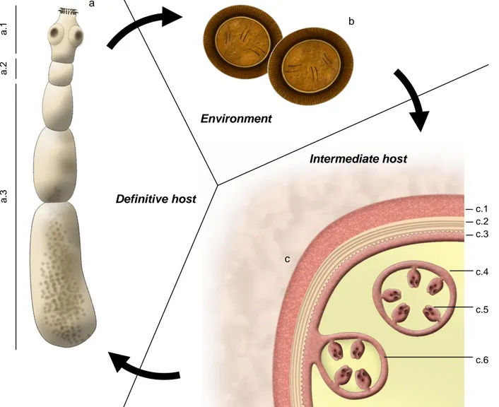
The adult stage of Echinococcus granulosus sensu lato is a small tapeworm whose length ranges from 2 to 7 mm. Body is composed by a scolex in the anterior end, followed by a neck and the strobila (see Figure 1-a). The scolex contains two rows of hooks (rostellum) and four muscular suckers (acetabula) that help parasite fixation to the intestine wall. The strobilaconsists of three to six proglottids. Proglottids mature progressively towards the posterior end and the last one is usually a gravid proglottid carrying eggs in uterus (Thompson & McManus, 2001; Eckert & Deplazes, 2004).
Echinococcus species lack digestive and respiratory systems, so the metabolic exchanges take
place through the tegument. This structure covers the body of the parasite and also has a protective role against host enzymes and immune responses (McManus, 2009).
Echinococcus sp. eggs are morphologically indistinguishable from other taeniids eggs. They are
ovoid, with approximately 30-36 μm in diameter, including inside the first larval stage (oncosphere or hexacant embryo harboring three pairs of hooks) (Roberts et al., 2009a). A layered wall surrounds the egg (see Figure 1-b). It consists of a thin outer capsule that is rapidly lost when eggs are released in the environment, a thick embryophore with a striated appearance and finally the oncosphere membrane which cannot be detected by light microscopy (Thienpont, Rochette & Vanparijs, 1986; Roberts et al., 2009a).
The eggs eventually develop into the next stage, the metacestode or hydatid cyst (see Figure 1-c). It is a fluid-filled bladder surrounded by an inner cellular layer (germinal layer) from where brood capsules arise through inward budding. In the inner surface of these brood capsules, the protoscoleces develop by asexual reproduction reaching numbers as high as hundreds of thousands in one single hydatid cyst (Thompson & McManus, 2001; Diaz et al., 2011a). Protoscoleces are only present in fertile cysts and each has the potential to evolve into a sexual mature adult parasite. They are invaginated immature forms of the adult worm and already present hooks and four suckers. Released protoscoleces and ruptured brood capsules precipitate in the hydatid fluid creating hydatid sand (David de Morais, 1998).
The structure of metacestodes is a key element to distinguish between different species of
Echinococcus. In E. granulosus, the hydatid cyst is a single chamber which grows concentrically.
Although some incomplete septa may develop, separating the primary cyst from secondary chambers, the metacestode is unilocular and asexual reproduction occurs internally (Thompson & McManus, 2001). If brood capsules rupture, daughter cysts develop, structurally similar to the primary cyst (Siracusano, Delunardo, Teggi & Ortona, 2012).
The outer surface of the germinal layer is covered with microtriches which are microvillus that increase the contact area, enhancing the nutrient uptake and other metabolic exchanges between the parasite and the intermediate host. This layer is also responsible for the synthesis of an external, acellular layer that separates the hydatid cyst from the host tissue. This laminated layer
6
has about 3 mm thickness in E. granulosus and protects metacestode against host immune system (Diaz et al., 2011a). Usually, as a result of this immune response, an adventitial layer is produced by the host surrounding the cyst (see Figure 1-c).
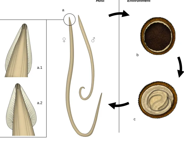
Figure 1 – Morphology of different stages of Echinococcus granulosus. (Original illustrations)
a b Environment Intermediate host Definitive host c
(a – adult stage; a.1 – scolex; a.2 – neck; a.3 – strobila; b – eggs in feces; c – metacestode; c.1 – adventitial layer; c.2. – laminated layer; c.3 – germinal layer; c.4 – daughter cyst; c.5 – protoscolex; c.6 – brood capsule)
2.1.3. Life Cycle
The life cycle includes a definitive mammalian host, usually a canid, carrying the adult egg-shedding stage, with low pathogenicity. The intermediate mammalian host, generally an ungulate, is infected by the eggs that develop into a larval stage (Eckert & Deplazes, 2004) (see Table 1). Transmission to the definitive host occurs after ingestion of organs infected with the metacestode. Pepsin action in stomach, as well as increased temperature and bile secretions in duodenum,
a .3 a .2 a .1 c.1 c.2 c.3 c.4 c.5 c.6
7
stimulate evagination of protoscoleces. The adult parasite develops, reaching maturity in four to six weeks in the anterior quarter of the small intestine, where it actively migrates to the crypts of Lieberkühn and attaches to the intestinal mucosa (Eckert et al., 2001a). Thousands of adult worms may be found in only one dog (David de Morais, 1998).
Gravid proglottids detach from strobila and are released with feces, contaminating the environment. Rupture of proglotiids can occur still in the intestine or eggs may be actively excreted out of the proglottids. Each proglotiid may contain 200-800 eggs (David de Morais, 1998). Prepatent period in dogs is about 45 days (European Scientific Counsel Companion Animal Parasites [ESCCAP], 2010).
After being ingested by a suitable intermediate host, eggs suffer the action of gastrointestinal enzymes. The oncosphere then releases itself from the embryophore before crossing the intestinal wall. This step is triggered by bile action, hooks movement and own lytic secretions (Eckert et al., 2001a, Siracusano et al., 2012). Through blood or lymph vessels, the oncosphere reaches internal organs. Lungs and liver are the most common location of hydatid cysts (Diaz et al., 2011a), although there is a broad spectrum of other possibilities, including spleen, kidneys, brain, muscles and heart (Eckert et al., 2001a; Infante Gil, 2005). Secondary Echinococcosis occurs when new cysts develop in new organs after rupture of an initial one (Eckert et al., 2001a).
If ingested by unsuitable intermediate hosts, some metacestodes lack production of protoscoleces, thus being called sterile/infertile metacestodes, in contrast with fertile ones. This intermediate hosts are considered aberrant or accidental hosts. They may be infected by the metacestodes, however these stages do not become fertile or the hosts do not interact with other animals from the life cycle. In both the former and the latter situation, there is no further transmission of the parasite and the cycle is not completed. Human beings are considered aberrant hosts (Eckert et al., 2001a).
2.1.4. Pathology and Immunity
Definitive hosts are infected with the adult stage of Echinococcus spp. This is called the intestinal form of Echinococcosis. Some histological changes may occur in response to parasite attachment (e.g. thickening of intestinal mucosa due to cellular infiltration or higher mucus production) but generally infection is asymptomatic in definitive hosts, although some degree of pruritus may develop if gravid proglotiids penetrate the anal glands (Eckert et al., 2001a).
Excretory/secretory (E/S) products are released from the parasite which stimulates an immune response by the definitive host. In experimentally infected dogs (n=3), Jenkins & Rickard (1986) could detect the presence of antibodies against protoscolex antigens at day 5 post-infection (pi) and against E/S scolex antigens at 10 days pi. Moreno et al. (2004) also detected a consistent increase in IgG against protoscolex antigens at least 14 days after experimental infection of six dogs. IgA and IgE were also detected but the results varied greatly among dogs. Although an immune response is mounted against parasites, studies failed to correlate IgG titers with worm
8
burden (Gasser, Lightowlers, Obendorf, Jenkins & Rickard, 1988; Moreno et al., 2004). Peyer patches are involved in immunity against infection, producing specific IgA and IgG (Deplazes, Thompson, Constantine & Penhale, 1994).
Overall, evidences exist of both humoral and cellular immune responses against E. granulosus infection in canid hosts. As reviewed by Torgerson (2006), data suggest some degree of protection against reinfection. Recently, Rossi et al. (2012) demonstrated that after a primary infection, dogs developed a markedly humoral immune response. In further trials, parasite burden was lower and the immune response was both cellular and humoral mediated.
Cystic Echinococcosis is the form of Echinococcosis in intermediate and aberrant hosts, caused by
E. granulosus. Growth of the metacestode is slow and several years are required to develop
clinical signs (10-12 months in pigs; 2-4 years in sheep) (Eckert et al., 2001a). Since the majority of farm animals are slaughtered in earlier ages, meat inspection in the abattoirs reveals a high percentage of subclinical infections. Clinical signs are rare and strongly depend on location, dimension and number of cysts and their relation with adjacent structures. In ruminants, the majority of cysts are found in lungs and liver (Eckert et al., 2001a; Correia, Beato, Parreira & Grácio, 2010). In Portugal, sheep harbor a higher percentage of cysts in liver, whereas in cattle the majority is in lungs (Correia et al., 2010). In pigs and horses, liver is the most commonly affected organ. Changes in liver function is the most common biochemical finding in these animals (Eckert et al., 2001a). Clinical signs have been found in horses with heavy infections (Eckert & Deplazes, 2004).
After infection of intermediate hosts, egg and oncosphere antigens are recognized and both humoral and T-cell mediated responses are mounted (Siracusano et al., 2012) which allow some control of the parasite as well as immunity to reinfection (Zhang, Ross & McManus, 2008). In vitro experiments demonstrated the antibody-mediated lytic activity of complement in sheep serum against oncospheres. Lower concentrations of serum from immune sheep were needed to destroy the oncospheres (Heath, Holcman & Shaw, 1994). Although cellular immunity is also mentioned, the mechanism is not yet fully understood (Zhang et al., 2008). As reviewed by Eckert & Deplazes (2004), the majority of experimental studies regarding immunity in intermediate hosts use parasitic burdens higher than those that would occur naturally. Hence, although some degree of immunity was demonstrated experimentally, the same should not happen in natural conditions, since high percentage of older animals is infected.
The laminated layer of the hydatid cyst induces a regulatory response, which attenuates host’s innate immune system. Ergo, involving the metacestode, host tissues produce an advential layer made from collagen fibers and few inflammatory cells, making a chronic evolution of the disease possible (Diaz et al., 2011b).
Fertility of cysts depends on the infective strain and species of intermediate host. In Portugal, in infections by E. granulosus sensu stricto, higher percentage of fertile cysts is found in sheep while
9
in cattle the majority are sterile (David de Morais, 1998; Beato, 2008; Correia et al., 2010). Paredes et al. (2011) found high concentration of bovine IgGs in the germinal layer of infertile cysts. According to the authors, fertility of cysts may be related in some degree with host’s immune response. Biding of antibodies to the germinal layer would prevent cellular division or differentiation and development of brood capsules, thus being responsible for the cyst infertility.
2.1.5. Diagnosis
There is a wide range of diagnostic tools available for Echinococcus granulosus. The choice for one method or the other depends on (i) the purpose of the study; (ii) the sample origin (definitive/intermediate host; animal/human; domestic animal/wildlife); (iii) whether it is in vitam or
post mortem diagnosis; (iv) the equipment and financial resources available.
Regardless of the chosen technique, for safety reasons, infective materials (intestines or fecal samples) should be previously frozen at -70º C for at least 4 days or at -80º C for 2 days (Eckert, Gottstein, Heath & Liu, 2001). Heating to 70 ºC for 12 hours is also a viable option. Personal protective equipment and laboratory decontamination must not be forgotten.
2.1.5.1. Diagnosis in Definitive hosts
Echinococcus spp. eggs can be found in fecal samples in both in vitam or post mortem diagnosis.
A concentration method with a saturated solution of zinc sulphate or sucrose can be used followed by microscopic examination. Mathis, Deplazes & Eckert (1996) developed a flotation and sieving method that allows higher sensitivity in recovering eggs from feces. As an example, a study in Lithuania with 240 dogs found prevalences of 14.2% with this technique against only 5.0% using a modified McMaster method (Bružinskaitė, Šarkūnas, Torgerson, Mathis & Deplazes, 2009).
Despite being quick, cheap and ideal for routine diagnosis, this method alone lacks sensibility as it can only detect patent infections and false negative results can appear due to low worm burden, irregular shedding of eggs or intact gravid proglotiids released in feces. Taking samples in different days, as well as analyzing higher amounts of feces may increase sensitivity (Torres, Pérez, Segovia & Miquel, 2001; Torgerson & Deplazes, 2009). In rare cases, proglotiids can be found in feces which may allow identification (Eckert et al., 2001a).
Purgation with arecoline hydrobromide was a common technique in the past (Kamiya, 2008; Deplazes, Knapen, Schweiger & Overgaauw, 2011). The drug, administered orally, acted as a parasympathomimetic agent that paralyzed the worms and stimulated intestinal smooth muscle motility. This eventually led to the elimination of the adult parasite in feces, which could later be identified (Eckert et al., 2001a). It had nearly 100% specificity but sensitivity could be lower than 65%. It was also a time-consuming method, with potential safety risks to animals, but also to humans since animals would be shedding adult worms and eggs (reviewed by Torgerson & Deplazes, 2009).
10
The gold standard technique for diagnosing both E. granulosus and E. multilocularis in carnivores is the Sedimentation and Counting Technique (SCT). Briefly, small intestine should be divided into five sections and incubated in physiological saline solution. After removal of intestine, the remaining liquid is left for successive sedimentation and decantation steps. Worms may be visualized and quantified using a stereomicroscope (Kamiya, 2008). Although this method does not need too much advanced equipment, is considered cost-effective, it is time-consuming and not suitable for a high number of samples. Concerning this, Umhang, Woronoff-Rhen, Combes & Boué (2011) recently described an adaption from this technique, the Segmental Sedimentation and Counting Technique (SSCT), where only two segments from the small intestine were screened. They found a sensitivity of 98.3% against 100% of SCT for E. multilocularis, just by screening the fourth segment combined with the first or second ones. Since E. granulosus has different location patterns in the gut, the screened segments have to be accessed for this species.
The Intestinal Scraping Technique (IST) is another necropsy-based method, in which scrapings from the intestinal mucosa in different locations are examined microscopically (Kamiya, 2008). Although it takes less time, a sensitivity of 78% was found for E. multilocularis, comparing with the SCT (Hofer et al., 2000). In the “Shaking in a Vessel” Technique (SVT), described by Duscher, Prosl & Joachim (2005), the small intestine is opened longitudinally and its content placed in a vessel filled with water, followed by shaking and decantation steps until the content is clear. The lid contains a mesh and the sediment here attached is screened with a stereomicroscope in several Petri dishes. Duscher et al. (2005) found a sensitivity of 96.2% for this method when searching for
E. multilocularis in foxes.
Worms of E. granulosus can be easily morphologically differentiated from E. multilocularis. However if immature stages or just scoleces are found, staining may be necessary for differential diagnosis. Rostellar hooks are often lost during freezing so they cannot be used to differentiate species (Eckert et al., 2001a).
More advanced techniques have been developed for diagnosis of Echinococcus species. Diverse coproantigen-ELISAs are available for detection of antigens in fecal samples from canids (Deplazes et al., 1992; Allan et al., 1992; Deplazes, Alther, Tanner, Thompson, Eckert, 1999). Some are specific for one species, whereas others detect both E. granulosus and E. multilocularis. They can be used both in live or dead hosts and allow the screening of high number of samples at the same time. Furthermore, prepatent infections can be detected. Deplazes et al. (1992) were able to detect positive samples 10-20 days pi. Nevertheless, false negative results may occur in areas with low prevalence, and also due to cross-reactivity with Taenia antigens (Christofi et al., 2002; Torgerson & Deplazes, 2009). For example, validation of a commercial ELISA kit revealed specificities of 98% in a group of helminth-free dogs but this value decreased to 80% in a group of
11
the same area, infected with Taenia species (Christofi et al., 2002). Another limitation is the impossibility to distinguish between current or past infections (Kamiya, 2008).
Serology methods to detect antibodies in serum from infected dogs are available, capable of giving positive results in just less than two weeks after infection (Moreno et al., 2004). However, as previously mentioned, no correlation was found between antibody titers and worm burden. Moreover, for epidemiological studies, especially in wild populations, this method presents serious limitations.
PCR-based techniques are highly specific and allow species and sometimes strain identification. They can be used in eggs recovered from feces. Since they are time-consuming and expensive, usefulness in large scale studies is questionable. When analysis is made in material collected from feces, this method will suffer from the same low sensitivity and limitations as the ones mentioned earlier for coprological techniques, namely detection of patent infections only. Furthermore, fecal inhibitors may interfere with PCR results (Torgerson & Deplazes, 2009). One way of overcoming this is if eggs are isolated through a sieving-flotation technique (Mathis et al., 1996) which, combined with PCR, presents sensitivity as high as 78% and specificity of 93% (Ziadinov et al., 2008). Several PCR-based techniques are now available for diagnosis in fecal samples (copro-PCR). Examples include methods for G1 (sheep) strain (Abbasi et al., 2003; Štefanić et al., 2004), multiplex-PCR to differentiate between E. granulosus, E. multilocularis and Taenia sp. with a specificity of 100% (Trachsel, Deplazes & Mathis, 2007), and a PCR/dot blot assay developed for different Taenia and Echinococcus species, including E. granulosus genotype 1 (Armua-Fernandez et al., 2011). Sequencing of amplified materials in multiplex-PCR can be used in species and strain differentiation for E. granulosus.
2.1.5.2. Diagnosis in Intermediate hosts
Since majority of infections are subclinical, post-mortem diagnosis is the best diagnostic tool for intermediate hosts. This is mainly done in slaughterhouses by visual inspection of organs, especially liver and lungs. However, prevalences collected from abattoirs may be underestimated since the majority of the individuals, namely sheep, are slaughtered in very young ages when hydatid cysts are still too small to be detected (Eckert & Deplazes, 2004; Kamiya, 2008).
Some studies carried out ultrasound screenings in lungs and liver from intermediate hosts. In Kenya, this method revealed a sensitivity of just 54.36% and a specificity of 97.64% against
post-mortem examination of 300 small ruminants (Sage et al., 1998).
Serology to detect antigens collected from hydatid fluid may be useful, especially in younger animals where cysts could be overlooked in post-mortem diagnosis. (Eckert et al., 2001a) However, since this methods lacks sensitivity, they are usually more suitable to identify the parasite in the overall herd, rather than for an individual diagnosis (Eckert & Deplazes, 2004).
12
Molecular techniques may be used for species and strain identification. A random amplified polymorphic DNA-PCR (RAPD-PCR) was developed to distinguish between species of
Echinococcus and between strains of E. granulosus (Scott & McManus, 1994). Also, restriction
fragment length polymorphism associated with PCR (RFLP-PCR) has been used successfully in distinguishing E. granulosus strains in cysts isolated from intermediate hosts (Beato, 2008).
2.1.6. Epidemiology
E. granulosus is the most widespread species in the genus, with a variety of life cycle patterns that
translates geographic, epidemiologic and cultural diversity. Echinococcus sp. evolved from an ancient population located in Northern Europe, where a predator-prey cycle between wolf and cervids was established (David de Morais, 1998). The growth of human civilization, colonization of different regions and domestication of ungulates and canids eventually led to the creation of multiple other life cycles. Presence of this parasite can now be found worldwide, except in some remote places as Greenland and in confined areas, mainly islands, where intensive control programs were mounted (e.g. Iceland, New Zealand, Tasmania) (Eckert & Deplazes, 2004).
The Mediterranean region is considered endemic for E. granulosus. Different factors are responsible for maintaining infection: (i) high tradition in extensive farming of sheep; (ii) close contact between human, dog and sheep in rural areas; (iii) abundance of stray dogs; (iv) home slaughter and feeding dogs with potentially infected organs; (v) lack of hygiene education and control programs (David de Morais, 1998; Eckert & Deplazes, 2004).
In the last decades, few studies accessed the prevalence and the transmission patterns of Echinococcosis in Portugal. In this section, a comprehensive review is presented about the epidemiological studies available. Since data are scarce, information from Spain is also presented whenever comparable conditions may exist.
2.1.6.1. Definitive Hosts
According to the current knowledge, domestic dog is the main definitive host for E. granulosus in Portugal, while sheep is the main intermediate host (David de Morais, 1998; Beato, 2008).
The prevalence of infection in dogs from Portugal, mentioned in the latest edition of the Manual on Echinococcosis in Humans and Animals by the WHO, refers to a study published by in 1970 (Eckert et al., 2001c). On that time, a prevalence of 10.44% was found in necropsies of 450 dogs from several locations in Portugal. Santarém revealed the highest prevalence (29.4% in just 17 animals). No significant differences were found between prevalences from the main geographic regions (North, Centre, Lisbon Metropolitan Area and South) (reviewed by David de Morais, 1998). David de Morais (1998) compiled almost all the studies done in Portugal about canine Echinocococcis since 1938 and less than 10 are cited. Although Alentejo is the region were most human cases are detected (David de Morais, 2010), prevalences found in dogs were lower than
13
5%. In this study, compiled by David de Morais (1998), however, authors admit not using sheep dogs, and since sample included animals from both rural and urban areas, real prevalence in key endemic areas may have been underestimated. Still, in another survey in Alentejo, only 1 case was found in 153 necropsied dogs (David de Morais, 1998). Further studies are needed, especially in regions where most human cases are reported.
Stray, sheep and hunting dogs are important in epidemiology, since they cover large areas and have contact with possibly infected intermediate hosts. In Spain, Garrudo Arias et al. (1999) found a prevalence of 2.12% of Echinococcus spp. in 754 stray dogs from the province of Extremadura, through necropsy. During a control program in La Rioja, Spain, Jiménez et al. (2002) considered stray dogs the most important reservoirs for Echinococcosus sp.. Likewise, in the province of Alava, Northern Spain, a higher prevalence of 14% (n = 726) was found in sheep dogs through coproantigen ELISA (Benito, Carmena, Joseph, Martínez & Guisantes, 2006) while in a previous study using necropsy just 0.5% of 1040 dogs from urban environments were positive (Benito et al., 2003, cited by Benito et al., 2006).
Previous studies failed in detecting infected foxes in Portugal or Spain (Carvalho-Varela & Marcos, 1993; Carvalho-Varela, Marcos & Grácio-Moura, 1993; Rodríguez & Carbonell, 1998; Criado-Fornelio et al., 2000; Eira, Vingada, Torres & Miquel, 2006; Martínez-Carrasco et al., 2007). Foxes do not seem to play an important role as definitive hosts for E. granulosus, except in specific ecosystems, e.g. Australia (Thompson & McManus, 2001).
E. granulosus sensu stricto (G1 strain) was already diagnosed through necropsy in the Iberian
Wolf (four positive cases in a group of 27 cadavers; three of them from provinces near the Spain-Portugal northern border) (Sobrino et al., 2006). No records exist about the Portuguese populations of the Iberian wolf.
2.1.6.2. Intermediate Hosts
Sheep, goats, cattle and pigs are known intermediate hosts for E. granulosus in Portugal. David de Morais (1998) compiled national statistical data about meat rejection in abattoirs during a period of over 20 years, between 1944 and 1968. The majority of infected sheep originated from the southern provinces of Portugal, with a prevalence of 1.7% in more than 4 million animals. Still, this number is thought to be an underestimation, since during those years several faults were pointed to meat inspection conditions and accuracy. The most recent publications do not provide clear statistical information regarding the infection of sheep. In the Zoonoses Report of the European Food Safety Authority (EFSA) from 2007, a prevalence of 9.4% is referred in a sample of just 32 animals. Prevalences of sheep infection are probably underestimated, since the majority is slaughtered with less than one year old, which makes diagnosis very difficult (Rojo-Vazquez et al., 2011).
14
Regardless the prevalences, sheep are considered important intermediate hosts for multiple reasons: (i) they are the most common ungulate in Portugal; (ii) extensive farming is common, with close contact between sheep and dog; (iii) this species feeds close to the soil, making infection easier; (iv) fertility of cysts seems to be higher in sheep than in cow (David de Morais, 1998; Beato, 2010; Correia et al., 2010).
During 1944-1968, pigs were the species most affected by hydatidosis with an overall prevalence of 4.6% (more than twice the value found for sheep). The majority of reported cases were from the Northern and Center regions, where prevalences of rejection were 8.9% and 6.3%, respectively (in a population of 11 487 595 animals) (David de Morais, 1998). In the following years, drastic reduction of pig population occurred in result of African Swine Fever foci in Portugal. Also intensive production of pigs largely replaced the previous systems. Overall, it resulted in a reduction of cases diagnosed, at least until 1983 (David de Morais, 1998). Recent studies in the Northern region, confirm that hydatidosis is still present among pigs in the North of Portugal. Prevalence of 8.1% was found in the District of Bragança, in 333 pigs from Bísaro breed (Freire et al., 2005). Also in Cantanhede preliminary data of a monitoring program found 5 animals harboring fertile cysts in a sample of 66 animals (Conceição, 2005). Nowadays, increasing popularity of extensive farming of pigs may give rise to new concerns and therefore this situation should be carefully monitored (Hernández Mira et al., 2008).
Bovine cases of hydatidosis were also mostly detected in the Northern region, with an overall prevalence of 4.3% in the Portuguese territory during 1944-1968 that decreased during 1968-1983 (David de Morais, 1998). The report from EFSA (2009) reveals a prevalence of less than 0.1% in a total of 174 834 animals, which is not a representative number of the total cattle slaughtered in Portugal. However, since majority of cysts found in cattle are sterile, this species does not seem to play an important role in the dissemination of the parasite (Beato, 2010).
According to the EFSA report published in 2011, 159 infected deer and 264 wild boars were reported in 2009 in Spain but these are only results from random inspections and no spatial distribution of infected cases is presented (Rojo-Vazquez et al., 2011). A recent work found a wild boar highly infected with fertile cysts from E. granulosus G1, the most common genotype infecting humans (Martín-Hernando, González, Ruiz-Fons, Garate & Gortazar, 2008). Since population of wild boar in the Iberian Peninsula is increasing, several recent works emphasize the importance that this species may play because they are important preys for Iberian wolf and also are available to dogs during hunting season (Martín-Hernando et al., 2008; Rojo-Vazquez et al., 2011).
2.1.6.3. Genetic Characterization
The few genetic characterization studies in Portugal revealed the presence of E. granulosus sensu stricto G1 and G3 (Beato, 2008) in cysts collected from sheep and cattle and also G7 (pig strain) in pigs (Castro et al., 2005). In Central Spain, Mwambete, Ponce-Gordo & Cuesta-Bandera (2004)
15
isolated G1 (sheep strain) hydatid cysts from sheep, cattle, humans, goats, pigs and wild boars, G4 (horse strain) from horses, and G7 from goats, pigs and wildboars. This study confirmed that G1 is the most important cause of hydatidosis in humans in this region and that the fertility of cysts in sheep was higher than in the other species, similarly to what happens in Portugal (Correia et al., 2010). Still, human infection with G7 was already recorded in several European countries (Rojo-Vazquez et al., 2011) and in a study in Austria the majority of Cystic Echinococcosis patients were infected with G7 genotype (Schneider, Gollackner, Schindl, Tucek & Auer, 2010).
In the Southern Portugal, the most important strain is the sheep strain, with higher number of human cases, whereas in the North, the pig strain seems more prevalent, with apparently low infectivity to humans (David de Morais, 2010).
2.1.6.4. Environmental Contamination
According to Gemmell, Roberts, Beard & Lawson (2001), biotic potential is “[…] the potential number of viable cysts which can be established in an intermediate host by an individual definitive host per day.” For Echinococcus sp. this is assumed to be relatively low compared to other taeniid species. Still, egg contamination in the environment is possible. Eggs are already infective when excreted in feces (Deplazes et al., 2011) and more than 8000 eggs can be shed, per day, in an average infected dog (Gemmell et al., 2001). Taeniid eggs are very resistant in the environment and they can still be infective until 1 year, sometimes longer, if kept in adequate temperatures (4 ºC to 15º C) and humidity levels (Gemmell et al., 2001; Thevenet et al., 2005). Blowflies can act as vectors, transporting the eggs through long distances (Deplazes et al., 2011). Eggs are very sensitive to desiccation, being destroyed after 4 days at 25% humidity and in 1 day after 0%. Extreme temperatures can also destroy them (death occurs in 5 min at 60-80º C) (Eckert et al., 2001b).
Taeniid egg contamination in public places was revealed in several studies worldwide. Prevalences may be low, especially due to the fact that coprology has low sensitivity for Echinococcus sp., as previously mentioned. More details will be given in the next chapter (Taenia spp.) about fecal contamination with taeniid eggs.
2.1.6.5. Hydatidosis in Humans
E. granulosus is an important zoonotic agent. Human infection is associated with poor hygiene
conditions. Theoretically, egg ingestion can occur after contact with dogs and their feces, contaminated water and food, but not much is known of which transmission pathway plays a more important role. Dog fur was found contaminated with eggs (Eckert & Deplazes, 2004) and in Portugal, similarly to other countries, possession of dogs was significantly higher in human patients with Cystic Echinococcosis (David de Morais, 2010). As reviewed by Deplazes et al. (2011), there
16
is a possibility of subcutaneous infection by taeniid eggs occurring through a solution of continuity in skin.
Human hydatidosis is usually asymptomatic in early stages. Clinical signs develop in a variable degree, depending on the same factors previously stated for intermediate hosts. More than half of primary human hydatidosis is located in liver (Eckert & Deplazes, 2004). David de Morais (2010) found that 85% of human cases in Portugal had hepatic cysts. Clinical signs are often liver-associated, e.g., cholestasis, hepatomegaly, portal hypertension and ascitis. Pulmonary and neurological signs have been described after cyst infection of respiratory system and brain, respectively. After rupture of cysts and release of hydatid fluid, strong anaphylactic reactions may occur (Thompson & McManus, 2001).
Diagnosis is usually made in older ages. David de Morais (2010) reviewed all the newly diagnosed human cases between 1979 and 2008 in Portugal, and approximately 70% were people older than 40 years old.
Recently, throughout the world several cases of emergence and reemergence of Echinococcosis are documented. In former Soviet Union countries, the decreased quality of meat inspection and veterinary activities may have resulted in increasing prevalences in humans and animals (Eckert & Deplazes, 2004). In Australia, recent prevalences found in dogs were unexpectedly high, and wildlife is supposed to play an important role in maintaining the infection pressure (Jenkins, Romig & Thompson, 2005). Also in China, high prevalences are found among humans (Eckert & Deplazes, 2004).
In 2009, 790 cases were notified in the European Union (EU), 11.3% less than in 2008. Seventy percent of these cases were from Bulgaria, Germany, Spain and Romania. Some countries have much higher incidences than Portugal, as will be presented further: incidence of 4.25/100 000 in Bulgaria (323 cases), 1.07/100 000 in Lithuania (36). Spain had 86 confirmed cases in 2009, with an incidence of 0.19/100 000 (EFSA, 2011). In Europe, however, some of the results shown include also E. multilocularis. As reviewed by Rojo-Vazquez et al. (2011), data from EFSA has several limitations, especially since notification does not always occur.
In the latest WHO/OIE Manual on Echinococcosis in Humans and Animals, the incidence of this disease in humans is Portugal is 2.2/100000 inhabitants but again this is an old reference, dating from 1944-1968. Recent data compiled by David de Morais (2010) and available at Direcção Geral de Saúde (DGS) (2010) were analyzed. During 1987-2007, 467 hydatid patients were officially notified, 76.9% from Alentejo, in the South of Portugal. David de Morais (2010) showed an increasing number of diagnosed cases in 1980s and 1990s. The highest values were found in the late 1980s / early 1990s and then a sudden decrease in the last decade with 112 cases diagnosed
17
between 2000 and 2008 (DGS, 2005; DGS, 2010). When confirmed, E. granulosus was the only species found (EFSA, 2009; EFSA, 2010).
As reviewed by David de Morais (2010), data shows that both in the 1980s (incidence 2.2 cases/100 000 inhabitants/year) and during 2003-2005 (0.1 cases/100 000 inhabitants/year) prevalences were too low to consider Portugal a hiperendemic country in its whole. According to the EFSA report published in 2011, the incidence of Echinococcosis confirmed cases in Portugal was 0.04/100 000 inhab. in 2009. These results are similar to the ones found in the previous four years: 9 cases (2005), 9 (2006), 10 (2007), 4 (2008). Although there seems to be a reduction in notified cases, is important to refer that many cases may not be notified and some, because they are asymptomatic, are never diagnosed.
The district of Évora, in Alentejo is the one with the highest rates of human hydatidosis, with an average incidence of 3.2/100 000 inhabitants/year between 2004 and 2008. The county of Alandroal has the highest in all country – 18.2 incidence /100 000 inhab./year (2004-2008) (David de Morais, 2010).
There are several environmental and socio-ecological factors that explain the higher prevalence of Human Hydatidosis in Alentejo. It is an area mainly inhabited by older people, with low educational levels, that strongly depend upon agriculture and livestock production and where hunting is an important economic activity (Hernández Mira et al., 2008). Sheep farming in extensive and semi-extensive systems is abundant and the contact between humans and dogs is high. Moreover, home slaughter still occurs and dogs are still fed with animal viscera.
2.1.7. Treatment and Control
Treatment of dogs should be made in areas of high prevalence at least each 6 weeks with praziquantel or epsiprantel (ESCCAP, 2010). For praziquantel, single treatment in the adequate dose of 5.0 mg/kg/bw (per os) or 5.7 mg/kg/bw (IM) revealed effective (Eckert et al., 2001a). Epsiprantel should be given at 5.5 mg/kg/bw (per os). Several experimental studies regarding immunity in dogs have been made but so far no vaccine is available (Kamiya, 2008)
Treatment of intermediate hosts is not common. Diagnosis is usually made post-mortem and when performed in vitam, it is not cost-effective (Kamiya, 2008). Treatment with mebendazole was studied in sheep and pigs, but long periods of daily treatment were needed (Eckert et al., 2001a). A recombinant vaccine (Eg95) was developed with almost 100% of protection achieved in sheep. It is also effective in goats and cattle. Two doses of the vaccine are injected subcutaneously in the neck with a one-month interval and immunity can last as long as 12 months (Heath, Jensen & Lightowlers, 2003). Vaccination, however, does not act upon already established cysts and so other control measures need to be taken in animals already infected. Passive immunity in colostrum was also achieved after vaccination (Eckert et al., 2001a).
18
As stated by Eckert & Deplazes (2004), baseline studies are important before implementing control programs, so that achievement of long-term results can be evaluated (e.g. prevalence and incidence in humans, dogs and livestock). Control measures are essential for managing dissemination. They should include public education and improvement of meat inspection and other veterinary-related activities, and focus should be made in individual protection of people working with infective material. Appropriate control of dog population is also important, namely correct deworming, dog registration and control of stray populations. Furthermore, attention should be paid to wildlife populations, since they can work as reservoirs.
Several control programs were held worldwide, e.g. New Zealand, Cyprus and South America (Gemmell et al., 2001b). Due to the epidemiological proximity, a successful control program held in La Rioja, Spain during 14 years is mentioned (Jiménez et al., 2002). Control measures targeted dogs, sheep and human populations. Dogs were treated regularly with praziquantel, covering all dog population but increasingly focusing on sheep dogs in later phases of the program. Some stray dogs were also killed to access the prevalence throughout the program. In local abattoirs, correct disposal of rejected organs was guaranteed. Furthermore, public health education was made on risk groups. After this program, human incidence was reduced from 19 to 4 newly diagnosed cases/ 100 000 inhabitants and prevalences in dogs and adult sheep decreased from 7.0 to 0.2% and 82.3 to 20.3%, respectively. A net benefit of more than 2 million USD was estimated after 14 years of this program.
Removing dead ungulates from fields is another important measure, especially since older animals harbor higher number of cysts, having a higher potential to spread the parasite (Torgerson, Ziadinov, Aknazarov, Nurgaziev & Deplazes, 2009).
Rough estimates of economic impact of Cystic Echinococcosis were determined by Budke, Deplazes & Torgerson (2006). Annual economic losses due to human cases were estimated over 300 million USD for Western Europe, USA, Canada, Australia and New Zealand and more than 700 million USD globally. Worldwide, livestock-associated economic losses accounted for more than 2 billion USD annually, with more than 100 billion just for liver condemnation alone. Although these are just estimates, still they translate high economic losses. In practice, effects on livestock also account for decreasing in milk production and animal body condition, which altogether has a huge impact, especially in poor communities and regions that strongly depend upon farming (Eckert & Deplazes). As stated by Budke et al. (2006) control measures are necessary and cost-effectiveness may support all the efforts.
After implementation of effective programs, permanent surveillance and other control measures must be maintained (Gemmell et al., 2001b). Furthermore, treatment of imported dogs should be mandatory; otherwise, infection levels will increase again.
19
Efforts in Portugal to control this disease may have begun in the late 1970s in the Southern region of the country (Borges Ferreira, 1979), where some campaigns against Echinococcosis began. These were based in public education and dog treatments. Dogs presented by their owners were treated with arecoline hydrobromide in the beginning and praziquantel in later years. Feces released from dogs were either taken for analysis or burnt. In 1991, after the end of these campaigns, the Fighting Group against Echinococcosis-Hydatidosis in the Counties of Elvas and Alandroal started its work. This is a group made by volunteers, mainly health care professionals that performed several screenings and sanitary education in Alentejo (Hernández Mira et al., 2008).
In 1996, a Control and Monitoring Program of Echinococcosis/Hydatidosis was approved and co-funded by the European Union in Portugal. Despite cessation of European funding, the program continued in Alentejo and later on in Algarve and Beira Interior. The activities include the notification of positive cases in meat inspection, followed by appropriate epidemiological inquiries and sanitary education, especially in schools. This disease is still part of the list of mandatory notifiable diseases in Portugal and OIE (Direção-Geral de Alimentação e Veterinária [DGAV], 2012). Also free deworming of dogs with praziquantel is performed during the annual Rabies vaccination campaigns in specific approved regions. Few results are available of this program, with no base-line studies done, since some years ago, which makes it difficult to understand the impact of these measures.