Identification of pathogens and virulence profile of
Rhodococcus equi
and
Escherichia coli
strains obtained from sand of parks
M.C. Fernandes
1, S. Takai
2, D.S. Leite
3, J.P.A.N. Pinto
1, P.E. Brandão
4, V.A.
Santarém
5, F.J.P. Listoni
1, A.V. Da Silva
6, M.G. Ribeiro
11Departamento de Higiene Veterinária e Saúde Pública, Faculdade de Medicina Veterinária e Zootecnia,
Universidade Estadual “Júlio de Mesquita Filho”, Botucatu, SP, Brazil. 2Department of Animal Hygiene, School of Veterinary Medicine and Animal Sciences,
Kitasato University, Japan.
3Departamento de Genética, Evolução e Bioagentes, Instituo de Biologia,
Universidade Estadual de Campinas, Campinas, SP, Brazil. 4Departamento de Medicina Veterinária Preventiva e Saúde Animal,
Universidade de São Paulo, São Paulo, SP, Brazil.
5Curso de Medicina Veterinária, Universidade do Oeste Paulista, Presidente Prudente, SP, Brazil. 6Departamento de Ciências Biológicas, Universidade Estadual de Feira de Santana, Bahia, Brazil.
Submitted: March 19, 2011; Approved: September 10, 2012.
Abstract
The identification of pathogens of viral (Rotavirus, Coronavirus), parasitic (Toxocaraspp.) and bac-terial (Escherichia coli, Salmonella spp.,Rhodococcus equi) origin shed in feces, and the virulence profile ofR. equiandE. coliisolates were investigated in 200 samples of sand obtained from 40 parks, located in central region of state of Sao Paulo, Brazil, using different diagnostic methods. From 200 samples analyzed, 23 (11.5%) strains ofR. equiwere isolated. None of theR. equiisolates showed a virulent (vapAgene) or intermediately virulent (vapBgene) profiles. Sixty-three (31.5%) strains ofE. coliwere identified. The following genes encoding virulence factors were identified in E. coli:eae,bfp,saa,iucD,papGI,sfaandhly. Phylogenetic classification showed that 63E. coli iso-lates belonged to groups B1 (52.4%), A (25.4%) and B2 (22.2%). NoE. coliserotype O157:H7 was identified. Eggs of Toxocara sp. were found in three parks and genetic material of bovine Coronavirus was identified in one sample of one park. NoSalmonellaspp. and Rotavirus isolates were identified in the samples of sand. The presence ofR. equi,Toxocarasp, bovine Coronavirus and virulent E. coli isolates in the environment of parks indicates that the sanitary conditions of the sand should be improved in order to reduce the risks of fecal transmission of pathogens of zoonotic poten-tial to humans in these places.
Key words:enteric pathogens, virulence, sand, feces, parks,E. coli,R. equi.
Introduction
Enteric pathogens are a major group of organisms re-lated to infections in humans and animals. These pathogens are resistant to adverse environmental conditions and are frequently transmitted by oral route due to fecal
contamina-tion of foods, water, vegetables and fruits (Acha and Szyfres, 2003).
Sandboxes are commonly found in parks all over the world, and are mainly used by children and adolescents. Access of companion animals, birds and occasionally live-stock, inadequate hygiene of the sandboxes, precarious hy-giene habits of children, and lack of knowledge about the
Send correspondence to M.G. Ribeiro. Disciplina de Doenças Infecciosas dos Animais Domésticos, Departamento de Higiene Veterinária e Saúde Pública, Faculdade de Medicina Veterinária e Zootecnia, Universidade Estadual Paulista “Julio de Mesquita Filho”, Caixa Postal 560, 18618-970 Botucatu, SP, Brazil. E-mail: [email protected].
risks posed by microorganisms eliminated in feces of ani-mals favor the transmission of pathogens of animal origin to humans in these places (Matsuo and Nakashio, 2005).
The present study investigated the presence of patho-gens of viral (Coronavirus, Rotavirus), parasitic (Toxocara spp.) and bacterial (E. coli,Salmonellaspp.,R. equi) origin eliminated in feces of animals, and the virulence profile of R. equiandE.coliisolates, obtained from the sand of parks located in the central region of state of Sao Paulo, Brazil.
Materials and Methods
Collection of samples
Two hundred samples of sand from 40 parks were an-alyzed. The strains were collected between 2008 and 2009. All parks were located in the central region of state of Sao Paulo, Brazil. After superficial dirt was removed, about 250 g of soil were collected 10-15 cm deep. Samples were placed in individual plastic bags and taken to laboratory un-der refrigeration (4-8 °C).
Culture, identification and storage of bacterial isolates
All samples were processed in the Laboratory of Mi-crobiology and Infectious Diseases of Animals, Depart-ment of Veterinary Hygiene and Public Health, School of Veterinary Medicine, UNESP - Universidade Estadual Paulista, Botucatu, Sao Paulo, Brazil. Samples were kept under refrigeration (4-8 °C) or frozen (-20 °C) until they were analyzed.
Samples (25 g) of feces from all parks were inocu-lated aseptically in 225 mL sterilized distilled water. After homogenization, 0.03 mL of material was inoculated in
defibrinated bovine blood agar (5%) and MacConkey agar forE. coliisolation. Plates were incubated at 37 °C for three days and were assessed every day for bacterial growth. Si-multaneously, 0.03mL of these samples were cultured in
NANAT selective media forR. equi(Takai et al., 1996). Microorganisms were identified by colony morphology, staining methods, and biochemical tests (Quinn et al., 1994). Isolates of bacterial origin were stored in Lignièris agar at 25 °C.
Identification ofSalmonellaspp.
Briefly, samples (25 g each) were inoculated into 250 mL of peptone water 1.0% (Oxoid) and incubated at 35 °C for 24 h. Aliquots of 0.1 mL and 1 mL were inocu-lated each into 10 mL of Rappaport-Vassiliadis (RV) (Oxoid) and Tetrathionate (TT) (Oxoid) broth, respec-tively, and incubated at 42 °C (RV) and 35 °C (TT) for 24 h. A loopful of each broth culture was inoculated simulta-neously in xylose-lysine-desoxycholate agar (XLD) (Oxoid) and bismuth sulfite agar (BS) (Oxoid), followed by incubation at 35 °C for 24 h. Colonies suggestive of Salmo-nellawere inoculated in triple sugar iron (TSI) and lysine
iron (LIA) agar slants. Tubes were incubated for 35 °C for 24 h. Colonies suggestive ofSalmonellaspp. in at least one of the culture media (TSI or LIA) were submitted to bio-chemical tests, agglutination test using polyvalent anti- Sal-monellaserum (Probac) (Quinn et al., 1994; Andrewset al., 1998), and serotype identification(Popoff and Le Mi-nor, 1992).
Virulence ofR. equi
Isolation of plasmid DNA was obtained by using an alkalin lysis method (Takaiet al., 2003). Target DNA for PCR amplification was based on the genes encoding a 15-17 kDa antigen (vapA gene), and a 20 kDa antigen (vapBgene) sequence. Plasmid DNA was digested with re-striction endonucleases (EcoRI, EcoT22I and HinddIII). Primer 1 (5’-GACTCTTCACAAGACGGT-3’) and primer 2 (5’-TAGGCGTTGTGCCAGCTA-3’) were used to de-tect virulent (vapAgene) strains and the 569-552 bp ex-pected product. Primer 3 (5’-AACGTAGTCGCGGTGAG AA-3’) and primer 4 (5’-ACCGAGACTTGAGCGACTA--3’) were used for intermediately virulent (vapBgene) iso-lates to detect the 1066-1048 bp expected product. Samples were submitted to 30 cycles of amplification as follows: de-naturation for 90 s at 94 °C, annealing for 1 min at 55 °C, and extension for 2 min at 72 °C (Takaiet al., 1996; Takai et al., 2003). Characterization of plasmid virulence was performed in Kitasato University, Japan.
E. coliserotypes and virulence factors
Sorbitol-negative O157:H7 serotypes were submitted to agglutination test with O157 and H7 sera (Probac). Ref-erence strains were E. coli FVL2 (sfa, pap, iucD, hly, cnf-1), FV35 (afa, iucD, cnf-1), J96 (papGII, papGIII), O157:H7 (vt1, vt2, eae), 2348/69 (eae, bfp, eaf), IANO (stb, lt1), EAEC O42 (eaec), B90 (cnf-2), FVL16 (cnf-1, hly, pap, sfa, iucD), ETEC13 (sta), EIEC (ipaH), supplied by the Laboratory of Bacterial Antigens, Campinas State University, Brazil.E. coliDH5astrain was used as a
nega-tive control. First, primers for virulence factor genes were determined individually using a template DNA from appro-priate positive and negative control strains. The presence of the following groups of genes were analyzed by PCR:papC andpapG alleles (P fimbria),sfaC/D(S fimbria),afaB/C (afimbrial adhesin),saa(self-agglutinating adhesin),iucD (aerobactin),cnf-1andcnf-2(cytotoxic necrotizing factor type 1 and 2),hly(a-hemolysin),vt1andvt2(verotoxins),
classifica-tion (chuA, yjaA, TspE4.C2) in groups A, B1, B2 and D was performed by PCR (Emödyet al., 2003).
Identification ofToxocaraspp.
Flotation-centrifugation with sodium nitrate (Na2NO3) 1.20 g/cm3 was used for the recovery of Toxocara spp. eggs. Centrifugation was performed at 2.500 rpm (679 g) for 5 min. After that, the supernatant of each tube was placed in microscope slides, covered with coverslips, and examined under a light microscope (10x). This process was repeated three times for each sample (Santarémet al., 1998).
Diagnosis of bovine coronavirus (BCoV)
Samples were tested for the presence of BCoV with a group II coronavirus specific RT-PCR assay targeted to the RNA-dependent RNA-polymerase gene (RdRp) with a 136-bp predicted product (Brandão et al., 2005). BCoV Kakegawa strain (Akashiet al., 1980) and PBS were used as positive and negative controls, respectively.
Diagnosis of rotavirus
Samples were analyzed for the presence of rotavirus 11-segment RNA using polyacrylamide gel electrophore-sis-PAGE (Herringet al., 1982). NCDV group A rotavirus strain was used as the positive control.
Statistical analysis
Chi-square test (Epi-Info, 6.4) was used to evaluate the differences in the presence of different pathogens in the parks, considering p <0.05 (Triola, 2005).
Results
The frequency of pathogens identified in samples of sand obtained from parks is shown in Table 1. There was no statistical difference (p >0.05) between the presence of the different pathogens in the parks sampled.
E. coli andR. equi strains were the most common pathogens isolated throughout the study. R. equi strains were isolated in 23 (11.5%) sand samples. None of theR. equiisolates showed virulent (vapAgene) or intermediately
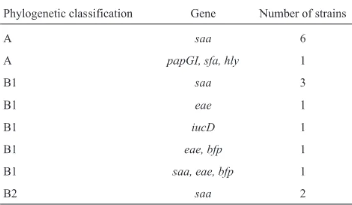
virulent (vapBgene) plasmid profiles. Sixty-three (31.5%) strains ofE. coliwere identified. The following virulence factor genes were identified in theE. colistrains:eae,bfp, saa,iucD,papGI,sfaandhly. Phylogenetic classification showed that the 63E. coliisolates belonged to groups B1 (52.4%), A (25.4%) and B2 (22.2%). NoE. coliserotype O157:H7 was identified (Table 2).
Eggs ofToxocaraspp. were recovered only in three of the parks.
Genetic material of bovine Coronavirus was identi-fied in one public park (Table 1), as suggested by the se-quencing analysis of the 136 bp amplicon obtained in one sample (data not show). NoSalmonella spp. or Rotavirus isolates were identified in the samples of sand.
Discussion
Rhodococcus equiis a well-recognized Gram posi-tive intracellular bacterium associated with different clini-cal manifestations in humans and animals. The organism is widespread in soil, particularly in feces of foals, other herbivores and their environment (Prescott, 1991). The virulence mechanism of this pathogen is related with the presence of virulence-associated plasmids-Vap (Takaiet al., 1991), and three levels of virulence are currently rec-ognized: virulent, intermediately virulent and avirulent (Takai, 1997). Virulent R. equi strains contain a large plasmid of 85-90 kb, responsible for encoding the 15-17-kDa antigens (VapA) that are considered the major causes of suppurative pneumonia in foals (Ribeiroet al., 2005). VapB or intermediately virulent isolates present 20-kDa antigens and a 79-100 kb plasmid (Takai, 1997). They are frequently observed in swine lymphadenitis (Ta-kaiet al., 1996) and patients infected by acquired immu-nodeficiency syndrome-AIDS (Takaiet al., 2003). In con-trast, avirulent strains show no evidence of eithervapAor vapBgenes. These strains are found in the soil of areas where foals are raised, in soil and/or sand of human dwell-ing, mainly in yards and parks, and in humans with rhodococcosis co-infected by AIDS virus (Takai et al., 1996; Takai, 1997).
Table 1- Identification of pathogens from fecal origin in sand of parks.
Brazil, 2010.
Pathogens Frequency p value
Isolates identified / total of specimens (%)
Escherichia coli 23/200 (11.5) 0.33
Rhodococcus equi 63/200 (31.5) 0.27
Toxocaraspp. 3/200 (1.5) 0.5
bovine Coronavirus 1/200 (0.5) 0.5
No= number.
p < 0.05 indicates statistical differences between microorganisms.
Table 2- Phylogenetic classification and virulence factors ofEscherichia colistrains isolated from sand samples of parks. Brazil, 2010.
Phylogenetic classification Gene Number of strains
A saa 6
A papGI, sfa, hly 1
B1 saa 3
B1 eae 1
B1 iucD 1
B1 eae, bfp 1
B1 saa, eae, bfp 1
All ourR. equi strains were classified as avirulent. These results are in agreement with similar study in Japan, which also reported the presence of avirulentR. equiin the soil of parks and yards (Takai et al., 1996). Avirulent strains have been frequently identified in the environment of domestic animals, particularly foals (Takai, 1997). Cur-rently, R. equi has emerged as a pulmonary pathogen among immunosuppressed patients, mainly those infected by AIDS virus (Acha and Szyfres, 2003). A recent survey ofR. equivirulence profile in 20 humans in Brazil showed 11 patients infected with avirulent strains (Ribeiroet al., 2011). Plasmid virulence of R. equi strains isolated in Brazil was characterized in foals (Ribeiroet al., 2005) and a dog (Fariaset al., 2007). The present study was the first in-vestigation in this country about virulence profile ofR. equi strains isolated from park sand. Beside the absence of viru-lent or intermediately viruviru-lentR. equistrains, the presence of this microorganism in the sand of parks constitutes a public health problem. This risk is particularly important to children and immunocompromised people, especially HIV-positive patients, because avirulentR. equimay cause the disease in immunosuppressed and non-immunosup-pressed patients (Takai et al., 2003), including in Brazil (Ribeiroet al., 2011).
E. coliis a very diverse species of bacteria found both in the intestinal tract of humans and animals, and in the en-vironment. The microorganism is classified in six different pathotypes based on enteric manifestations, as follows: enterotoxigenic (ETEC), enteropathogenic (EPEC), ente-roinvasive (EIEC), enterohemorrhagic (EHEC), enteroag-gregative (EAEC), and diffusely adherent (DAEC). Patho-genic manifestations of E. coli are closely related with different virulence factors, including enterotoxins, cyto-toxins, fimbriae, adhesins, and iron chelation mechanisms (Kaperet al., 2004).
Geneeaeencodes intimin, which mediates the inti-mate attachment of EPEC and EHEC to epithelial cells, and stimulates mucosal immune response and intestinal crypt hyperplasia. Geneeaeandbfpwere found in three strains isolated from the sand of parks in the present study, and are generally related with atypic enteropathogenicE. coli. This class ofE. coliEPEC causes diarrhea in children younger one year of age, mainly in emerging countries (Kaperet al., 2004). In Brazil, there was a case of concurrent infection of a child and a dog by enteropathogenicE. colithat showed eaegene and was isolated from feces (Rodrigues et al., 2004). Atypic EPEC isolated from parks constitutes a pub-lic health risk, especially for children and immunosup-pressed humans.
P fimbriae are known to contribute forE. coli patho-genesis by promoting colonization of host tissues and stim-ulating injurious inflammatory response in the host (Kuehn et al., 1994). PapG adhesin is located on P fimbria. Three classes of PapG (PapGI, PapGII and PapGIII) are recog-nized: papG Class I are predominantly found in fecal
strains; allele GII in strains involved in pyelonephritis and bacteremia cases; and allele GIII in isolates responsible for cystitis cases in humans and animals (Bergsten et al., 2005). S pillus is constituted of subunits: sfaS subunit me-diatesE. coliinteraction with intestinal and other epithelial cells.sfaS gene is associated with human pyelonephritis, meningitis and sepsis (Fériaet al., 2001). Haemolysin is a pore-forming cytotoxin that lyses erythrocytes, leukocytes, and endothelial and epithelial cells of mammals. Thehly genes are frequently found in extraintestinalE. coli infec-tions in humans and animals (Johnsonet al., 1991). One of our isolates harbored the genes that encodepapGI,sfaand hly. The identification of these virulence factors in a same isolate may be explained by the presence of a pathogenicity island (PAI), which enhances the infectivity of the microor-ganism. PAIs have been frequently found inE. coli respon-sible for human extraintestinal infections (Kurazonoet al., 2000). In Brazil, genes that encodepapGadhesins, as well ashlyandsfagenes, were found inE. colistrains isolated from pyometra, urinary tract infections, and feces of dogs (Siqueira et al., 2009). Free access of dogs to parks in-creases the risk of human infection with virulentE. coli. These animals may act as reservoirs, harboring pathogenic strains with virulence factors such aspapG,hly, and sfa genes.
Iron is essential for bacterial metabolism.E. coliuses this ion for the transport and storage of both electrons and oxygen, and for DNA synthesis (Emödy et al., 2003). Growth of bacteria under restricted iron concentrations make them use successfully competitive mechanisms to obtain this ion from the host. Aerobactin is the most effec-tive iron chelation system employed byE. colifor iron ac-quisition, mediated by iuc genes types A, B, C and D (Griffiths, 1997). In humans, this virulence factor is inti-mately associated with urinary infections and septicemia (Torreset al., 2001). TheiucDgenes were found in only one isolate of our study. Currently,iuc genes have been found in dogs with pyometra (Cooganet al., 2004), urinary tract infections, and in dog feces in Brazil (Siqueiraet al., 2009). Like other virulence factors, the presence ofiucD gene inE. colistrains isolated from sand represents a risk to the population visiting these parks.
The presence of a self-agglutinating adhesin (saa) in E. colihas been previously described (Patonet al., 2001). Virulence of this adhesin to humans and domestic animals remains unclear. However,saagene was found in 19.0% of E. colistrains obtained from the sand of parks in the current study. This result suggests that further studies should be carried out in order to investigate the role of this adhesin as anE. colivirulence factor.
isolates obtained from the sand of parks were classified in A and B1 groups. Although these groups are predominantly related with non-pathogenicE. colistrains, these results in-dicate fecal contamination of the environment.
Toxocariasis is a cosmopolitan parasitic zoonosis. Toxocara spp. is one of the most common parasites of young dogs and cats. Eggs of the parasite are frequently shed in large amounts in the feces of companion animals. Toxocariasis in human occur by spread of the larvae, lead-ing to different clinical forms of disease. Clinical manifes-tations involve serious neurological, ophthalmologic, pul-monary, and/or cutaneous signs (Acha and Szyfres, 2003). The presence of eggs ofToxocaraspp. in the sand of parks have been reported in several countries (Dubnáet al., 2007; Matsuo and Nakashio, 2005), including in Brazil(Santarém et al., 1998). Three parks had positive samples for eggs of this parasite. These results suggest environmental contami-nation by feces of companion animals and indicate risk of toxocariasis to humans that use these parks, particularly children.
Coronavirus infections in animals were reviewed elsewhere (Brandãoet al., 2001). In Brazil, previous stud-ies have identified Coronavirus in feces of cattle and dogs with and without diarrhea (Brandãoet al., 2005, 2007). Identification of bovine Coronavirus in the sand from parks in Brazil is uncommon, although it also represents fecal contamination of the environment.
Rotavirus was detected in the feces of domestic ani-mals (Rodriguezet al., 2004; Ruizet al., 2009) and chick-ens (Villarrealet al., 2006) with and without diarrhea in Brazil. Likewise, differentSalmonellaspp. serotypes were detected in feces of livestock (Ribeiroet al., 2010), birds and chickens (Hoferet al., 1997) in this country. None of sand samples collected in our parks showed Rotavirus and Salmonellaspp. In contrast, an epidemiological study in-volving human patients with salmonellosis in several Euro-pean countries revealed that one the major risk factors for the disease was the access of children up to four years of age to the sand of parks (Doorduyn et al., 2006). These findings indicate that similar studies must be performed in other regions in Brazil in order to investigate the occur-rence ofSalmonellaspp. and Rotavirus in the sand of parks. Despite the absence ofSalmonellaspp. and Rotavirus in the samples analyzed, these pathogens should be included in microbiological testing required to determine the sanitary conditions of the sand used in parks, as they may be shed in the feces of birds and domestic animals.
The identification ofR. equi,E. coliEPEC, bovine Coronavirus, andToxocaraspp. are indicators of fecal con-tamination of the sand of the parks sampled. Concon-tamination may have been caused by feces from domestic animals (Takaiet al., 1996), birds (Prescott, 1991), or contaminated shoes of people who visit these places. Our results suggest the need to introduce control measures to prevent contami-nation of the sand by pathogens eliminated in animal feces.
In fact, the risks of the transmission of pathogens shed in animal feces in parks may reduce if access of domestic ani-mals to these places is prevented, fecal material is daily re-moved from the sand, sand is periodically tested for sani-tary quality and replaced with material of known origin, and people are continuously educated on hygiene habits be-fore using parks.
The presence ofR. equi,E. coliEPEC,Toxocaraspp. and bovine Coronavirus identified in parks studied indi-cates environmental contamination by microorganisms found in feces of domestic animals, birds, and/or contami-nated shoes of people. These results represent a risk for the transmission of pathogens with zoonotic potential to hu-mans in these places, particularly to children.
Acknowledgments
This work was supported by FAPESP - Fundação de Amparo à Pesquisa do Estado de São Paulo, Brazil (2007/57781-3)
References
Acha PN, Szyfres B (2003) Zoonoses y Enfermidades Trans-misibles Comunes al Hombre y los Animales. 3rded., Orga-nización Panamericana de la Salud, Washington., vol. 1, 398 pp.
Akashi H, Inaba Y, Miura Y, Tokuhisha S, Sato K, Satoda K (1980) Properties of a coronavirus isolated from a cow with epizootic diarrhea. Vet Microbiol 5:265-276.
Andrews WH, June GA, Sherrod PS, Hammack TS, Amaguana RM (1998) Salmonella. In: Andrews, W.H.; June, G.A.; Sherrod, P.S.; Hammack, T.S.; Amaguana, R.M. (eds). Bac-teriological Analytical Manual. Food and Drug Administra-tion, Gaithersburg, pp 5001-5020.
Aranda KRS, Fagundes-Neto U, Scaletsky ICA (2004) Evalua-tion of Multiplex PCRs for diagnosis of infecEvalua-tion wit diar-rheagenicEscherichia coliand Shighella spp. J Clin Micro-biol 42:5849-5853.
Bergsten G, Wullt B, Svanborg C (2005)Escherichia coli,
fim-briae, bacterial persistence and host response induction in the human urinary tract. Int J Med Microbiol 295:487-502. Blanco M, Blanco JE, Blanco J, Alonso MP, Balsalobre C,
Mou-rino M, Madrid C, Juárez A (1996) Polymerase chain reac-tion for detecreac-tion of Escherichia coli strains producing cytotoxic necrotizing factor type 1 and type 2 (CNF1 and CNF2). J Microbiol Methods 26:95-101.
Blanco M, Blanco JE, Alonso MP, Mora A, Balsalobre C, Munoa F, Juárez A, Blanco J (1997) Detection of pap, sfa and afa adhesin-encoding operons in uropathogenic Escherichia colistrains: relationship with expression of adhesions and
production of toxins. Res Microbiol 148:745-755.
Brandão PE, Gregori F, Villarreal LYB, Rosales CAR, Soares RM, Jerez JA (2005) A nested polymerase chain reaction to bovine coronavirus diagnosis. Virus Rev Res 10:45-49. Brandão PE, Gregori F, Heinemann MB, Lima CHA, Rosales
CAR, Ruiz VLA (2001) Animal coronaviruses. Virus Rev Res 6:7-13.
Bo-vine coronavirus (BCoV). Arq Bras Med Vet Zootec 59:1074-1076.
Clermont O, Bonacorsi S, Bingen E (2000) Rapid and Simple De-termination of the Escherichia coli Phylogenetic Group. Appl Environ Microbiol 66:4555-4558.
Coogan JÁ, Oliveira CM, Faustino M, Moreno AM, Von Sydow AC, Melville PA, Benites N.R. (2004) Estudo micro-biológico de conteúdo intra-uterino de cadelas com piometra e pesquisa de fatores de virulência em cepas deEscherichia coli. Arq Inst Biol. São Paulo, 71:513-515.
Doorduyn Y, Van Den Brandhof WE, Van Duynhoven YT, Wannet WJ, Van Pelt W (2006) Risk factors forSalmonella
Enteritidis e Typhimurium (DT104 and DT104) infections in the Netherlands: predominant roles for raw eggs in Enteritidis and sandboxes in Typhimurium infections. Epidemiol Infect 134:617-626.
Dubná S, Langrová I, Jankovská I, Vadlejch J, Pekár S, Nápravník J, Fechtnera J (2007) Contamination of soil withToxocara
eggs in urban (Prague) and rural areas in the Czech Repub-lic. Vet Parasitol 144:81-86.
Emödy L, Kerényi M, Nagy G (2003) Virulence factors of uropa-thogenicEscherichia coli. Int J Antimicrob Agents 22:29-33.
Farias MR, Takai S, Ribeiro MG, Fabris V (2007) Cutaneous pyogranuloma in a cat caused by virulentRhodococcus equi
containing an 87 kb type I plasmid. Aust Vet J 85:29-31. Féria C, Machado J, Correia JD, Gonçalves J, Gaastra W (2001)
Distribution of papG alleles among uropathogenic
Esche-richia coliisolated from different species. FEMS Microbiol
Lett 202:205-208.
Griffiths E (1997) Iron and the virulence ofEscherichia coli.In: Sussman, M. (Ed.).Escherichia coliMechanisms of
Viru-lence. Cambridge University Press, Cambridge, pp 331-371. Herring AJ, Inglis NF, Ojeh CK, Snodgrass DR, Menzies JD (1982) Rapid diagnosis of rotavirus infection by direct de-tection of viral nucleic acid in silver-stained poliacrylamide gels. J Clin Microbiol 16:473-477.
Hofer E, Silva Filho SJ, Reis EMF (1997) Prevalência de Soro-vares deSalmonellaisolados de aves no Brasil. Pesq Vet Bras 17:55-62.
Johnson JR (1991) Virulence factors inEscherichia coliurinary tract infection. Clin Microbiol Rev 4:80-128.
Kaper JB, Nataro JP, Mobley HLT (2004) Pathogenic Esche-richia coli. Nature Reviews 2:23-140.
Karkkainen UM, Kauppinen J, Ikaheimo R, Katila ML, Siitonen A (1998) Rapid and specific detection of three different G adhesin classes of P-fimbriae in uropathogenicEscherichia coli by polymerase chain reaction. J Microbiol Methods 34:23-29.
Kuehn MJ, Jacob-Dubuisson F, Dodson K, Slonim L, Striker R, Hultgren SJ (1994) Genetic, biochemical, and structural studies of biogenesis of adhesive pili in bacteria. Methods Enzymol 236:282-306.
Kurazono H, Yamamoto S, Nakano M, Fair GB, Terai A, Chai-cumpa W, Hayashi H (2000) Characterization of a putative virulence island in the chromosome of uropathogenic
Esche-richia coli possessing a gene encoding a
uropathogenic-specific protein. Microbial Pathogen 28:183-189.
Matsuo J, Nakashio S (2005) Prevalence of fecal contamination in sandpits in public parks in Sapporo City, Japan. Vet Para-sitol 128:115-119.
Paton, AW, Srimatone P, Woodrow MC, Paton JC (2001) Charac-terization of Saa, a novel autoagglutinating adhesin pro-duced by locus of enterocyte effacement-negative Shiga toxigenicEscherichia colistrains that are virulent for hu-mans. Infect Immun 69:6999-7009.
Popoff MY, Le Minor L (1992) Formules antigéniques des séro-vars deSalmonella. Centre Collaborateur OMS de réference
et de recherches pour lesSalmonella.
Prescott JF (1991) Rhodococcus equi: an animal and human
pathogen. Clin Microbiol Rev 4:20-34.
Quinn PJ, Carter ME, Markey BM, Carter GR (1994) Clinical Veterinary Microbiology. Wolfe, London, 648 pp. Ribeiro MG, Takai S, Vargas AC, Mattos-Guaraldi AL, Camello
TCF, Ohno R, Okano H, Silva AV (2011) Identification of virulence associated plasmids in Rhodococcus equiin hu-mans with and without acquired immunodeficiency syn-drome in Brazil. Am J Trop Med Hyg 85:510-513. Ribeiro MG, Seki I, Yasuoka K, Kakuda T, Sasaki Y, Tsubaki S,
Takai S (2005) Molecular epidemiology of virulent
Rhodococcus equifrom foals in Brazil: virulence plasmids
of 85-kb type I, 87-kb type I, and a new variant, 87-kb type III. Comp Immunol Microbiol Infect Dis 28:53-61. Ribeiro MG, Fernandes MC, Paes AC, Siqueira AK, Pinto JPAN,
Borges AS (2010) Caracterização de sorotipos em linhagens do gêneroSalmonellaisoladas de diferentes afecções em
animais domésticos. Pesq Vet Bras 30:155-160.
Rodrigues J, Thomazini CM, Lopes CAM, Dantas LO (2004) Concurrent infection in a dog and colonization in a child with a human enterophatogenic Escherichia coli clone. J Clin Microbiol 42:1388-1389.
Rodriguez CAR, Brandão PE, Ferreira F, Gregori F, Buzinaro MG, Jerez JA (2004) Improved animal rotavirus isolation in MA-104 cells using different trypsin concentrations. Arq Inst Biol. São Paulo, 71:437-441.
Ruiz VLA, Brandão PE, Gregori F, Rodriguez CAR, Souza SLP, Jerez JA (2009) Isolation of rotavirus from asymptomatic dogs in Brazil. Arq Bras Med Vet Zoot 61:996-999. Santarém VA, Sartor IF, Bergamo FMM (1998) Contaminação
por ovos deToxocaraspp em parques e praças públicas de Botucatu, SP, Brasil. Rev Soc Bras Med Trop 31:529-532. Schmidt H, Knop C, Franke S, Aleksic S, Heesemann J, Karch H
(1995) Development of PCR for screening of enteroag-gregativeEscherichia coli. J Clin Microbiol 33:701-705.
Siqueira AK, Ribeiro MG, Leite DS, Tiba MR, Moura C, Lopes MD, Prestes NC, Salerno T, Silva AV (2009) Virulence fac-tors inEscherichia colistrains isolated from urinary tract in-fection and pyometra cases and from feces of dogs. Res Vet Sci 86:206-210.
Takai S, Sekizaki T, Ozawa T, Sugawara T, Watanabe W, Tsu-baki S (1991) Association between a large plasmid and 15-to 17-kilodal15-ton antigens in virulentRhodococcus equi.
In-fect Immun 59:4056-4060.
Takai S (1997) Epidemiology ofRhodococcus equiinfections: A
review. Vet Microbiol 56:167-176.
Takai S, Fukunaga N, Ochiai S, Sakai T, Sasaki Y, Tsubaki S (1996) Isolation of virulent and intermediately virulent
Rhodococcus equifrom soil and sand on parks and yards in
Japan. J Vet Med Sci 58:669-672.
Kirikae T (2003) Molecular epidemiology ofRhodococcus equi of intermediate virulence isolated from patients with and without Acquired Immune Deficiency Syndrome in Ching Mai, Thailand. J Infect Dis 188:1717-1723.
Torres AG, Redford P, Welch RA, Payne SM (2001) TonB-dependent systems of uropathogenic Escherichia coli: aerobactin and heme transport and TonB are required for virulence in the mouse. Infect Immun 69:6179-6185. Triola MF (2005) Introdução à Estatística. 9thed. LTC, Rio de
Ja-neiro, 682 pp.
Villarreal LYB, Uliana G, Valenzuella C, Chacón JLV, Sain-denberg ABS, Sanches AA, Brandão PE, Jerez JA, Ferreira AJP (2006) Rotavirus detection and isolation from chicken with and without symptoms. Braz J Poultry Dis 8:187-191. Yamamoto S, Terai A, Yuri K, Kurazono H, Takeda Y, Yoshida O
(1995) Detection of urovirulence factors inEscherichia coli
by multiplex polymerase chain reaction. FEMS Immunol Med Microbiol 12:85-90.