Novel Mitochondrial Electron Transport-Chain Inhibitors
as Potential Antimalarial Agents
Tiago Correia de Oliveira Rodrigues
DOUTORAMENTO EM FARMÁCIA
(QUÍMICA FARMACÊUTICA E TERAPÊUTICA)
2010
Novel mitochondrial electron transport-chain inhibitors as
potential antimalarial agents
Tiago Correia de Oliveira Rodrigues
Tese orientada pela Professora Doutora Francisca Lopes
Dissertação apresentada à Faculdade de Farmácia da Universidade de Lisboa, com vista à obtenção do grau de Doutor em Farmácia (Química Farmacêutica e Terapêutica)
Este trabalho foi desenvolvido sob orientação da Professora Doutora Francisca Lopes, no iMed.UL (Research Institute for Medicines and Pharmaceutical Sciences) da Faculdade de Farmácia da Universidade de Lisboa.
O trabalho foi financiado pela Fundação para a Ciência e Tecnologia através da bolsa de doutoramento SFRH/BD/30689/2006 e do projecto PTDC/SAU-FCT/098734/2008.
This work was developed under scientific guidance of Dr. Francisca Lopes, at iMed.UL (Research Institute for Medicines and Pharmaceutical Sciences), Faculty of Pharmacy, University of Lisbon.
The work was financially supported by Fundação para a Ciência e Tecnologia, through the doctoral grant SFRH/BD/30689/2006, and project PTDC/SAU-FCT/098734/2008.
“But, on the other hand, every one who is seriously involved
in the pursuit of science becomes convinced that a spirit is manifest in the laws of the Universe - a spirit vastly superior to that of man, and one in the face of which we with our modest powers must feel humble.”
Albert Einstein (1879-1955)
My PhD studies spanned roughly four years and many people crossed my path along the way. Several have proven essential for the completion of my work and I am greatful for the time they have made available to help me out, sometimes crippling their own work.
I want to sincerely thank my supervisor Dr. Francisca Lopes and Professor Rui Moreira for giving me the opportunity to do research in medicinal chemistry in the first place, while I was still a young undergraduate student. I would also wish to express my gratitute for presenting me with such an enthralling and challenging project, and with that, enabling me to learn a lot more on medicinal chemistry. Thank you so much for your sympathy, encouragement and guidance during all these years, and enlightening me through all your knowledge. Thanks are also owed for allowing me to try out my own ideas, which was great, even if they ended up in nothing.
I would also like to acknowledge Dr. Rita C. Guedes and Dr. Daniel dos Santos for introducing me to the computational chemistry world, which was key in the attempt of making a hybrid medicinal chemist out of me. Thank you so much for your patience, especially in the beginning, since I had no background in Linux whatsoever. The tools that I was taught were more than I had ever expected and our fruitful discussions helped me reach new hights.
I am also in debt with those whose work was undispensable to the completion of this project. I would like to thank Professor Phil J. Rosenthal and Dr. Jiri Gut (UCSF) for the antiplasmodial testing.
To Professor Paul M. O’Neill (University of Liverpool), Professor Steve Ward, Dr. Nick Fisher and Dr. Giancarlo Biagini (LSTMH) for their collaboration regarding the ongoing biochemical studies and insightful critics and suggestions.
To Dr. Filipa P. da Cruz, Dr. Miguel Prudêncio and Dr. Maria M. Mota (IMM) for the tests regarding the liver stage and the cytotoxicity assays.
To Dr. Maria do Rosário Bronze and Dr. Isabel Joglar for the mass spectra.
To RIAIDT at Santiago de Compostela, for performing the elemental analysis and mass spectroscopy experiments.
To Dr. Buno Dacunha Marinho (University Santiago Compostela), for performing the X-Ray crystallography of my compounds, and his help in the preparation of the first paper.
I would also want to thank all the technical staff, Sr. Francisco and Lena, for helping me out with the material and ordering me reagents. I cross my fingers for Franscisco’s endeavour in becoming an EuroMillions winner ends shortly.
This project involved quite a lot of lab work and uncountable hours amid smelly solvents. I was fortunate to come across many funny and pleasant labmates; João, Ana, Rita and others, without whoom, life at the lab would have been utterly bleak. I thank your interest in my work, abundant discussions and suggestions, and for encouraging me when the molecules didn’t react the way they were supposed to… or at least in my mind.
I wish to express my deepest gratitute to my friends. Several pages would be necessary to go into details, but thanks are due to Helder for his friendship, and among other things, letting me play games on his laptop, discussing about science, UFOs and life in the outter Universe with me, while some of the reactions were refluxing downstairs. I am greatly in debt with you for listening about what was bothering me, and giving me your advice for both scientific and personal matters. I would also like to thank Rute, Su and Jalmira for their support and friendship. The list goes way beyond the limits of this section but rest assured that all of those that I did not mention have not been forgotten.
Acknowledgements are also due to my family in general, to whom this thesis is dedicated, particularly the loving memory of my ancestors; to my parents, for their love and continuous support in all its forms. I certainly wouldn’t be here if you didn’t encourage me to thrive in studies since I was very young, and motivating me whenever I was feeling miserable. I believe this is the result of a serious dedication to studies, and for that, this is also for you. I hope I have made you proud! I also thank my always little sister for her love and believing in me. She’s been a focus in my life from the day she was born and I held her in my arms. There’s more to life than pursuing PhD studies and you are the accurate example of how I feel about it.
I also thank my parents and brother-in-law for their love and always receiving me open-heartedly, for their understanding when I was absent working, and for cheering me up with their good-moods.
also thank the endless guidance and support; for giving me His hand and shedding light on me, especially in the darkest and seemingly unbearable moments of life. A million words are not enough to express my gratitude!
Finally, but certainly not least, I wish to thank my beloved wife Cláudia for her love and unconditional support; for being keystone. I wouldn’t have got this far if it weren’t for you. This is not only dedicated to you, but also your own accomplishment!
GENERAL INDEX ... i
INDEX OF FIGURES... v
INDEX OF SCHEMES ...viii
INDEX OF TABLES... xi
ABSTRACT ...xiii
RESUMO ... xiv
LIST OF ABBREVIATIONS AND SYMBOLOGY... xv
CHAPTER 1 1. INTRODUCTION ... 1
1.1OVERVIEW OF MALARIA AND MITOCHONDRIAL DRUG TARGETS... 1
1.2THE ELECTRON TRANSPORT-CHAIN PATHWAY... 2
1.3CYTOCHROME BC1 INHIBITORS... 3 1.3.1 1,4-Naphthoquinones... 5 1.3.2 4(1H)-Quinolones... 12 1.3.3 Acridones... 14 1.3.4 Acridinediones... 16 1.3.5 4(1H)-Pyridones... 17 1.3.6 (E)-β-Methoxyacrylates... 20 1.3.7 Chalcones... 20 1.3.8 8-Aminoquinolines... 22 1.3.9 Miscellaneous... 22
1.4AIMS OF THE THESIS... 25
CHAPTER 2 2. PYRIDONIMINE SCAFFOLD ... 29
2.1.2 Molecular geometry of 4-pyridonimines... 29
2.1.3 Frontier orbital energies and densities... 35
2.1.4 Molecular electrostatic potentials (MEP)... 38
2.2MOLECULAR DOCKING...41
2.2.1 Brief overview on molecular docking... 41
2.2.2 In silico cytochrome bc1 model validation... 42
2.2.3 Docking of atovaquone, clopidol, and 2.1 into the active site.... 43
2.2.4 De novo structure-based design of 4-pyridonimines... 44
2.3SYNTHESIS...51
2.3.1 Rationale for Mannich-base 4-pyridonimines... 51
2.3.2 4-Pyridinamines... 52
2.3.3 4-Pyridonimines... 54
2.3.4 Rationale for structure-based 4-pyridonimines... 61
2.3.5 Intermediates for structure-base 4-pyridonimines... 61
2.3.6 4-Pyridonimines... 72 2.4CONCLUSIONS...81 CHAPTER3 3. QUINOLONIMINE SCAFFOLD ...85 3.1RATIONALE...85 3.2SYNTHESIS...86 3.2.1 4,7-Dichloro-2-methylquinoline... 86
3.2.2 Quinolinium salt intermediates... 87
3.2.3 1-Nitro-4-phenoxybenzene intermediates... 88 3.2.4 4-Phenoxyanilines intermediates... 90 3.2.5 4-Quinolonimines... 91 3.3CONCLUSIONS...106 CHAPTER 4 4. CHROMONE SCAFFOLD ...109
4.2SYNTHESIS... 109
4.2.1 Retrosynthetic analysis of flavones... 109
4.2.2 4-Phenoxybenzonitrile and 4-phenoxybenzoic acid intermediates... 110
4.2.3 Flavones... 112
4.2.4 Retrosynthetic analysis of isoflavones... 121
4.2.5 Attempted synthesis of isoflavones... 122
4.2.6 Antiplasmodial activity and molecular docking... 124
4.2.7 Anti-liver activity and cytotoxicity... 126
4.3CONCLUSIONS... 128
CHAPTER 5 5. VIRTUAL SCREENING STUDIES... 131
5.1BRIEF OVERVIEW ON VIRTUAL SCREENING... 131
5.23D-PHARMACOPHORE MODEL GENERATION AND SCREENING... 132
5.3RECEPTOR-BASED VIRTUAL SCREENING... 136
5.3ANTIPLASMODIAL ACTIVITY... 137
5.4CONCLUSIONS... 142
CHAPTER 6 6. CONCLUSIONS AND PERSPECTIVES... 145
CHAPTER 7 7. EXPERIMENTAL SECTION ... 151
7.1REAGENTS AND SOLVENTS... 151
7.2CHROMATOGRAPHY... 151
7.3EQUIPMENT... 151
7.4SYNTHESIS... 152
7.4.1 Mannich-base side chain... 152
7.4.2 4-(Pyridin-4-ylamino)phenols... 153
7.4.6 Structure-base designed 4(1H)-pyridonimines... 175
7.4.7 Intermediates of 4(1H)-quinolonimines... 182
7.4.8 4(1H)-Quinolonimines... 185
7.4.9 Intermediates of flavones... 191
7.4.10 Flavones... 195
7.4.11 Intermediates for isoflavones... 200
7.5COMPUTATIONAL APPROACH...201
7.5.1 Quantum mechanical calculations... 201
7.5.2 Molecular docking and virtual screening... 202
7.6HEMATIN BINDING STUDIES...204
APPENDICES Appendix 1...209
APPENDIX 1.1ENERGY-MINIMIZED STRUCTURES...209
APPENDIX 1.2DOCKING POSE OF THE SYNTHESIZED MANNICH-BASE 4-PYRIDONIMINES...211
APPENDIX 1.3X-RAY DATA FOR COMPOUND 2.8...212
Appendix 2...213
APPENDIX 2.1X-RAY DATA FOR COMPOUND 3.22...213
APPENDIX 2.2HEMATIN TITRATION WITH 4-QUINOLONIMINES...214
Appendix 3...218
APPENDIX 3.1PHARMACOPHORE MODEL VALIDATION,RMSD ...218
APPENDIX 3.2MOE DATABASE: TOP 100 LIGANDS...219
APPENDIX 3.3ZINC DATABASE: TOP 100 LIGANDS...224
Figure 1.1 Mitochondrial electron transfer chain enzymes and the interplay with PfDHODH from
pyrimidine biosynthesis. (Adapted from http://sites.huji.ac.il/malaria/). ...3
Figure 1.2 Cytochrome bc1 complex. Image generated from PDB 1KYO, using PyMol [43, 46]. ...4
Figure 1.3 Atovaquone docked at the oxidation site of the yeast bc1 complex [58]. ...6
Figure 1.4 Structure of antimycin A and cytochrome bc1 interactions [164]. ...24
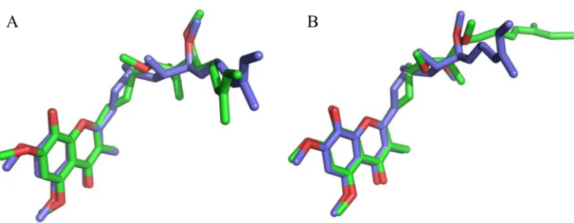
Figure 2.1 In silico optimized (E)-2.8 (green) superimposed with VMD 1.8.6 [182] to the crystallized atomic coordinates (red, RMSD = 0.31Å)………... 34
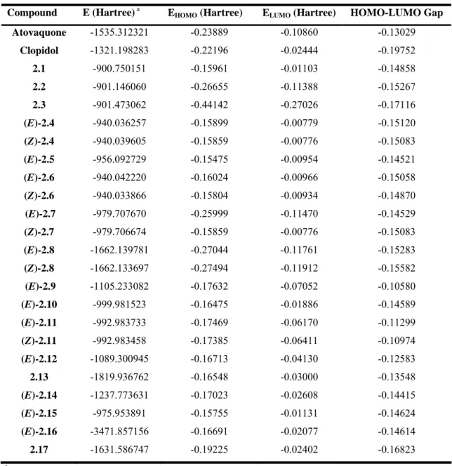
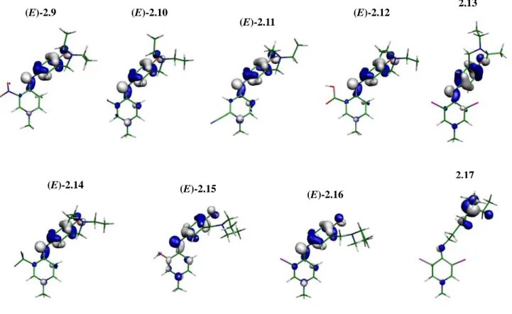
Figure 2.2 LUMOs of atovaquone, clopidol and compounds 2.1-17……… 36
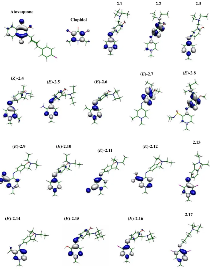
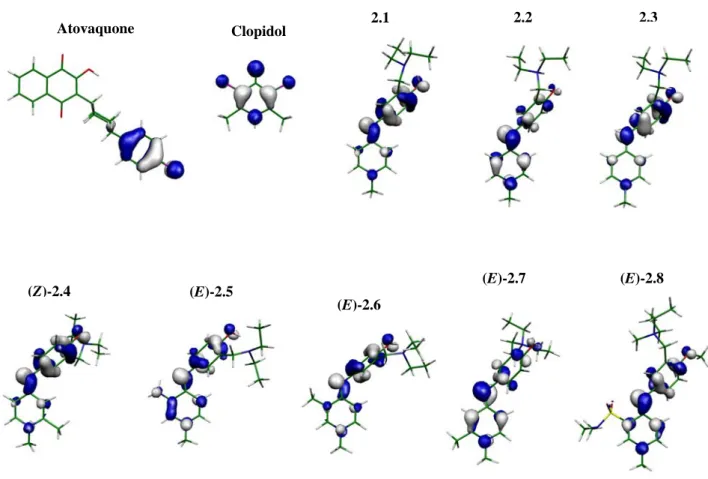
Figure 2.3 HOMOs of atovaquone, clopidol and compounds 2.1-17………37
Figure 2.4 MEPs of atovaquone, clopidol, GW844520 and compounds 2.1-17………... 39
Figure 2.5 Binding poses of stigmatellin. In blue the crystallized structure and in green the docking prediction: (A) ChemScore; (B) GoldScore……… 42
Figure 2.6 Docking poses of (A) atovaquone; (B) clopidol (two possibilities); and (C) 2.1. The molecular electrostatic potential of the Qo pocket calculated through the APBS formalism is also displayed: in red the negative potential (dark red is -15 kT/e) and in blue the neutral potential……….44
Figure 2.7 Binding mode of GW844520………... 44
Figure 2.8 Docking pose of (A) 2.18 and (B) 2.19……… 45
Figure 2.9 Docking poses of (A) 2.20; (B) 2.21; (C) 2.22; (D) 2.23; (E) 2.24; (F) 2.25; (G) 2.26; (H) 2.27; (I) 2.28………. 47
Figure 2.10 Docking poses of (A) 2.29; (B) 2.30; (C) 2.31; (D) 2.32; (E) 2.33; (F) 2.34; (G) 2.35; (H) 2.36; (I) 2.37; (J) 2.38; (K) 2.39; (L) 2.40………..49
Figure 2.11 Docking poses for compounds 2.41-45……….. 50
Figure 2.12 ORTEP view of the molecular structure of 2.8, showing the labelling of all non-hydrogen atoms. Displacement ellipsoids for non-non-hydrogen atoms are shown at the 50% probability level. Hydrogen atoms have been omitted for clarity……… 59
Figure 2.13 2D NOESY spectrum of 2.123………...73
Figure 2.14 Fragmentation pattern for 4-pyridonimines, exemplified by 2.129………... 76
Figure 2.15 Docking poses of (A) clopidol (purple), and GW844520 (blue); (B) 2.130 (purple), and 2.131 (blue)……… 79
percentage of control (MeOH) inhibition. The cytotoxicity was measured in fluorescence (dots) from the Alamar Blue test and is also given as percentage of control. Primaquine was tested at 5 μM. All concentrations were tested and missing bars account for the total suppression of parasite load……….80
Figure 3.1 Predicted LogP for the 4-pyridonimine and 4-quinolonimine scaffolds. ... 85 Figure 3.2 Fragmentation pattern for 4-quinolonimines, exemplified by 3.28 and 3.29... 93 Figure 3.3 ORTEP view of the molecular structure of 3.22, showing the labelling of all
non-hydrogen atoms. Displacement ellipsoids for non-non-hydrogen atoms are shown at the 50% probability level. ... 94
Figure 3.4 Docking poses of 3.20, 3.22 and 3.27, with mesh highlighting the volume and shape of
the ligands inside the Qo binding site of cytochrome bc1... 97
Figure 3.5 Antiplasmodial activity of compounds 3.20-31 at 10 μM (black bars) and 2 μM (grey
bars) against liver stage P. berghei. The luminescence (bars) is given as percentage of control (MeOH) inhibition. The cytotoxicity was measured in fluorescence (dots) from the Alamar Blue test and is also given as percentage of control. Primaquine was tested at 5 μM. All concentrations were tested and missing bars account for the total suppression of parasite load... 98
Figure 3.6 (A) Spectroscopic changes in the Soret band (400 nm) when hematin is titrated with
increasing concentrations of chloroquine (20 ºC, apparent pH 5.5, HEPES buffer with 40% DMSO); (B) Absorbance of chloroquine under the same experimental conditions as (A). ... 101
Figure 3.7 A) Spectroscopic changes in the Soret band (400 nm) when hematin is titrated with
increasing concentrations of clopidol (20 ºC, apparent pH 5.5, HEPES buffer with 40% DMSO). ... 103
Figure 3.7 (cont.) (B) Variation of absorbance of hematin at 400 nm as a function of clopidol
concentration. The solid line represents the best fit curve for the 1:1 stoichiometry model. The curve was corrected for dilution and absorbance of the ligand. ... 104
Figure 3.8 (A) Spectroscopic changes in the Soret band (400 nm) when hematin is titrated with
increasing concentrations of 3.28 (20 ºC, apparent pH 5.5, HEPES buffer with 40% DMSO). ... 104
Variation of absorbance of hematin at 400 nm as a function of 3.28 concentration. The solid line represents the best fit curve for the 1:1 stoichiometry model. The curve was corrected for dilution and absorbance of the ligand...105
Figure 4.1 Docking poses of (A) stigmatellin; (B) 4.12, 4.13, 4.15, 4.16 and 4.31. ...125 Figure 4.2 Antiplasmodial activity of compounds 4.12-21 and 4.31-32 against the liver stage of P.
berghei. The luminescence (bars) is given as percentage of control (DMSO) inhibition.
The cytotoxicity was measured in fluorescence (dots) from the Alamar Blue test and is also given as percentage of control. The compounds were tested in two concentrations: 10 μM (black bars), 2 μM (grey bars) and primaquine was tested at 5 μM. Compound
4.34 was only tested at 10 μM. ...127
Figure 4.3 Dose-response curve of luminescence intensity, as a function of the logarithm of
compound concentration. The red markers refer to logIC50...128
Figure 5.1 The ligand-receptor interactions for GW844520, 1.113; strong hydrophobic interactions
can be seen between the side chain and hydrophobic aminoacid residues. Water molecules have not been included in docking calculations, but are likely to intervene as hydrogen-bond mediators...132
Figure 5.2 Chemical structures of the training set selected for the pharmacophore modeling. ...134 Figure 5.3 (A) shows the pharmacophore model used to screen the ZINC database; (B) shows the
model used for the MOE database. Green spheres represent hydrophobic regions, orange represents aromatic regions, blue is a hydrogen-bond acceptor and its projection and purple represents hydrogen-bond donor and its projection...134
Figure 5.4 Virtual screening protocol breakdown...136 Figure 5.5 Structures of compounds selected from the virtual screening protocol. ...138 Figure 5.6 Docking poses for selected compounds: (A) 5.6; (B) 5.7; (C) 5.10; (D) 5.11...141 Figure 5.6 (cont.) Docking poses for selected compounds: (E) 5.12; (F) 5.20; (G) 5.21; (H) 5.23.
...142
Figure A1.1 Energy-minimized strutures of atovaquone, clopidol and compounds 2.1-17. ...209 Figure A1.2 Docking poses of 2.4, 2.6-8. ...211
Scheme 2.1 Retrosynthetic analysis for Mannich-base derivatives. ... 51
Scheme 2.2 Synthesis of pyridin-4-amines 2.51-58. Reagents and conditions: (i) DEA, CH2O,
EtOH, reflux 24h (ii) DEA, CH2O, EtOH, reflux 48-72h (iii) HCl 6N, reflux overnight (iv) 4-chloropyridine, EtOH, reflux... 52
Scheme 2.3 Synthetic pathway to compounds 2.1 and 2.4-8. Reagents and conditions: (i) a) dry
THF or DMF, NaH, rt b) MeI; (ii) dry THF, alkyl iodide, rt or reflux; (iii) EtOH, 2.49, reflux; (iv) DMF, MeI; (v) NaOH, rt... 55
Scheme 2.4 Retrosynthetic analysis of target 4-pyridonimines. ... 61 Scheme 2.5 Synthetic pathway to compounds 2.72-80. Reagents and conditions: (i) dry benzene or
toluene, PPh3, reflux. ... 62
Scheme 2.6 Synthetic pathway of compounds 2.81-104. Reagents and conditions: (i) a) dry
benzene, n-BuLi, N2, b) aldehyde, rt or reflux; (ii) NaOH, CH2Cl2, aldehyde, rt; (iii) NaOH, CH2Cl2, aldehyde, MW. ... 63
Scheme 2.7 Mechanism for in situ generation of H2... 68
Scheme 2.8 Synthetic pathway to compounds 2.103-110. Reagents and conditions: (i) CH2Cl2,
MeOH, TES, Pd-C 10%, rt. ... 68
Scheme 2.9 Synthetic pathway to compounds 2.113-118. Reagents and conditions: (i) dry toluene,
TfOMe or TfOEt, rt. ... 70
Scheme 2.10 Synthetic pathway to compound 2.119. Reagents and conditions: (i) NaOH, reflux;
(ii) toluene, EtOTf, TEA, rt. ... 71
Scheme 2.11 Synthetic pathway for compound 2.121 and 2.122. Reagents and conditions: (i)
CH3CN, TMSI, N2, reflux; (ii) CH2Cl2, MeOH, NBS, rt, light... 72
Scheme 2.12 Synthetic pathway for compound 2.123. Reagents and conditions: (i) a) DMF, NaH b)
EtI. ... 72
Scheme 2.13 Synthetic pathway to compound 2.124-137. Reagents and conditions: (i) EtOH, TEA,
aniline, reflux... 73
Scheme 2.14 Synthetic pathway to compound 2.138. Reagents and conditions: (i) CHCl3, mCPBA,
reflux (ii) EtOH, TEA, aniline, reflux. ... 75
chloroaniline, 110 ºC (ii) 150 ºC. ...86
Scheme 3.3 Synthetic pathway to compound 3.1. Reagents and conditions: (i) POCl3, reflux. ...87
Scheme 3.4 Attempted synthesis of 3.7. ...88 Scheme 3.5 Retrosynthetic analysis for the phenoxyanilines...88
Scheme 3.6 Synthetic pathway to compounds 3.8-13. Reagents and conditions: (i) DMF, Na2CO3,
CuI, reflux. ...89
Scheme 3.7 Synthetic pathway to compounds 3.14-19. Reagents and conditions: (i) CH2Cl2,
MeOH, TES, Pd-C 10%, rt; (ii) Sn, HCl, reflux. ...90
Scheme 3.8 Synthetic pathway to compounds 3.20-32. Reagents and conditions: (i) EtOH, TEA,
aniline, reflux. ...91
Scheme 3.9 Simulated glutathione attack to afford 3.33. Reagents and conditions: (i) TEA, MeOH,
rt. ...96
Scheme 3.10 Models fitted to the experimental curves. ...100
Scheme 4.1 Retrosynthetic analysis of target flavones...110
Scheme 4.2 Synthetic pathway to compounds 4.1-4. Reagents and conditions: (i) DMF, Na2CO3,
CuI, nuclophile, reflux; (ii) DMF, Na2CO3, nucleophile, reflux. ...111
Scheme 4.3 Synthetic pathway to compounds 4.9. Reagents and conditions: (i)
2-hydroxyacetophenone, DCC, CH2Cl2, DMAP, rt (ii) a) SOCl2, reflux; b) 2-hydroxyacetophenone, CH2Cl2, DMAP, rt; (iii) a) CH2Cl2, TEA, rt; b) ClCO2Et, rt; c) 2-hydroxyacetophenone, DMAP, rt; (iv) 2-hydroxyacetophenone, dry pyridine, rt.
...113
Scheme 4.4 Synthetic pathway for compounds 4.11. Reagents and conditions: (i) Dry pyridine,
3-(trifluoromethyl)benzoyl chloride, DBU, reflux...114
Scheme 4.5 Synthetic pathway to compound 4.28. Reagents and conditions: (i) Sn, HCl, EtOH,
reflux; (ii) dry pyridine, acetic anhydride, reflux; (iii) dry pyridine, DBU, [1,1’-biphenyl]-4-carbonyl chloride, reflux (iv) Similar to (iii), MW...116
Scheme 4.6 Synthetic pathway to compounds 4.29 and 4.30. Reagents and conditions: (i) NBS,
AIBN, benzene, reflux 2 h; (ii) NBS, AIBN, benzene, reflux 24 h; (iii) MeOH, MeONa, reflux. ...116
Scheme 4.7 Synthetic pathway to compounds 4.31-33. Reagents and conditions: (i) NBS, benzoyl
peroxide, CCl4, reflux; (ii) NBS, ZrCl4, CCl4, rt; (iii) MeOH, MeONa, reflux. ...117
Scheme 4.10 Synthetic pathway to compounds 4.35 and 4.36. Reagents and conditions: (i) DMSO,
I2, MW; (ii) DMSO, I2, reflux; (iii) PdCl2, AcONa, AcOH, AIBN; (iv) H2O2 30%, NaOH, EtOH... 120
Scheme 4.11 Synthetic pathway to compound 4.37. Reagents and conditions: (i) TEA, TBTU,
NH(Me)OMe, rt; (ii) dry THF, LiAlH4, -5 ºC; (iii) EtOH, NaOH, acetophenone, rt. ... 120
Scheme 4.12 Retrosynthetic analysis of isoflavones. ... 121 Scheme 4.13 Synthetic procedure for 4.40. Reagents and conditions: (i) DMF-DMA, 95 ºC; (ii)
CHCl3, pyridine, I2, rt. ... 122
Scheme 4.14 Synthetic procedure for 4.41. Reagents and conditions: (i) DMF, CuI, 4-chlorophenol
or 4-fluorophenol, Na2CO3, reflux... 122
Scheme 4.15 Synthetic procedure for compounds 4.42 and 4.43. Reagents and conditions: (i)
4-(fluorophenyl)boronic acid, DME, H2O, Na2CO3, Pd-C, 45 ºC; (ii) 4-chlorophenol, CuI, Na2CO3, DMF, reflux. ... 123
Scheme 4.16 Alternative pathway to 4.43, using the same reactions as in Scheme 4.15. ... 123 Scheme 4.17 Synthetic procedure for compounds 4.45 and 4.46. Reagents and conditions: (i) DMF,
Na2CO3, CuI, 3-(trifluromethoxy)phenol, reflux; (ii) a) dry THF, n-BuLi, borate trisiopropyl, -78 ºC b) HCl 6N, rt; (iii) a) dry THF, Mg, N2, I2, reflux b) triisopropyl borate, -78 ºC c) HCl 6N, rt. ... 123
Table 1.1 In vitro activity of Kigelia pinnata compounds [86]. ...10 Table 1.2 Antiplasmodial in vitro activity of thiophenonaphthoquinone compounds against the BHz
26/28 chloroquine-resistant strain [87]...10
Table 1.3 In vitro activity of 1,4-naphtoquinone compounds [88]...11
Table 1.4 Antiplasmodial activity of ferrocenyl 1,4-naphthoquinone compounds [89]...12
Table 1.5 Antiplasmodial activities of 4(1H)-quinolones [99]...13
Table 1.6 Antiplasmodial activities of acridone series [100]. ...15
Table 1.7 Antiplasmodial activities of 4(1H)-pyridones: influence of side chain on activity [19]. ....17
Table 1.8 Antiplasmodial activities of phenoxyaryl-4(1H)-pyridones [19]. ...19
Table 1.9 Antiplasmodial activities of β-methoxyacrylate against the K1 strain [131]...20
Table 1.10 Antiplasmodial activities of selected chalcones [134]. ...21
Table 2.1 Structures of compounds 2.1-17 (only the (E) conformer is explicitly included)...31 Table 2.2 Selected electronic properties...32 Table 2.3 Selected angle and dihedral angles...33 Table 2.4 Structures and GoldScores of compounds 2.20-28. ...46 Table 2.5 Structures and GoldScores of compounds 2.29-40. ...49 Table 2.6 Structures and GoldScores of compounds 2.41-45. ...51 Table 2.7 Synthesis of key intermediates 2.51-58...53
Table 2.8 Reaction conditions for the SNAr reactions and synthesis of 2.59. ...53
Table 2.9 Synthesis of compounds 2.62-69...55 Table 2.10 Synthesis of compounds 2.1 and 2.4-8. ...56 Table 2.11 Yields of the several species isolated from the alkylation of 2.56. ...57 Table 2.12 Antiplasmodial activity of 4-pyridonimines containing a Mannich-base side chain, 2.1, 2.4 and 2.6-8. ...60 Table 2.13 Phosphonium salts synthesized. ...62 Table 2.14 Comparison of Wittig reaction methods, to acquire 2.81-83. ...64 Table 2.15 Reaction of nitro-substituted benzyltriphenyl phosphonium salts with aldehydes under
standard PTC conditions at room temperature (Method B) and microwave-assisted synthesis (Method C). ...65
Table 2.19 Structure of compounds 2.103-110... 69 Table 2.20 Structure of compounds 2.111 and 2.112. ... 70 Table 2.21 Structure and obtained yields for compounds 2.113-118. ... 71 Table 2.22 Structure of 4-pyridonimines and yields... 74 Table 2.23 Antiplasmodial activity against P. falciparum W2 and FCR3 strains. ... 78
Table 3.1 Structure and yields of 3.4-6. ... 87 Table 3.2 Structure and yileds of 3.8-13. ... 89 Table 3.3 Structure and yields for compounds 3.14-19. ... 90 Table 3.4 Structure and yields of compounds 3.20-32. ... 92
Table 3.5 Effect of R2-R4 and X substitutions in compounds 3.20-31 on the antiplasmodial activity
against P. falciparum W2 strain and association constants (binding to FPIX in 1:1 stoichiometry). ... 95
Table 4.1 Structure and yields of compounds 4.1-4. ... 111 Table 4.2 Structure and yields of compounds 4.5-8. ... 112 Table 4.3 Structure and yields (over 4 steps) of compounds 4.12-21 under standard heating
conditions and MW-assisted synthesis. ... 115
Table 4.4 Substituent effect on the antiplasmodial activity, against the W2 strain, of compounds 4.12-21, 4.31, 4.32. ... 124
Table 5.1 Biological data for compounds selected from virtual screening... 139
Table 7.1 Conditions for the synthesis of 2-(diethylaminomethyl)paracetamol... 152 Table 7.2 Conditions for the synthesis of 4-(pyridin-4-ylamino)phenols... 153 Table 7.3 Conditions for the synthesis of 2.53. ... 154 Table 7.4 Conditions for the synthesis of 2.59. ... 157
The bc1 complex is an attractive a validated drug target in the fight against malaria. The mitochondrial electron transport-chain, in which this complex is involved, is fundamental in
Plasmodium sp.. The parasites do not possess the requested enzymatic machinery to salvage
pyrimidines from their metabolism and, therefore, have to perform de novo pyrimidine biosynthesis to enable their survival. Blockage of this pathway leads to their death. The present work focused on the development of novel inhibitors with structural similarity to known bc1 complex antagonists. Also, this work aimed at delivering novel leads for drug development.
4-Pyridonimines with extended lipophilic side chains showed potential as isosteric replacements for 4(1H)-pyridones. The structure of those compounds was derived from structure-based design and they were active in vitro against P. falciparum. The most active compound presented an IC50 of
ca. 1 μM, and the mode of action was hypothesized through docking studies.
A series of 4-quinolonimines was also prepared. Those presented enhanced antiplasmodial activity in comparison to the previous set of compounds, with IC50s ranging from 0.5 to 1 μM. These also showed outstanding activity against the liver stage of P. berghei. Despite the mechanism of action not being clear at the moment, the compounds demonstrated to bind to hematin. However, the docking studies in the Qo site of the bc1 complex also showed a good fit of the compounds.
Flavones were also synthesized with the aim of optimizing the antiplasmodial activity of stigmatellin. All compounds showed modest activity against both blood and liver stages, with the most active compound presenting an IC50 of 6 μM against P. falciparum W2 strain.
Finally, the virtual screening study that was performed allowed the discovery of novel scaffolds with antiplasmodial activity. A combination of ligand- and receptor-based approaches was successful in retrieving 7 active compounds out of the 23 that were purchased. One of them presented an IC50 of 2 μM in vitro.
KEYWORDS: Cytochrome bc1; 4-pyridonimine; 4-quinolonimine; flavone; molecular docking;
O complexo bc1 é um alvo terapêutico atractivo e validado na luta contra a malária. A cadeia transportadora de electrões, em que este complexo está envolvido, é fundamental em parasitas do género Plasmodium sp.. Os parasitas não possuem as enzimas necessárias para reciclar as pirimidinas vindas do metabolismo e, por isso, necessitam de sintetizá-las de novo, de forma a permitir a sobrevivência do parasita. O bloqueio desta via metabólica conduz à morte sua morte. O presente trabalho incidiu no desenvolvimento de novos inibidores com semelhança estrutural a antagonistas conhecidos do complexo bc1. De igual forma, este trabalho focou-se na descoberta de novos protótipos para o desenvolvimento de novos antimaláricos.
As 4-piridoniminas com cadeias lipofílicas longas mostraram potencial como isósteros das 4(1H)-piridonas. A estrutura dos primeiros foi derivada de estudos de docking molecular e apresentaram actividade in vitro contra P. falciparum. O composto mais activo possui um IC50 de aproximadamente 1 μM e o seu modo de acção foi posto em hipótese por docking molecular.
Uma série de 4-quinolomininas foi também preparada. Estas mostraram ser mais activas que a série de compostos anteriores, com IC50 entre 0,5 e 1 μM, tendo mostrado também excelente actividade contra a fase hepática de P. berghei. Apesar do mecanismo de acção não ser claro neste momento, os compostos mostraram ligar-se à hematina. Contudo, os estudos de docking molecular no sítio Qo do complexo bc1 podem, igualmente, justificar as actividades obtidas.
Foi sintetizada uma série de flavonas com o intuito de optimizar a actividade antiplasmódica da estigmatelina. Todos os compostos obtidos mostraram actividade modesta contra as fases sanguínea e hepática, com o composto mais activo a apresentar um IC50 de 6 μM contra a estirpe W2 de P.
falciparum.
Finalmente, o estudo de screening virtual que foi efectuado permitiu a descoberta de novos núcleos com actividade antiplasmódica. A combinação de um método aplicando, de forma faseada, a informação de ligandos e do receptor resultou na obtenção de 7 compostos activos, de um total de 23 comprados. Um dos compostos apresentou um IC50 de 2 μM in vitro.
PALAVRAS-CHAVE: Citocromo bc1; 4-piridonimina; 4-quinolonimina; flavona; docking
Δψm Mitochondrial Electrochemical Gradient
AcOEt Ethyl acetate
ADMET Absorption, Distribution, Metabolization, Excretion, Toxicity AIBN Azobisisobutyronitrile
ATP Adenosine Triphosphate B3LYP Becke 3 Lee, Yang, Parr br.d Broad doublet br.s Broad singlet
C Cysteine
COSY Correlation Spectroscopy CTH Catalytic Transfer of Hydrogen D Aspartic Acid
d Doublet
dd Doublet of doublets ddd Doublet of doublet of doublets DCC Dicyclohexylcarbodiimide DBU 1,8-Diazabicycloundec-7-ene DEA Diethylamine
DEPT Distortionless Enhancement by Polarization Transfer DFT Density Function Theory
DMAP Dimethylaminopyridine DME Dimethoxyethane DMF N,N-Dimethylformamide DMF-DMA N,N-Dimethylformamide-Dimethylacetal DMSO Dimethylsulfoxide dq Doublet of quartets dt Doublet of triplets E Glutamate EI Electronic Impact Eq. Equivalent EtOH Ethanol
FPIX Ferriprotoporphyrin IX
G Glycine
GFP Green Fluorescent Protein
GOLD Genetic Optimization for Ligand Docking GSK GlaxoSmithKline
H Histidine
HOMO Highest Occupied Molecular Orbital
HMQC Heteronuclear Multiple Quantum Coherence HTS High-Throughput Screening
I Isoleucine IR Infra-Red
ISP Iron-Sulfur Protein
J Coupling constant
K Lysine
Kass Association constant
Kd Dissociation constant
L Leucine
logSw Log Solubility in water
LUMO Lowest Unoccupied Molecular Orbital
M Methionine
m Multiplet
mCPBA Meta-chloroperbenzoic acid
MeOH Methanol
MeONa Sodium Methoxide
MEP Molecular Electrostatic Potential MOE Molecular Operating Environment mp Melting point
mtETC Mitochondrial Electron Transport-Chain MW Microwaves
NADH Nicotinamide Adenine Dinucleotide NBS N-bromosuccinimide
NOESY Nuclear Overhauser Effect Spectroscopy
P Proline
PDB Protein Data Bank
PfNDH2 Alternative type II NADH Dehydrogenase PfDHODH Dihydroorotate Dehydrogenase
PPA Polyphosphoric acid PTC Phase Transfer Catalysis
Py Pyridine
q Quartet
Q.E. Quinine Equivalent Qi Ubiquinone reduction site Qo Ubiquinol oxidation site RMSD Root Mean Square Deviation rt Room temperature
s Singlet
SAR Structure-Activity Relationships SDH Succinate:ubiquinone oxireductase SNAr Nucleophilic Aromatic Substitution SN2 Bimolecular Nucleophilic Substitution
T Threonine
t Triplet
TBTU O-(Benzotriazol-1-yl)-N,N,N’,N’-tetramethylauronium tetrafluoroborate
td Triplet of doublets TEA Triethylamine TES Triethylsilane THF Tetrahydrofuran
TLC Thin Layer Chromatography TMSI Trimethylsilane tt Triplet of triplets V Valine VS Virtual Screening W Tryptophan Y Tyrosine
CHAPTER 1
1. INTRODUCTION
1.1 Overview of malaria and mitochondrial drug targets
Malaria remains a major infectious disease. Approximately half of the world’s population is at risk of infection, with an estimated 300 million new cases annually, and 1 million deaths, mostly children under the age of five [1-3]. Malaria is most prevalent in tropical areas and 90% of all cases occur in Africa, where the death toll is the highest among endemic regions [4, 5]. Five species from the genus Plasmodium cause infection in humans, with Plasmodium falciparum being the most virulent, followed by P. vivax.
The life cycle of malaria parasites is complex and multi-staged. It includes an asexual cycle in humans and a sexual cycle in the Anopheles mosquito. In humans, it can be further distinguished into a liver and an erythrocytic stage [6]. During the blood meal, the mosquito transfers sporozoites into the blood stream, which conceal from the host immune system by invading the hepatocytes. They convert to trophozoites, and divide into several schizonts. After rupture of the hepatocytes, the merozoites are released into the blood stream. These invade the erythrocytes and mature into a trophozoite. Then, the matured trophozoites divide into schizonts, and the merozoites are released into the blood stream to invade other red blood cells. With the rupture of the blood cells, parasitic waste and cell debris are released, causing the clinical symptoms of the disease. After a number of asexual life cycles, the merozoites eventually develop into sexual forms, which are transferred to the mosquito during another blood meal. These gametocytes undergo sexual reproduction within the mosquito mid-gut, producing sporozoites, which finally migrate to the salivary glands and are ready for a new infection [6].
The swift emergence of multi-drug resistant strains is currently impairing both prophylaxis and chemotherapy. Thus, there is an urgent need to find novel drugs, for both known and new drug targets, that might overcome the clinical resistance to marketed antimalarials [7-10].
Pyrimidine biosynthesis has long been known as a potential target for antimalarial chemotherapy [11, 12] and presents a set of attractive drug targets. In higher organisms, the electron transport-chain is composed of four enzyme complexes, located in the inner mitochondrial membrane:
a) NADH:ubiquinone oxidoreductase, complex I;
b) Succinate:ubiquinone oxidoreductase, complex II, succinate dehydrogenase or SDH; c) Ubiquinol:cytochrome c oxidoreductase, complex III or cytochrome bc1;
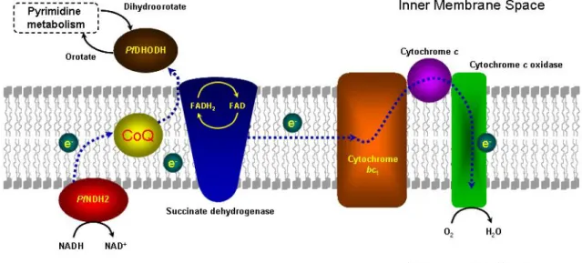
Complexes II through IV are conserved in Plasmodia, but an alternative type II NADH dehydrogenase (PfNDH2) replaces complex I. Additionally, other oxidoreductases, such as dihydroorotate dehydrogenase (PfDHODH) are present in the mitochondria and display an important role in de novo pyrimidine biosynthesis.
1.2 The electron transport-chain pathway
Unlike many eukaryotic cells, malaria parasites obtain almost all their ATP via glycolysis rather than oxidative phosphorylation in the mitochondrion [14, 15]. Additionally, sequencing of the malarial genome has revealed that genes encoding enzymes from the pyrimidine biosynthetic pathway have been conserved, whereas those responsible for salvaging pyrimidines have not [16]. Thus, malaria parasites rely completely on the de novo pyrimidine biosynthesis, essential for the formation of nucleic acids, glycoproteins and phospholipids. Despite its low activity, the mitochondrial electron transport-chain (mtETC) is responsible for maintaining an electrochemical gradient (Δψm) across
the mitochondrial membrane, as well as a constant pool of ubiquinone for pyrimidine biosynthesis [17]. Therefore, the shutdown of the mtETC completely arrests crucial metabolic pathways within the microorganism, rendering these enzymes valid and attractive drug targets. Furthermore, these enzymatic complexes have proven to be structurally different from the homologous human enzymes, which gave rise to the recent interest from both academia and pharma industry [18-20].
Three drug targets have been exploited for the discovery of selective inhibitors: PfNDH2, SDH and cytochrome bc1. The biochemistry, including mechanistic details for these enzymes have been reviewed elsewhere [21, 22]. In short, PfNDH2 catalyses the electron transfer from NADH to ubiquinone in a ping-pong fashion, to maintain a constant pool of NAD+ for reductive metabolic pathways such as glycolysis and the tricarboxylic acid cycle [23]. On the other hand, SDH feeds electrons to complex III, which are ultimately transferred to the final complex [24].
PfDHODH is the fourth enzyme in the de novo biosynthesis of pyrimidines and catalyses the
oxidation of dihydroorotate to orotate at the outer side of the inner mitochondrial membrane. The pair of electrons abstracted from dihydroorotate in this oxidation step is transferred through the flavin mononucleotide co-factor to ubiquinone, that was generated at the bc1 complex [25-27]. Moreover, it is thought that the main metabolic function of the mtETC is to regenerate the ubiquinone necessary for the final step of pyrimidine biosynthesis [28]. Figure 1.1 shows these pathways.
Figure 1.1 Mitochondrial electron transfer chain enzymes and the interplay with PfDHODH from pyrimidine
biosynthesis. (Adapted from http://sites.huji.ac.il/malaria/).
1.3 Cytochrome bc
1inhibitors
Cytochrome bc1 represents the only enzyme complex common to almost all respiratory electron transfer-chains, from Archaea and Bacteria to Eukarya, and its structure has been extensively studied [29, 30]. Cytochrome bc1 consists of 11 different polypeptides, three of which display catalytic functions: cytochrome b, cytochrome c1 and the Rieske protein, or iron-sulfur protein (ISP), due to the iron-sulfur cluster present in it, Figure 1.2 [31]. The ISP is highly mobile and evidence suggests that this feature is crucial for the activity of the complex [32-35].
To date, the modified proton-motive Q cycle mechanism provides the most satisfactory model that accounts for electron transfer coupled to the proton translocation through cytochrome bc1. This is thoroughly reviewed elsewhere [21, 22, 34, 36-39]. Briefly, ubiquinol produced by dehydrogenases upstream to the bc1 complex binds to the oxidation site (Qo) where it is involved in the release of two protons, along with the loss of two electrons into the intermembrane space. Each electron follows a separate path, reducing two different acceptors: a) heme bL located in cytochrome b and b) iron-sulfur cluster in the head domain of the Rieske protein. Next, heme bL reduces heme bH, also located within cytochrome b, which further recycles the electron through the reduction of ubiquinone to ubiquinol at the reduction site (Qi). Meanwhile, the reduced ISP transfers an electron to the heme c group in cytochrome c1. This transfer is accomplished via a conformational shift, during which the histidine acceptor residue at the head group of the ISP rotates, allowing close
contact of the iron-sulfur cluster with heme c. This change is due to the shortening in length of the hinge segment of the ISP [21, 40].
The complex III from numerous organisms has been crystallized with several ligands bound to the oxidation and reduction sites, providing further insight into the complex function [31, 39, 41-45].
Figure 1.2 Cytochrome bc1 complex. Image generated from PDB 1KYO, using PyMol [43, 46].
Cytochrome bc1 has been the major drug target in the mtETC, and its inhibitors can be classified into four groups according to their binding points. Group I, which includes β-methoxyacrylates, bind to the Qo site blocking electron transfer from ubiquinol to the ISP and electron transfer onto the bL centre. Group II, which include hydroxyquinone derivatives, also bind to the Qo site, inhibiting electron transfer from the ISP to cytochrome c1 as well as electron transfer onto the bL centre. Group III include Qi site inhibitors, responsible for blocking electron transfer from the bH centre to ubiquinone. Finally, a fourth group of chromone inhibitors also block the Qo site, but with different properties from those of groups I and II [47].
1.3.1 1,4-Naphthoquinones
Currently, atovaquone, 1.1, is the only drug targeting the bc1 complex in clinical use [48-50]. However, high levels of resistance, related with point mutations in cytochrome b, have been observed for this drug. Consequently, in an attempt to improve its efficiency and decrease the mutation rate, the drug is now used in combination with proguanil. Mutations are predominantly restricted to a highly conserved ‘PEWY’ region, that helps recognition of ubiquinol, and is implied in the electron transfer within the Qo site [13]. The most prevalent point mutations conferring clinical failure of atovaquone have been assigned to codon 268. A specific change of the in vivo wild-type tyrosine to asparagine or serine, Y268N/S, was found to increase the IC50 800-10,000 fold, as a consequence of an altered fit and binding [51-53]. However, despite being sufficient, the Y268 mutation seems not to be necessary for treatment failure [51]. Mutation from methionine to isoleucine on residue 133, M133I, and from leucine to phenylalanine on residue 271, L271F, has generated resistance in vitro and can also be identified in several Plasmodia [54-56]. Other mutations in positions 258, 267, 272 and 280 have also resulted in a 1,000-fold increase of the drug’s IC50 value [8, 57].
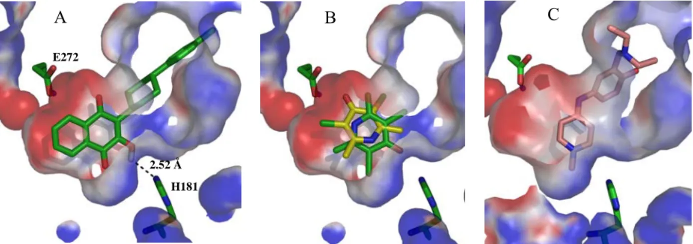
Due to the lack of a crystallized bc1 complex from malaria parasites, molecular modeling studies regarding atovaquone binding to the bc1 complex have been carried out with the homologous enzyme from Saccharomyces cerevisiae, because of the high sequence homology [58]. Atovaquone is a competitive inhibitor for ubiquinol that results in collapse of the parasitic mitochondrial membrane potential, but with no effect on the mammalian counterpart [17]. It binds when the soluble domain of the Rieske protein is proximal to cytochrome b and interacts directly with the ISP. This prevents mobilization to cytochrome c1 and, consequently, impairs the Δψm[14, 49,
58, 59]. It has been predicted that this drug forms a hydrogen bond between the hydroxyl group on the
naphthoquinone ring of the inhibitor and H181 at the ISP. A second water-mediated hydrogen bond between the carboxyl group of E272 and a carbonyl group from the quinone system is also expected, Figure 1.3 [58, 60, 61]. Other putative contact residues are I119, F123, Y126, M133, V140, I141, I144, I258, P260, F264, F267, Y268, L271, V284, L285 and L288 [59, 62, 63].
Atovaquone displays broad antiprotozoal activity, in the low nanomolar range, on several development stages of plasmodia. Moreover, synergism of atovaquone and other naphthoquinones with tetracyclines, dihydrofolate reductase inhibitors, and 4(1H)-pyridones has been reported for the W2 and D6 strains [64-67].
1,4-Naphthoquinones have long been known to possess antiplasmodial activity. Hydrolapachol,
1.2, for instance, was first reported in the 1940s, and in the same decade the antiplasmodial
the isoalkyl side chain of 1.2, by insertion of methylene groups, increased the antimalarial activity in ducks, to a maximum at C9. Longer side chains decreased activity. The same trend could be observed for the n-alkyl series and other related naphthoquinones. Compound 1.3 is almost twice as potent as its isoalkyl and n-alkyl counterparts. On the other hand, introduction of a ring into the side chain, e.g. 1.4, shifted the activity peak to compounds with a higher number of carbons in the side chain, C10 or C11. When two rings are present, as in 1.5-7, the maximum quinine equivalent, Q.E. - the ratio of dose of quinine, given in mg/kg, and that of the drug under assay which cause the first sharp drop in parasitemia in relation to untreated controls [68] - shifts to C12 or C13. All trans diastereomers are more potent than their cis isomers, and compound 1.7 with a Q.E. equal to 15.3 is the most potent molecule. Moreover, the 2-OH group seems indispensable for activity as loss of activity was observed with several other substituents: OMe, SH, Me, H, Cl and NHCOMe. The same trend was obtained when substitutions were made in the side chain, and in the naphthoquinone ring. Methyl groups reduced the activity of the compounds when introduced in the core scaffold
[68-73].
Figure 1.3 Atovaquone docked at the oxidation site of the yeast bc1 complex [58].
Metabolism studies of naphthoquinones 1.3 and 1.4 demonstrated the oxidation of the side chains, yielding metabolites with significantly reduced antimalarial activity. In fact, compound 1.4 is rapidly metabolized, and any suppressive activity observed for this compound is due to its long-lived metabolite 1.8. Thus, hydroxyl groups have been introduced in the following series, resulting in compounds metabolically more stable, despite having lower activity [70, 74].
However, it was noted that an increase in the number of carbon atoms could compensate for any drop of activity, and the introduction of an aryl moiety afforded 1.9, which was stable and as active as 1.4. With these features in mind, a series of hydroxylated naphthoquinones were built, yielding lapinone 1.10 and other naphthoquinones alike, which displayed high activity and antirespiratory effect [70, 74-76].
Due to the emergence of chloroquine resistant strains in the 1960s, a renewed interest in naphthoquinones emerged. The synthesis of 3-cyclohexylalkyl and adamantyl 2-hydroxy-1,4-naphthoquinone derivatives afforded compounds with good antimalarial activities against P.
berghei. Using chloroquine as a control for suppression of malaria, over 28 days at 5 mg/kg, similar
results were obtained for 25 mg/kg of 1.11, and menoctone, 1.12, derivatives, while decreased activity was observed for the 3-(ω-cyclohexylnonyl) homologue 1.13. For the adamantyl series, activities were lower, and compound 1.14 cured mice at 40 mg/kg for a week, but at half dose only two of five mice were cleared from parasitemia. Indeed, for 1.14 relapse was noted after 14 days [77,
In another study, a series of sixty four 1,4-naphthoquinone derivatives were assayed for curative activity on P. berghei. From these, compounds 1.15 and 1.16 displayed good antimalarial activity
[79].
In 1981, the ongoing effort to find efficient and metabolism-resistant molecules led to the discovery of parvaquone, 1.17, and its cis-dicyclohexyl analogue, 1.18; these were equipotent, and ten times more active than their template menoctone. Introduction of oxygen and nitrogen atoms into the cycloalkyl substituent reduced activity [80], while subsequent exploration of the cyclohexyl substituent afforded BW58C, 1.19, a broad spectrum antiprotozoal. This was over 5,600 times more potent than 1.12, over 1300 times more potent than 1.17, and approximately 650-fold more active than chloroquine in in vitro assays. However, in P. yoelii infected mice, BW58C was only four times as active as chloroquine, with and ED50 equal to 1.19 mg/kg, 7 x p.o. The diastereomer mixture of 1.19 also showed activity on P. cynonolgi and no apparent recrudescence was noted. Further studies revealed that it also had prophylactic activity against P. berghei. Nonetheless, in humans, the tert-butyl group is rapidly hydroxylated to a 1000-fold less active metabolite and further development was discontinued. Replacement of the tert-butyl by a 4-chlorophenyl group affords atovaquone, 1.1 [81, 82]. Several structural modifications on atovaquone have been examined, either to improve activity or the formulation properties of tablets / i.v. dosage forms. Thus, derivatization of the hydroxyl group in 1.1 as a phosphate or carbamate yielded compounds that were shown to be useful for both treatment and prophylaxis of malaria [83-85].
More recently, a series of 2-hydroxy-1,4-naphthoquinones derived from rhinacanthin, 1.20, showed potent antiplasmodial activity. The optimum side chain length was found to have C13, 1.21, or C15, 1.22, with IC50 values of 32 and 30 nM, respectively, against the K1 multi-drug resistant strain. It was also noted that the α-methyl substituent in the ester moiety was beneficial for antiplasmodial activity, when compared to its β and α-ethyl counterparts. Furthermore, the geminal methyl groups in the propyl chain are pivotal for the activity in this series. Compound 1.21 provided also specific Qo site inhibition, IC50 = 79.6 ± 3.41 nM, against the homologous yeast bc1 complex, and poor inhibition of the rat enzyme, IC50 = 2,495 ± 820 nM [18].
Additionaly, a series of four unique naphthoquinones isolated from the rootbark of Kigelia
pinnata demonstrated useful antiplasmodial activity.
2-(1-Hydroxyethyl)naphtho[2,3-b]furan-4,9-dione, 1.23, was found to be the most active molecule, with IC50 values of 627 nM and 718 nM against the K1 and T9-96 P. falciparum strains, respectively, Table 1.1. Isopinnatal, 1.24, kigelinol,
1.25, and isokigelinol, 1.26, exhibited lower activities, especially the latter two. Moreover, despite
the cytotoxicity of these compounds, the antiplasmodial activity was not due to in vitro cytotoxicity, as the selectivity indexes were of at least 10. The study also suggested that furanonaphthoquinones possessed much less affinity to parasitic mitochondria when compared to naphthoquinones, and that minor changes in furanonaphthoquinones would favour accumulation in the parasitic mitochondrial membrane. This would eventually increase the activity [86].
Table 1.1 In vitro activity of Kigelia pinnata compounds [86]. Antiplasmodial Activity IC50 (nM) Compound K1 strain T9-96 strain 1.23 627 718 1.24 763 1,552 1.25 16,660 15,200 1.26 15,200 11,930
Similarly, fully synthetic thiophenonaphthoquinones 1.27-32 displayed moderate to good in
vitro activity against P. falciparum at 0.2 μM, but were not active in vivo, Table 1.2 [87].
Table 1.2 Antiplasmodial in vitro activity of thiophenonaphthoquinone compounds against the BHz 26/28
chloroquine-resistant strain [87]. Compound R1 R2 % infection reduction at 0.2 μM 1.27 H H 55 1.28 H 8-OMe 7 1.29 H 5-OMe 78 1.30 H 6-OMe 78 1.31 H 7,8-di-OMe 51 1.32 2-NO2 H 45
A series of amino-1,4-naphthoquinones was also tested for antiplasmodial activity and 2-amino-3-chloro-1,4-naphthoquinone, 1.33, was the most potent compound with an IC50 of 180 nM against the W2 strain, Table 1.3. The presence of a primary amino group in R1 appeared to be essential for activity, since substitution of that group for a halogen, 1.34, decreased activity by 260-fold, while inclusion of other amino groups at R2, 1.35-37, rendered compounds with only modest antiplasmodial activity [88].
Table 1.3 In vitro activity of 1,4-naphtoquinone compounds [88].
Antiplasmodial activity IC50 (nM) Compound R1 R2 R3 R4 R5 R6 W2 strain D6 strain 1.33 NH2 Cl H H H H 180 920 1.34 Cl Cl H H H H 43,260 43,980 1.35 Cl N-pyrrolidino H H H H 9,630 36,650 1.36 Cl N-morpholino H H H H 31,960 115,140 1.37 H N-anilino H H H H 47,670 63,970
Given that the use of metal complexes capable of enhancing the activity of biological compounds has become a relevant strategy, a small library of ferrocene derivatives of 1,4-naphthoquinone was built, incorporating a modified side chain of 6-8 carbons. Those displayed moderate antiplasmodial activity. The IC50 of compounds 1.38-40 was 3 to 6-fold higher than that of atovaquone, hinting that the ferrocene unit is damaging to activity, Table 1.4. Thus, it was suggested that this series do not act at the bc1 complex level [89].
Table 1.4 Antiplasmodial activity of ferrocenyl 1,4-naphthoquinone compounds [89]. IC50 (μM) Compound R1 3D7 strain Dd2 strain 1.38 (CH2)5CH3 5 ± 0.4 2.5 ± 0.3 1.39 (CH2)6CH3 2.5 ± 0.3 5 ± 0.4 1.40 (CH2)7CH3 6.25 ± 1.5 6 ± 1.25 Atovaquone 0.6 ± 0.2 0.7 ± 0.35 1.3.2 4(1H)-Quinolones
4(1H)-Quinolones are also valuable antiprotozoal scaffolds, acting on the mitochondrial electron transport-chain [11, 90-92]. Much work, directed at improving both the antimalarial activity and the solubility of endochin, 1.41, in water has been carried out. Structural modifications include carbonates, N-oxides, Mannich-bases and esters. Introduction of the N-hydroxyl group in the endochin molecule (BD26235), 1.42, for example, resulted in increased water solubility and in improved antimalarial activity [90, 91, 93]. However, substitution of the alkyl side chain for a cyclopentyl group, or substitution of the methoxy group by a chlorine atom, results in appreciable loss of activity [94]. A separate study yielded more promising results, with some alkenylquinolones,
1.43-45, displaying activity or curative properties on infected mice. Compound 1.45 had activity
comparable to that of endochin, with an IC50 of 5.7-16.6 nM, and displayed no cross resistance with some marketed antimalarials [95]. Additionally, elimination of the double bond in conjugation with the quinolone ring destroyed activity [96]. Compound 1.42, on the other hand, proved to be non-toxic and was curative in infected chicks. BE11382, 1.46, increased the mean survival time in parasite infected mice, suggesting that a substituent at C7 of 3-carboethoxy-4(1H)-quinolones may be beneficial [97].
More recently, in a search for new scaffolds that comply with the structural features of the 4(1H)-quinolones, an in silico pharmacophore model was employed to screen virtual libraries of compounds [98]. Also, based on the structures of WR 194,905, 1.47, and WR 197,236, 1.48, a small set of compounds was synthesised and important structure activity relationships were drawn.
Simple quinolones without a long alkyl side chain yielded IC50 values of 2.5 μM, 1.49, 2.3 μM,
1.50, and 325 nM, 1.51, while compounds with longer side chains were active in the low nanomolar
range, e.g. IC50 1.2 nM for 1.52. Moreover, evidence suggests a mechanism of action similar to that of atovaquone, as significant cross resistance was attained against the Tm90-C2B strain. A 3-trifluorohexyl group also improved antimalarial activity by 70-fold over the corresponding unsubstituted counterpart, e.g. 1.53 vs. 1.50 and, more interestingly, no cross resistance to atovaquone was observed. Regarding the metabolism, this substituent may also be interesting since its terminal location is expected to block cytochrome P450 mediated oxidation. Other structure activity relationships that can be drawn are: substitution of 7-OMe for 7-OH reduces activity, and lengthening of the haloalkyl side chain increases it. This class of compounds also showed synergism with 4(1H)-pyridones and inhibition of oxygen consumption at the bc1 complex level [99]. A summary of antiplasmodial activities can be found in Table 1.5.
Table 1.5 Antiplasmodial activities of 4(1H)-quinolones [99].
IC50 (nM)
Compound
D6 strain Dd2 strain Tm90-C2B strain
1.49 > 2,500 > 2,500 > 2,500 1.50 2,300 1,260 1,290 1.51 325 303 750 1.52 1.2 1.2 270 1.53 32 30 66 1.54 680 390 1,360 1.55 1.25 1.44 4.7 1.56 7.3 5.5 26.6 Endochin 3.2 2.8 17.4 Atovaquone 0.3 0.5 5,090
1.3.3 Acridones
Related acridones have also demonstrated potent antiplasmodial activity, Table 1.6 [100, 101]. These reveal structure activity trends identical to the quinolones; those containing a longer side chain and terminal CF3 groups exhibit higher potencies, with IC50s as low as ~1 pM, 1.78. The ring nitrogen is also critical for activity, given the observed decrease in potency by over 50,000-fold when the nitrogen is replaced by oxygen; the xanthone 1.88 has an IC50 of 16 μM. It was also noted that alkylation of the nitrogen resulted in drop of activity, to a lesser extent, i.e. 1.85 [100].
Table 1.6 Antiplasmodial activities of acridone series [100]. IC50 (nM) Compound R1 R2 R3 D6 strain Dd2 strain 1.57 H H H 2,000 2,500 1.58 H 2-NH2 H 50,000 50,000 1.59 H 2-OMe H 329 271 1.60 H 3-OMe H 283 224 1.61 H 2-OH H 9,000 4,400 1.62 H 3-OH H 2,000 504 1.63 6-Cl 2-OMe H 45 65 1.64 6-Cl 2-OH H 190 260 1.65 6-Cl 2-O(CH2)4CH2Br H 70 152 1.66 6-Cl 2-O(CH2)4CH2Cl H 46 40 1.67 6-Cl 2-O(CH2)5NMe2 H 67 95 1.68 6-Cl 3-OMe H 76 192 1.69 6-Cl 3-OH H 2,200 9,200 1.70 6-Cl 3-O(CH2)4CH2Br H 27 54 1.71 6-Cl 3-O(CH2)4CH2Cl H 12 13 1.72 6-Cl 3-O(CH2)3CF3 H 1.0 1.2 1.73 6-Cl 3-O(CH2)4CF3 H 0.3 0.5 1.74 H 3-O(CH2)4CF3 H 0.5 0.3 1.75 6-Cl 3-O(CH2)5CF3 H 0.06 0.07 1.76 6-Cl 2-O(CH2)5CF3 H 10 15 1.77 6-Cl 3-O(CH2)4CF2CF3 H 0.02 0.02 1.78 6-Cl 3-O(CH2)4CF(CF3)2 H 0.0015 0.0008 1.79 H 2-O(CH2)5CF3 H 36 49 1.80 H 3-O(CH2)5CF3 H 0.43 0.015 1.81 H 4-O(CH2)5CF3 H 446 515 1.82 6-Cl 3-O(CH2)7CF3 H 0.16 0.17 1.83 6-Cl 3-O(CH2)7CH2OH H 2.2 3.5 1.84 6-Cl 3-O(CH2)10CF3 H 0.023 0.025 1.85 6-Cl 3-O(CH2)4CF3 Me 4,000 3,500 1.86 6-NO2 3-O(CH2)5CF3 H 3.2 5.8 1.87 6-NH2 3-O(CH2)5CF3 H 0.018 0.025
1.3.4 Acridinediones
Acridinediones are another class of known potent antimalarials [102-104], that have been reported to inhibit the parasite respiratory pathway, causing a reduction of oxygen consumption. Acridinediones are predicted to block the bc1 complex, but small changes into their structure affect not only their potency, but also their mechanism of action. While some inhibit the bc1 complex others inhibit hematin polymerization [20, 64, 105, 106]. (S)-WR 249685, 1.89, and racemic floxacrine,
1.90, are two selective bc1 complex inhibitors for P. falciparum. Their IC50s for the enzyme are in
the nanomolar range and consistent with whole cell growth inhibition. Additionally, data suggests mild cross resistance with atovaquone, associated to bc1 complex mutations. This results in an increase of the IC50s, which is an indication of Qo site blocking [20]. Compounds that lack the N-hydroxyl present in 1.90 have reduced antiplasmodial activity in general; N-allyl derivatives display modest activity, while the N-alkyl acridinediones are inactive. Replacement of the ketone function at the 1-position by an imine, afforded derivatives with comparable activities, and longer side chains on the imine moiety improved activity. Compounds 1.91 and 1.92 were curative in doses as low as 5 mg/kg in mice. Commonly, an aryl moiety at C3 is required for high potency, as alkyl substituents either yield compounds that are devoid of antiplasmodial activity or are marginally active. Electron withdrawing groups in the C3-aryl moiety also enhance potency, whereas electron donating groups diminish it. Interestingly, a methyl group at C2′ of the aromatic ring is not deleterious, as opposed to bulkier substituents which decrease the potency. A chlorine located at C7 is also important for activity, but the lack of any substituents in positions C5 and C8 affords molecules with lowered effectiveness [107].
1.3.5 4(1H)-Pyridones
The novel class of 4(1H)-pyridones is based on clopidol, 1.93. Clopidol is long known for its antiplasmodial (curative at 160 mg/kg) and anticoccidal activities through inhibition of mitochondrial respiration [108, 109]. However, its poor solubility in several solvents led to the synthesis of various derivatives [108, 110]. Currently, GlaxoSmithKline (GSK) is developing a series of clopidol analogues with more lipophilic side chains in an effort first disclosed in 1991 [111-113]. Compared to its lead, the n-octyl derivative 1.94 has enhanced activity in vitro, but is inactive in
vivo due to metabolic degradation of the side chain, Table 1.7. Introduction of side chains less prone
to metabolism, 1.95-99, improved not only the in vitro, but also in vivo activities [19].
Table 1.7 Antiplasmodial activities of 4(1H)-pyridones: influence of side chain on activity [19].
Compound R P. falciparum T9-96 IC50 (nM) P. yoelii ED50 (mg/kg) 1.93 Cl 20,000 40 1.94 n-C8H17 4,000 > 60 1.95 Ph 11,000 22 1.96 2,500 20 1.97 50 0.6 1.98 400 0.7 1.99 60 0.6 Atovaquone 3 0.03
Further structure-activity relationship (SAR) analysis on derivatives containing the 3(4’-phenoxy)phenyl side chain was carried out. A halogen at C5,either chlorine or bromine, leads to more potent derivatives. Though, other electron withdrawing substituents at that position do not improve activity, and electron donating moieties result in a significant increase of the IC50, Table 1.8. Variation within the terminal aryl moiety does not influence activity significantly, and the phenoxy side chain is best positioned at 3′ or 4′; a much reduced activity is observed for the 2′
analogue, i.e. 1.120. The methyl groups at C2 and C6 also appear to be critical, and significant loss of activity is achieved upon their withdrawal or their replacement by a trifluoromethoxy group. The
N-oxide derivatives are 10-fold less active than their pyridone counterparts, e.g. 1.125 vs. 1.104 and
1.126 vs. 1.102 [19].
In addition to potent activity against erythrocytic stages of malaria, these compounds showed in
vitro and in vivo activity against liver stages, making them amenable to prophylaxis [19, 114].
ADME studies on compound GW844520, 1.113, revealed a half-life adequate for short duration of oral therapy, activity against resistant isolates, and no cross resistance with atovaquone, among other features. However, according to the MMV 2005 annual report its development was discontinued due to toxic properties [19, 115, 116]. GW308678, 1.110, was then selected for further development, but unfortunally showed recrudescence at any dose up to 32 mg/kg [117]. Since 2006, two further patents from GSK have disclosed structural modifications of the lead compound: compounds with biaryl or related side chains at C3, and those containing modified side chains instead of methyl groups at C2 or C6 display promising in vitro antiplasmodial activities for advanced development [118, 119]. The MMV portfolio for the second quarter of 2010 includes one of those compounds, GSK932121, 1.127, in phase I of clinical trials [120].
Table 1.8 Antiplasmodial activities of phenoxyaryl-4(1H)-pyridones [19]. P. falciparum IC50 (nM) Compound R1 R2 R3 X Isomer R4 T9-96 3D7A P. yoelii ED50 (mg/kg) 1.100 Me Me H Br 4-OAr H 150 N.A. 4 1.101 Me Me H Br 4-OAr 4-F 40 N.A. 0.6 1.102 Me Me H H 4-OAr 4-Cl 250 N.A. 2.5 1.103 Me Me H Cl 4-OAr 4-Cl 60 N.A. 1.7 1.104 Me Me H Br 4-OAr 4-Cl 40 N.A. 0.3 (5) 1.105 Me Me H Cl 4-OAr 3-Cl 30 N.A. > 5 1.106 Me Me H Br 4-OAr 3-Cl 30 N.A. 3.9 1.107 Me Me H H 4-OAr 4-CF3 500 N.A. 1.3 1.108 Me Me H Cl 4-OAr 4-CF3 60 N.A. 0.6 1.109 Me Me H Br 4-OAr 4-CF3 30 N.A. 0.3 (1.1) 1.110 Me Me H Cl 4-OAr 3-CF3 30 N.A. 0.2 (0.2-0.6) 1.111 Me Me H Br 4-OAr 3-CF3 30 N.A. 0.6 (3.6) 1.112 Me Me H H 4-OAr 4-OCF3 160 160 > 5 1.113 Me Me H Cl 4-OAr 4-OCF3 30 5 0.2 (0.4-1.3) 1.114 Me Me H Br 4-OAr 4-OCF3 30 8 0.3 (0.2-0.5)
1.115 Me Me H CF3 4-OAr 4-OCF3 N.A. 30 N.A.
1.116 Me Me H NO2 4-OAr 4-OCF3 N.A. 30 N.A.
1.117 Me Me H OMe 4-OAr 3-CF3 N.A. 300 N.A.
1.118 Me Me H 4-OAr 4-OCF3 N.A. 1,290 N.A.
1.119 Me Me H Br 3-OAr 4-OCF3 N.A. 7 N.A.
1.120 Me Me H Br 2-OAr 4-OCF3 N.A. 400 N.A.
1.121 H Me H Br 4-OAr 4-OCF3 N.A. 200 N.A.
1.122 Me H H Br 4-OAr 4-OCF3 N.A. 110 N.A.
1.123 Me CF3 H Br 4-OAr 4-OCF3 N.A. > 1,000 N.A.
1.124 H CF3 H Br 4-OAr 4-OCF3 N.A. > 1,000 N.A.
1.125 Me Me OH Br 4-OAr 4-Cl 450 N.A. ~ 1,000
1.126 Me Me OH H 4-OAr 4-Cl 2,200 N.A. > 1,000
![Figure 1.2 Cytochrome bc 1 complex. Image generated from PDB 1KYO, using PyMol [43, 46]](https://thumb-eu.123doks.com/thumbv2/123dok_br/15186050.1016391/40.892.214.634.285.678/figure-cytochrome-complex-image-generated-pdb-using-pymol.webp)
![Figure 1.3 Atovaquone docked at the oxidation site of the yeast bc 1 complex [58] .](https://thumb-eu.123doks.com/thumbv2/123dok_br/15186050.1016391/42.892.280.569.569.901/figure-atovaquone-docked-oxidation-site-yeast-bc-complex.webp)
![Table 1.8 Antiplasmodial activities of phenoxyaryl-4(1H)-pyridones [19] . P. falciparum IC 50 (nM) Compound R 1 R 2 R 3 X Isomer R 4 T9-96 3D7A P](https://thumb-eu.123doks.com/thumbv2/123dok_br/15186050.1016391/55.892.119.815.197.1070/table-antiplasmodial-activities-phenoxyaryl-pyridones-falciparum-compound-isomer.webp)