ORIGIN
AL RESEAR
CH
Protective efects of exercise against sepsis-induced
energy metabolism dysfunction in skeletal muscle of
rats
Efeitos protetores do exercício contra a disfunção do metabolismo energético induzida por sepse
na musculatura esquelético de ratos
Efectos del ejercicio contra la disfunción del metabolismo energético inducida por sepsis en la
musculatura esquelética de ratones
Carla Werlang-Coelho1,2,3, Glauco Adrieno Westphal2, Felipe Dal-Pizzol4,
Emilio Luiz Streck4, Eliezer Silva1,5
Mailing address: Carla Werlang Coelho – Rua Paulo Malschitzki, 10 – Campus Universitário – CEP 89219-710 – Joinville (SC), Brazil – E-mail: [email protected] – Financing source: Fundação de Apoio à Pesquisa do Estado de Santa Catarina (FAPESC) – Conlicts of interest: nothing to declare – Presentation: Mar. 2014 – Accepted for publication: Apr. 2015 Approved by the Brazilian College of Animal Experimentation and Ethics Committee of UNIVILLE, Protocol No. 008/08 – COEA.
Study developed at Universidade da Regiao de Joinville (UNIVALLE), Joinville (SC), Brazil.Paulista (UNESP) - Marília (SP), Brazil.
1Postgraduate Program in Anesthesiology of the School of Medicine of Universidade de São Paulo – São Paulo (SP), Brazil.
2Physical Education and Medicine Department of Universidade da Região de Joinville (UNIVILLE) – Joinville (SC), Brazil.
3Chemistry Department of Universidade do Estado de Santa Catarina (UDESC-Joinville) – Joinville (SC), Brazil.
4Experimental Pathophysiology Laboratory of Universidade do Extremo Sul Catarinense (UNESC) – Criciúma (SC), Brazil.
5Intensive Care Unit of Hospital Israelita Albert Einstein – São Paulo (SP), Brazil.
ABSTRACT | We evaluated effects of aerobic
physical preconditioning on general performance and energy metabolism in skeletal muscle of septic rats. Forty-eight 10-wk-old male Wistar rats were randomly assigned to either Untrained or Trained groups. Aerobic exercise training protocol (AETP) consisted of an 8-week treadmill program. After AETP, performance was evaluated by graded treadmill and functional ambulation testing. Afterwards animals from both groups were randomly assigned to Sham or CLP surgery (cecal ligation and perforation), resulting in the following groups: Sham untrained (ShamU), CLP untrained (CLPU), Sham trained (ShamT), and CLP trained (CLPT). Two days after surgery, animals repeated the ambulation test, and were euthanized after this. Diaphragm, soleus and plantaris muscles were harvested. Mitochondrial electron transport chain enzyme (METC) and creatine kinase (CK) activity were measured. AETP led to significant improvement in performance of distance run and in skeletal muscle function of the Trained group. Forty-eight hours after surgery the CLPT group was able to maintain similar muscle performance as Sham groups. Dysfunction was shown in the diaphragm in METC complexes I and II-III and in locomotive soleus muscles in complex I; CK enzyme activity was significantly increased in sedentary 133
CLPU group in soleus and plantaris muscle, but in the diaphragm there was only a tendency (p=0.07). CLPT animals that were submitted to AETP avoided all these negative results. Taken together our results provide evidence of the positive effects obtained with an aerobic physical preconditioning program on METC and CK enzyme activity related to the diaphragm and locomotive muscles mitigating sepsis-induced energy metabolism dysfunction.
Keywords | Sepsis; Muscle, Skeletal; Energy Metabolism; Exercise.
RESUMO | Foram avaliados os efeitos do
(CLPT). Dois dias após a cirurgia, os animais repetiram o teste de deambulação e logo após foram sacriicados. O diafragma e os músculos sóleo e plantar foram colhidos. A atividade da enzima da cadeia mitocondrial transportadora de elétrons (METC) e da creatina quinase (CK) foi medida. A AETP levou a uma melhoria signiicativa no desempenho em corridas de longa distância e na função da musculatura esquelética do grupo treinado. Quarenta e oito horas após a cirurgia, o grupo CLPT foi capaz de manter um desempenho muscular semelhante ao dos grupos Sham. Foi mostrada disfunção no diafragma nos complexos METC I e II-III e nos músculos sóleos locomotivos no complexo I; a atividade da enzima CK sofreu aumento signiicativo no grupo CLPU sedentário em músculo sóleo e plantar, mas no diafragma havia apenas uma tendência (p=0,07). Os animais CLPT que foram submetidos ao AETP evitaram todos estes resultados negativos. Tomados em conjunto, nossos resultados fornecem evidências dos efeitos positivos obtidos com um programa de pré-condicionamento físico aeróbico em relação à atividade das enzimas METC e CK relacionada ao diafragma e aos músculos locomotivos atenuando a disfunção do metabolismo energético induzida por sepse.
Descritores | Sepse; Músculo Esquelético; Metabolismo Energético; Exercício.
RESUMEN | En este estudio se analizó los efectos del
preacondicionamiento físico aeróbico en el rendimiento general y en el metabolismo energético de la musculatura esquelética de ratones sépticas. Se seleccionaron aleatoriamente 48 ratones Wistar con 10 semanas de edad, asignados en los grupos “entrenado” y “no entrenado”. El protocolo de entrenamiento del
ejercicio aeróbico (AETP) constituyó por un programa de tapiz rodante de ocho semanas. Tras el AETP se evaluó el rendimiento a través de tapiz rodante y de pruebas de deambulación funcional. En seguida, se dividieron aleatoriamente los ratones de ambos grupos en Sham o cirugía CLP (ligadura cecal y perforación), teniendo como resultados los grupos: Sham no entrenado (ShamU), CLP no entrenado (CLPU), Sham entrenado (ShamT) y CLP entrenado (CLPT). Dos días después de la cirugía, se repitió la prueba de deambulación en los animales y, en seguida, se los sacriicaron, recolectando el diafragma y los músculos sóleo y plantar. Se midió la actividad de las enzimas de la cadena mitocondrial de transporte de electrones (METC) y de la creatina quinasa (CK). El AETP tuvo una mejora signiicativa en el rendimiento en carreras de larga distancia y en la función de la musculatura esquelética del grupo entrenado. Cuarenta y ocho horas tras la cirugía, el grupo CLPT mantuvo un rendimiento muscular semejante al de Sham. Se mostró una disfunción en el diafragma en los complejos METC I y II-III así como en los músculos sóleos locomotores del complejo I; sufrió un aumento la actividad de la enzima CK en el grupo CLPU sedentario con músculo sóleo y plantar, pero en el diagrama hubo sólo una tendencia (p=0,07). Los animales CLPT que fueron sometidos al AETP no tuvieron estos resultados negativos. Los resultados mostraron indicios de efectos positivos obtenidos por preacondicionamiento físico aeróbico sobre la actividad de las enzimas METC y CK relacionada al diafragma y a los músculos locomotores, disminuido, así, la disfunción energética inducida por sepsis.
Palabras clave | Sepsis; Músculo Esquelético; Metabolismo Energético; Ejercicio.
INTRODUCTION
Patients with sepsis-induced multiple organ dysfunction (MOD) often experience muscle fatigue in both locomotive and respiratory muscles1,2. Muscle
fatigue prolongs the stay in the intensive care unit, mainly because of prolonged weaning from the ventilator, and the recovery after a period of intensive care treatment3,4. Muscle fatigue arises largely because
the muscle is incapable of producing energy during contraction. As mitochondria are the main producers of cellular energy and skeletal muscle and comprise 50-60% of body cell mass, these organelles and tissues play a key role in the pathogenesis of muscle dysfunction and fatigability in sepsis5.
he Krebs cycle and the electron transport chain occur in the matrix and in the inner mitochondrial membrane, respectively, and are responsible for more than 90% of adenosine triphosphate (ATP) generation. Researches have suggested that during sepsis there is structural injury of mitochondria in various systemic organ tissues and skeletal muscle6,7.
In addition, several animal models of sepsis and critical illness have shown mitochondrial derangements and the subsequent disruption of energy metabolism in skeletal muscle and other tissues6,8,9
, conirming
In contrast to sepsis, exercises, as a chronic contractile activity, produce muscle mitochondrial biogenesis11.
his adaptation leads to a signiicant change in aerobic energy metabolism and corresponding improvements in resistance to fatigue12. It is important to point out that
skeletal muscle is a highly malleable tissue, capable of considerable metabolic and morphological adaptations in response to repeated bouts of contractile activity (i.e. exercise)13. Highly speciic adaptations are induced in
muscles by contractile activity, and depend on the type of exercise (i.e. resistance vs. endurance), its frequency, intensity and duration14. It has been well established
that chronic endurance exercise results in a change in expression of a wide variety of gene products, leading to a change in the muscle phenotype and enhancing resistance to fatigue15,16. here is high correlation
between this enhanced resistance and increase in the mitochondrial density and enzyme activity of muscles, referred to as “mitochondrial biogenesis”17.
herefore, the improvement in the number of mitochondria and their function in the skeletal muscle before sepsis insult may avoid the occurrence of aerobic metabolism dysfunction in both locomotive and diaphragm muscles during the sepsis syndrome. In fact, treadmill exercise before sepsis induction in an animal model was associated with attenuation of septic inlammatory responses and mitigation of organ
damage18,19. Furthermore, our group demonstrated
that exercise training before sepsis stimulus enabled avoiding oxidative stress and protect locomotive skeletal muscle from damage20. In the present study, we tested
the hypothesis that endurance training before sepsis inducement could produce phenotypic adaptations that would confer protection of the diaphragm and locomotive muscles against sepsis-induced energy metabolic dysfunction. hus, the purpose of this study was to evaluate the efects of aerobic physical preconditioning acquired through endurance training on the general performance and on energy metabolism in both the diaphragm and locomotive muscles of rats with sepsis induced by cecal ligation and perforation (CLP).
METHODOLOGY
Animals
Adult male Wistar rats (70 days old) were obtained from the breeding colony of Universidade do Vale de
Itajai (UNIVALI). herats were maintained in a light-dark cycle (12:12hr) in temperature controlled (22 °C) environment with free access to standard laboratory chow (20% protein, 70% carbohydrate and 10% lipid, from Nuvital Nutrientes) and tap water. Initially, after one week of adaptation, the animals were randomly assigned to untrained (n=24) and trained (n=24) groups. Upon completion of the eight weeks of aerobic exercise training protocol, animals from the trained group were randomly assigned to Sham (fake) or CLP surgery (cecal ligation and perforation). he untrained group was subjected to the same surgical procedures. After this phase, there were the following groups: Sham trained (ShamT; n=7), CLP trained (CLPT; n=17), Sham untrained (ShamU; n=7) and CLP untrained (CLPU; n=17). All rats were euthanized two days post Sham and CLP surgery. his study was conducted in accordance with the ethical principles in animal research adopted by the Colégio Brasileiro de Experimentação Animal (COBEA) and approved by the Ethics Committee of Universidade da Região de Joinville (UNIVILLE), protocol No. 008/08 – COEA.
Graded treadmill exercise test
Before the irst exercise test, rats were conditioned to treadmill exercises over a period of a week (10 minutes of exercise per session). During the graded treadmill exercise test, rats were placed on the treadmill and allowed to acclimatize for at least 15 minutes. he intensity of theexercise was then increased by 3m/min (6-33m/min) every 3 minutes at 0% inclination until exhaustion (the point of maximum running speed). he graded treadmill exercise test was performed before the exercise training and then during the 4th and 8th week
of exercise training. Exercise capacity was estimated by the total distance run, correlated with skeletal muscle capacity, which is a method used for detecting exercise intolerance. hus, exercise capacity was evaluated using a graded treadmill exercise protocol as previously described21.
Skeletal muscle functional assessment
the average was computed from three attempts. he size of the rat (naso-anal length) was also measured and the relationship between the average stride length and size of the rat was determined, generating an ambulation index22.
Aerobic exercise training protocol
he aerobic exercise training protocol consisted of an 8 week program of running on a motorized treadmill (KT-4000 model INBRAMED, RS, Brazil) for 5 days a week during 60 minutes at 60% of the maximum running speed obtained in the graded treadmill test, as described in other study21. All untrained rats were
exposed to treadmill exercises (5 minutes) three times a week to become familiarized with the exercise protocol and handling.
CLP Surgery
he animals from both untrained and trained groups were subjected to the surgical procedure 72 hours after the last treadmill exercise test, as previously described23.
For the CLP surgery, the rats were anesthetized with ketamine (80mg/kg), administered intraperitoneally. Under aseptic conditions, a laparotomy with a 3cm midline was performed to allow the exposure of the cecum with the adjoining intestine. he cecum was tightly ligated with a 3.0 silk suture at its base, below the ileocecal valve, and was perforated once with a 14-gauge needle. he cecum was then gently squeezed to extrude a small amount of feces from the perforation site before being returned to the peritoneal cavity. he laparotomy was then closed with 3.0 silk sutures. All animals received antibiotics (ceftriaxone 30mg/kg and clindamycin 25mg/kg) starting 6 hours after CLP and then every 6 hours up to 24 hours after CLP. Afterwards, they were all returned to their cages with free access to food and water. In the Sham-operated group, rats were submitted to all surgical procedures and received isotonic saline solution immediately after the surgery. hey also received antibiotics, but the cecum was not ligated or perforated.
All survivor animals were included as subjects of this study and euthanized two days after the surgery, and their soleus, plantaris and diaphragm muscles were harvested for posterior analysis. he sepsis survival rate in this model was of approximately 60%.
Mitochondrial electron transport chain enzymes activity
Skeletal muscles were homogenized (1:10, wt/vol) in SETH bufer (250mM sucrose, 2mM EDTA, 10mM Trizma base, 50IU/ml heparin, pH 7.4) for determining the mitochondrial respiratory chain enzyme activities (complexes I, II, II–III, and IV). NADH dehydrogenase (complex I) was evaluated according to the method described by the rate of NADH-dependent ferricyanide reduction at 420nm24. he activities of succinate –
DCIP oxidoreductase (complex II) – and succinate – cytochrome c oxidoreductase (complex II-III) – were
determined according to the method of Fischer25.
Complex II activity was measured by following the decrease in absorbance due to the reduction of 2,6-DCIP at 600nm. Complex II-III activity was measured by cytochrome c reduction from succinate. he activity of cytochrome c oxidase (complex IV) was assayed by following the decrease in absorbance due to the oxidation of previously reduced cytochrome c at 550nm26.
Creatine kinase enzyme activity
Creatine kinase activity was measured in skeletal muscles homogenates pretreated with 0.625mM lauryl maltoside. he reaction mixture consisted of 60mM Tris–HCl, pH 7.5, containing 7mM phosphocreatine, 9mM MgSO4 and approximately 0.4-1.2µg protein in a inal volume of 100µL. After 15min of preincubation at 37° C, the reaction was started by the addition of 0.3µmol of ADP plus 0.08µmol of reduced glutathione. he reaction was stopped after 10min by the addition of 1µmol of p-hydroxymercuribenzoic acid. he creatine formed was estimated according to the colorimetric
method of Hughes27. he color was developed by
the addition of 100µL 2% α-naphtol and 100µL
0.05% diacetyl in a inal volume of 1mL and read spectrophotometrically after 20min at 540nm.
STATISTICAL ANALYSIS
he data are presented as means and standard error of the means (mean±SEM). Two-way ANOVA with
post hoc testing by Duncal was used to compare the
Statistical signiicance was considered achieved when the p-value was <0.05.
RESULTS
Exercise performance
After 8 weeks of aerobic exercise training protocol, the trained group showed a greater increase in performance in distance run and improvement in skeletal muscle function measured through the ambulation test. (Figure 1).
Energy metabolism
Related enzyme activity of mitochondrial electron transport chain (METC) and creatine kinase (CK): 48 hours after surgery, the diaphragm, soleus and plantaris muscles were harvested from all experimental groups for biochemical analyses.
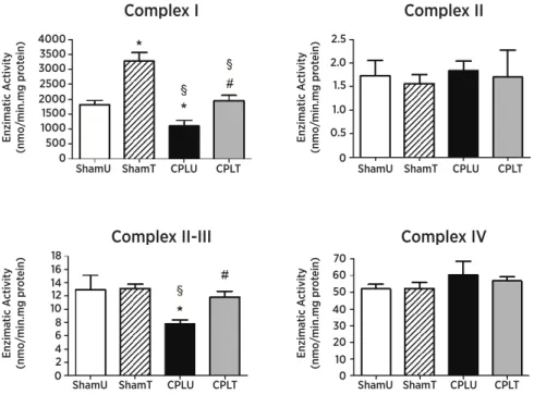
As shown in Figure 2, the enzyme activities of complexes I and II-III from the diaphragm muscle of the rats in the CLPU group were signiicantly decreased compared with all the experimental groups (p<0.05). However, these complex activities from the diaphragm muscle of the group of trained rats (CLPT)
Untrained n=24
Trained Untrained Trained
n=24 n=24 n=24
600
500
400
300
200
100
0
0.6 0.7 0.8
0.5
0.4 0.3 0.2 0.1 0
*p<0.05 vs. untrained
Figure 1. The capacity of exercise tolerance represented by the maximum distance covered in maximal exercise test (A) and the strength of skeletal muscles represented by the ambulation index accomplished in ambulation test (B) in Wistar rats from trained and untrained groups in the pre-surgical time interval. The tests were performed after eight weeks of training protocol on treadmill. Data are presented as mean±SEM and were analyzed using the Student’s t test
4000 3500 3000 2500 2000 1500 1000 500 0
2.5
2.0
1.5
1.0
0.5
0
16 18
14 12 10 8 6 4 2 0
70 60 50
40 30 20 10 0 ShamU
Enzima
tic A
ctivit
y
(nmo/
min.mg pr
ot
ein)
Enzima
tic A
ctivit
y
(nmo/
min.mg pr
ot
ein)
Enzima
tic A
ctivit
y
(nmo/
min.mg pr
ot
ein)
Enzima
tic A
ctivit
y
(nmo/
min.mg pr
ot
ein)
ShamT
Complex I Complex II
Complex II-III Complex IV
CPLU CPLT ShamU ShamT CPLU CPLT
ShamU ShamT CPLU CPLT ShamU ShamT CPLU CPLT
*p<0.05 vs. ShamU; #p<0.05 vs. CLPU; §p<0.05 vs. ShamT
physical training, Peruchi et al10. investigated whether
sepsis induced by CLP could modify the activity of mitochondrial enzymes evaluated by comparing the METC of the diaphragm with that of the quadriceps. At 48 hours after CLP, only the diaphragm showed signiicant decrease in the four METC complexes, which led the authors to conclude that this appeared to be secondary to early oxidative stress and it was correlated with decreased muscle contractile force10. In
our experiment, we used the same experimental sepsis induction model23, the same 48 hour period and the
diaphragm, but we evaluated the soleus and plantaris muscles in addition before making the comparisons. As our goal was to study the efects of training on sepsis-induced skeletal myopathy, various skeletal muscles were evaluated, representing diferent locations and predominance of muscle iber types: (1) the diaphragm, with predominantly oxidative muscle iber (central), (2) the soleus, also with predominantly oxidative (peripheral), and (3) the plantar, with predominantly glycolytic (peripheral). his is particularly important due to muscle adaptations being dependent on the characteristics of the types of muscles, and the type of training33.
In our study, the activity of the METC complexes and CK enzyme was evaluated. Regarding the METC enzymes in the diaphragm, our results corroborate the indings of Peruchi et al.10, which showed a signiicant
decrease in the diaphragm METC complexes10. In the
trained group with sepsis (CLPT), the METC enzyme activity of the diaphragm was maintained within the normal limits. Whereas, the plantaris muscle, similarly to the quadriceps with regard to the prevalence of type II ibers (glycolytic), showed results difering from those of Peruchi et al.10, which showed an increase in enzyme
activity of the METC group complexes with induced sepsis. Regarding the plantaris muscle of the CLPU group, our results showed a decrease in the activity of METC complexes, which was more prominent in complexes II-III, with p=0.064. When this situation of sepsis was assessed in the soleus muscle, with a predominance of type I ibers (oxidative), a statistically signiicant decrease in the activity of complex I was shown.
he exercise protocol used in this study was of predominantly aerobic type21 and was characterized
by moderate and long duration. Many studies have demonstrated the adaptation mechanisms of moderate exercise, such as improved metabolism,
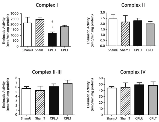
were able to maintain values similar to those of the control group (ShamU). In the locomotive muscle, as was the case in the soleus muscle from the CLPU group, the enzyme activity of complex I was shown to be signiicantly decreased (p<0.05) in comparison with those of the ShamU and ShamT groups. Moreover, the CLPT group was able to achieve a value similar to that of the ShamU group (Figure 3). As shown in Figure 4,the enzyme activity of complex II-III from the locomotive muscle (plantaris) in the CLPU group was decreased (p=0.06) in comparison with that of the ShamU group, however, there was statistically signiicant decrease only when compared with the ShamT group (p<0.05).
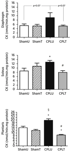
Figure 5 shows the enzyme activity by CK levels in the muscles of the diaphragm, soleus and plantaris. he CLPU group showed signiicantly increased activity in the soleus (B) and plantaris (C) when compared with the ShamU group (p<0.05). In addition, there was an increase in the diaphragm (A) CK enzymatic activity in comparison with the ShamU group, but the statistical test showed a p value of 0.07. he CLPT group presented CK activity values similar to those of the ShamU group in all muscles studied (Figure 5 A, B, C).
DISCUSSION
In the present research, we tested whether exercise training before the induction of sepsis would prevent energy metabolism dysfunction in the diaphragm and locomotive muscles. We found that diaphragm and locomotive muscles are sensitive to the positive efects of exercise training against energy metabolism dysfunction. Furthermore, we showed not only that the locomotive muscle plantaris is more resistant to aerobic metabolism dysfunction than the soleus, but also that exercise training could improve the energy metabolism and muscle strength in the septic groups. herefore, our study reveals that there were protective efects of exercise training against sepsis-related muscle energy metabolism dysfunction. In fact, we have recently showed that sepsis induces mitochondrial electron transport chain dysfunction in the diaphragm muscle10, and in another study, we also
3000 2500 2000 1500 1000 500 0
2.5 3.0
2.0 1.5 1.0 0.5 0.0
7 8
6 5 4 3 2 1 0
60
50
40
30 20
10
0 ShamU
Enzima
tic A
ctivit
y
(nmo/
min.mg pr
ot
ein)
Enzima
tic A
ctivit
y
(nmo/
min.mg pr
ot
ein)
Enzima
tic A
ctivit
y
(nmo/
min.mg pr
ot
ein)
Enzima
tic A
ctivit
y
(nmo/
min.mg pr
ot
ein)
ShamT
Complex I Complex II
Complex II-III Complex IV
CPLU CPLT ShamU ShamT CPLU CPLT
ShamU ShamT CPLU CPLT ShamU ShamT CPLU CPLT
1200 1400
1000 800 600 400 200 0
5 6
4
3
2
1
0
14 16 18 20
p=0.64
12 10 8 6 4 2 0
60
50
40
30
20
10
0 ShamU
Enzima
tic A
ctivit
y
(nmo/
min.mg pr
ot
ein)
Enzima
tic A
ctivit
y
(nmo/
min.mg pr
ot
ein)
Enzima
tic A
ctivit
y
(nmo/
min.mg pr
ot
ein)
Enzima
tic A
ctivit
y
(nmo/
min.mg pr
ot
ein)
ShamT
Complex I Complex II
Complex II-III Complex IV
CPLU CPLT ShamU ShamT CPLU CPLT
ShamU ShamT CPLU CPLT ShamU ShamT CPLU CPLT
*p<0.05 vs. ShamU; #p<0.05 vs. CLPU; §p<0.05 vs. ShamT
Figure 3. Mitochondrial electron transport chain enzyme activity (METC) of the soleus muscle 48 hours after surgical procedures. The animals were divided into four groups: ShamU (n=7), ShamT (n=7), CLPU (n=9) and CLPT (n=9). The data are presented as mean±SEM and were compared between groups by two-way analysis of variance (ANOVA) with post hoc Duncan
*p<0.05 vs. ShamU; #p<0.05 vs. CLPU; §p<0.05 vs. ShamT
Regular endurance exercise is a widely recognized modality for the general improvement of strength and metabolic function28,29. Our data show that after eight
weeks of exercise training, the tolerance to exercise and strength of skeletal muscles were signiicantly improved in the trained group (p<0.05).
It has been well described in the literature that mitochondrial dysfunction was implicated in organ dysfunction pathogenesis6. Within the context of sepsis,
mitochondrial dysfunction has been demonstrated in the liver30, brain structures31, heart32 and skeletal
muscle10. In a similar experimental design, but without
reduction in oxidative stress and increase in the
antioxidant enzyme system14,20. In discussing
mitochondrial function, which is directly related to aerobic metabolism, our results support the claim that training improves and/or maintains the aerobic metabolism of the diaphragm muscles even after the induction of sepsis, thereby managing to preserve mitochondrial function.
Metabolic compensation is capable of increasing the enzyme CK system of high-energy phosphates, which plays a central role in the metabolism of tissues such as muscle, which consume large amounts of energy34.
Furthermore the elevation of CK may be an indicator of muscle damage35. Our results indicated signiicant
exacerbation of CK enzyme activity in the soleus, plantaris and diaphragm of the CLPU group, suggesting that there was metabolic compensation and there may have been microlesions in the muscle.
Taken together, our results provide evidence of the positive efect of aerobic physical preconditioning obtained with an aerobic exercise training program in mitigating sepsis-induced skeletal muscle energy metabolism dysfunction. herefore, the behavior of the diaphragm, which was more susceptible to dysfunction in METC complexes I and II-III, was shown to be diferent, and the locomotive muscle behavior was more related todysfunction in CK activity. hus, the clinical implications of these results in septic patients suggest that people who exercise regularly may have less muscle metabolic disorders.
Further studies are warranted to gain better understanding of the mechanisms underlying the diferential regulation of skeletal metabolic dysfunction, taking into consideration muscles composed of diferent types of ibers and with diferent functional characteristics.
REFERENCES
1. Bolton CF. Neuromuscular manifestations of critical illness. Muscle Nerve. 2005;32(2):140-63.
2. Letter MACJ, Schmitz PI, Visser LH, Verheul FA, Schellens RLLA, Op Coul DA, et al. Risk factors for the development of polyneuropathy and myopathy in critically ill patients. Crit Care Med. 2001;29(12):2281-6.
3. Lanone S, Taille C, Boczkowski J, Aubier M. Diaphragmatic fatigue during sepsis and septic shock. Intens Care Med. 2005;31(12):1611-7.
4. Laghi F, Tobin MJ. Disorders of the respiratory muscles. Am J Resp Crit Care Med. 2003;168(1):10-48.
14 16
p=0.07 p=0.07
12 10 8 6 4 2 0
Diaphr
agm
CK (nmol/
min.mg pr
ot
ein)
ShamU ShamT CPLU CPLT
14 16
12 10 8 6 4 2 0
Soleus
CK (nmol/
min.mg pr
ot
ein)
ShamU ShamT CPLU CPLT
1416 18 2022 24 26
12 10 8 6 4 2 0
Plan
taris
CK (nmol/
min.mg pr
ot
ein)
ShamU ShamT CPLU CPLT
*p<0.05 vs. ShamU; #p<0.05 vs. CLPU; §p<0.05 vs. ShamT
5. Fredriksson K, Rooyackers O. Mitochondrial function in sepsis: respiratory versus leg muscle. Crit Care Med. 2007;5(Supl 9):S449-53.
6. Crouser ED, Julian MW, Blaho DV, Pfeifer DR. Endotoxin-induced mitochondrial damage correlates with impaired respiratory activity. Crit Care Med. 2002;30(2):276-84. 7. Porta F, Takala J, Weikert C, Bracht H, Kolarova A, Lauterburg
BH, et al. Efects of prolonged endotoxemia on liver, skeletal muscle and kidney mitochondrial function. Crit Care. 2006;10(4):R118.
8. Brealey D, Karyampudi S, Jacques TS, Novelli M, Stidwill R, Taylor V, et al. Mitochondrial dysfunction in a long-term rodent model of sepsis and organ failure. Am J Physiol. 2004;286(3):R491-7.
9. Fredriksson K, Hammarqvist F, Strigard K, Hultenby K, Ljungqvist O, Wernerman J, et al. Derangements in mitochondrial metabolism in intercostal and leg muscle of critically ill patients with sepsis-induced multiple organ failure. Am J Physiol Endocrinol Metab. 2006;291(5):E1044-50. 10. Peruchi BB, Petronilho F, Rojas HA, Constantino L, Mina F, Vuolo
F, et al. Skeletal muscle electron transport chain dysfunction after sepsis in rats. J Surg Res. 2011;15;167(2):e333-8.
11. Hood DA. Invited review: contractile activity-induced mitochondrial biogenesis in skeletal muscle. J Appl Physiol. 2001;90(3):1137-57.
12. Hood DA, Saleem A. Exercise-induced mitochondrial biogenesis in skeletal muscle. Nutr Metab Cardiovasc Dis. 2007;17(5):332-7.
13. Bassel-Duby R, Olson EN. Signaling pathways in skeletal muscle remodeling. Annu Rev Biochem. 2006;75:19-37. 14. Powers SK, Criswell D, Lawler J, Ji LL, Martin D, Herb RA, et al.
Inluence of exercise and iber type on antioxidant enzyme activity in rat skeletal muscle. Am J Physiol. 1994;266(2 Pt 2):R375-80.
15. Boveris A, Navarro A. Systemic and mitochondrial adaptive responses to moderate exercise in rodents. Free Radical Bio Med. 2008;44(2):224-9.
16. Cofey VG, Hawley JA. The molecular bases of training adaptation. Sports Med. 2007;37(9):737-63.
17. Hood DA, Adhihetty PJ, Colavecchia M, Gordon JW, Irrcher I, Joseph AM, et al. Mitochondrial biogenesis and the role of the protein import pathway. Med Sci Sports Exer. 2003;35(1):86-94.
18. Chen HI, Hsieh SY, Yang FL, Hsu YH, Lin CC. Exercise training attenuates septic responses in conscious rats. Med Sci Sports Exer. 2007;39(3):435-42.
19. Araujo CC, Silva JD, Samary CS, Guimaraes IH, Marques PS, Oliveira GP, et al. Regular and moderate exercise before experimental sepsis reduces the risk of lung and distal organ injury. J Appl Physiol. 2012;112(7):1206-14.
20. Coelho CW, Jannig PR, Souza AB, Fronza Jr H, Westphal GA, Petronilho F, et al. Exercise training prevents skeletal muscle damage in an experimental sepsis model. Clinics (Sao Paulo, Brazil). 2013;68(1):107-14.
21. Ferreira JC, Rolim NP, Bartholomeu JB, Gobatto CA, Kokubun E, Brum PC. Maximal lactate steady state in running mice: efect of exercise training. Clin Exp Pharmacol Physiol. 2007;34(8):760-5.
22. Kennel PF, Fonteneau P, Martin E, Schmidt JM, Azzouz M, Borg J, et al. Electromyographical and motor performance studies in the Pmn mouse model of neurodegenerative disease. Neurobiol Dis. 1996;3(2):137-47.
23. Ritter C, Andrades M, Frota Junior ML, Bonatto F, Pinho RA, Polydoro M, et al. Oxidative parameters and mortality in sepsis induced by cecal ligation and perforation. Intens Care Med. 2003;29(10):1782-9.
24. Cassina A, Radi R. Diferential inhibitory action of nitric oxide and peroxynitrite on mitochondrial electron transport. Arch Biochem Biophys. 1996;328(2):309-16.
25. Fischer JC, Ruitenbeek W, Berden JA, Trijbels JM, Veerkamp JH, Stadhouders AM, et al. Diferential investigation of the capacity of succinate oxidation in human skeletal muscle. Clin Chim Acta. Int J Clin Chem. 1985;153(1):23-36.
26. Miro O, Cardellach F, Barrientos A, Casademont J, Rotig A, Rustin P. Cytochrome c oxidase assay in minute amounts of human skeletal muscle using single wavelength spectrophotometers. J Neurosci Method. 1998;80(1):107-11. 27. Hughes BP. A method for the estimation of serum creatine
kinase and its use in comparing creatine kinase and aldolase activity in normal and pathological sera. Clin Chim Acta. Int J Clin Chem. 1962;7(5):597-603.
28. Noble EG, Milne KJ, Melling CW. Heat shock proteins and exercise: a primer. Appl Physiol Nutr Metab. 2008;33(5):1050-65.
29. Powers SK, Jackson MJ. Exercise-induced oxidative stress: cellular mechanisms and impact on muscle force production. Physiol Rev. 2008;88(4):1243-76.
30. Crouser ED. Mitochondrial dysfunction in septic shock and multiple organ dysfunction syndrome. Mitochondrion. 2004;4(5-6):729-41.
31. Comim CM, Rezin GT, Scaini G, Di-Pietro PB, Cardoso MR, Petronilho FC, et al. Mitochondrial respiratory chain and creatine kinase activities in rat brain after sepsis induced by cecal ligation and perforation. Mitochondrion. 2008;8(4):313-8.
32. Joshi MS, Julian MW, Huf JE, Bauer JA, Xia Y, Crouser ED. Calcineurin regulates myocardial function during acute endotoxemia. Am J Respir Crit Care Med. 2006;173(9):999-1007.
33. Adhihetty PJ, Irrcher I, Joseph AM, Ljubicic V, Hood DA. Plasticity of skeletal muscle mitochondria in response to contractile activity. Exp Physiol. 2003;88(1):99-107.
34. Wallimann T, Wyss M, Brdiczka D, Nicolay K, Eppenberger HM. Intracellular compartmentation, structure and function of creatine kinase isoenzymes in tissues with high and luctuating energy demands: the ‘phosphocreatine circuit’ for cellular energy homeostasis. Biochem J. 1992;281 ( Pt 1):21-40.