Androgen receptor is expressed in murine choroid plexuses and downregulated by 5?-dihydrotestosterone in male and female mice
80
0
0
Texto
(2) Androgen receptor is expressed in murine choroid plexuses and downregulated by 5α-dihydrotestosterone in male and female mice.. Master Thesis in Biochemistry Project supervised by: Cecília Santos, PhD.
(3) This scientific project is exclusive property of Universidade da Beira Interior, and its contents are from the responsibility of the author (C. Henrique Alves). ________________________________________________________________ III.
(4) Acknowledgments. Acknowledgments First, I like to thank to all the good friends, Ana Martinho, Ana Ramalhinho, Claudio, Olga, Susana, Eugénia, Telma, that every days work with me, in the laboratory, making all easier, and funny. To my family for the friendship and support, they are always present when I need. A great kiss to my dear Sandra, she knows.... A big hug to the people “in the house” Nuno Macedo, Ricardo Eiras, Pedro Varão. A special acknowledgment to Professor Cecília Santos, that made this master thesis possible.. ________________________________________________________________ IV.
(5) Abbreviations Index. Abbreviations Index . Aβ - β-amyloid peptide AD - Alzheimer’s disease AF1 - Activation function 1 AF2 - Activation function 2 AGE - Advanced glycation products APP - Aβ precursor protein AR - Androgen receptor BDNF - Brain derived nerve factor b2mgb - β-2 microglobulin CNS - Central nervous system CPs - Choroid plexuses CSF - Cerebrospinal fluid DBD - DNA-binding domain DHEA - Dehydroepiandrosterone DHT - 5α-dihydrotestosterone ELISA - Enzyme-linked immunoadsorbent assay ERβ - Estrogen receptor β ERK - Extracellular signal regulated kinase ERT - Estrogen replacement therapy GDNF - Glial cell line-derived neurotrophic factor GDX - Gonadectomy Hsp - Heat shock protein ISH - In situ hybridization IHC - Immunohistochemistry LBD - Ligand binding domain MAPK - Mitogen activated protein kinase MCI - Mild cognitive impairment NGF - Nerve growth factor NFT - Neurofibrillary tangles NLS - Nuclear localization signal NTD - N-terminal domain PBS - Phosphate buffered saline PCR - Polymerase chain reaction PFA - Paraformaldehyde PNS - Peripherical nervous system PR - Progesterone receptor PREG - Pregnenolone PROG - Progesterone PVDF - Polyvinylidene diflouride P450c11β - P450scc and 11 β -hydroxylase sAPPα - Soluble APP-α SARMS - Selective androgen receptor modulators SP - Senile plaques SSC - Saline sodium citrate TBS - Tris buffered saline ________________________________________________________________ V.
(6) Abbreviations Index . TGF-β - Transforming growth factor β TTR - Transthyretin 17β-HSD - 17β-hydroxysteroid dehydrogenase 3β-HSD - 3β-hydroxysteroid dehydrogenase-isomerase. ________________________________________________________________ VI.
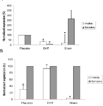
(7) Figure Index. Figure Index Figure 1: Morphological illustration of CPs within the lateral ventricle......................... 4 Figure 2: The neurosteroidogenic pathway. .................................................................. 13 Figure 3: Sex steroid synthesis in the CNS. .................................................................. 14 Figure 4: Structural organization of the human AR gene and protein .......................... 22 Figure 5: ISH using a specific rat AR cRNA riboprobe................................................ 32 Figure 6: Western blot analysis of 20μg of total protein extracted from male and female rat CPs, and female liver (L) using an antibody against the C-terminal region (C19)........................................................................................................................... 33 Figure 7: Immunostaining of rat CPs using an antibody against the AR C-terminal region (C- 19) ......................................................................................................... 34 Figure 8: Comparison of AR expression in male and female CPs from mice by Real Time PCR. .............................................................................................................. 35. ________________________________________________________________ VII.
(8) Table Index. Table Index Table 1: Polypeptides synthesized in CPs............................................................6 Table 2: Pathological changes of CPs in AD.......................................................8. ________________________________________________________________ VIII.
(9) Index. Index I. Resumo.............................................................................................................. 1 I. Abstract ............................................................................................................. 2 II. Introduction...................................................................................................... 3 1) Alzheimer’s Disease..................................................................................... 3 2) Choroid Plexus ............................................................................................. 3 2.1) Structure ................................................................................................ 3 2.2) Function................................................................................................. 4 2.3) Ageing of the choroid plexus ................................................................ 7 2.4) Implication in Alzheimer’s disease ....................................................... 8 2.5) Neuroprotective molecules produced by CPs and regulated by androgens................................................................................................................ 10 3) Neurosteroids ............................................................................................. 12 3.1) Neurosteroids synthesis....................................................................... 12 3.2) Testosterone and ageing ...................................................................... 14 3.3) Implication of testosterone in neuroprotection.................................... 15 3.4) Testosterone and Alzheimer’s disease ................................................ 17 4) Androgen receptor...................................................................................... 21 4.1) Gene and protein structure .................................................................. 21 4.2) Function and physiologic roles ........................................................... 23 4.3) Implication in neurodisease / Alzheimer’s disease ............................. 24 III. Objectives ..................................................................................................... 26 IV. Material and methods ................................................................................... 27 1) Animals ...................................................................................................... 27 2) DHT stimulation experiment...................................................................... 27 3) AR mRNA detection .................................................................................. 28 3.1) RNA isolation...................................................................................... 28 3.2) RT- PCR.............................................................................................. 28 3.3) ISH ...................................................................................................... 29 4) AR protein analysis .................................................................................... 29 ________________________________________________________________.
(10) Index 4.1) Antibodies ........................................................................................... 29 4.2) Western blot ........................................................................................ 30 4.3) IHC ...................................................................................................... 30 5) Analysis of the AR transcription response to DHT treatment by Real Time PCR............................................................................................................................. 31 V. Results ........................................................................................................... 32 1) AR mRNA and protein are present in CPs epithelial cells......................... 32 2) DHT down-regulates AR expression in CPs of male and female mice ..... 34 VI. Discussion .................................................................................................... 36 VII. Conclusions and Future Perspectives.......................................................... 39 VIII. References.................................................................................................. 40 IX. Annexes - Protocols ..................................................................................... 58 1) In situ Hybridization Protocol (Paraffin sections)...................................... 58 2) Western blot ............................................................................................... 65 3) Immunohistochemistry ............................................................................... 68. ________________________________________________________________.
(11) I. Resumo. I. Resumo A doença de Alzheimer (AD) é um dos maiores problemas de saúde pública, nos países desenvolvidos. É caracterizada por uma deteorização progressiva da memória e das funções corticais superiores, culminando numa degradação total das actividades intelectuais e mentais. As características neuropatológicas da doença são: a deposição extracelular de placas de amilóide, constituídas por fibrilhas do péptido amilóide β (Aβ),. a formação de agregados neurofibrilares, e a perda de neurónios. Estudos. recentes comprovam que alterações morfológicas e funcionais nos plexos coróideu (CPs) estão associadas à AD. Os CPs formam uma barreira única entre o sangue periférico e o líquido cefalorraquidiano (CSF). Vários péptidos envolvidos no processo de neuroprotecção, tais como, o factor de crescimento neural (NGF), o factor β de crescimento e transformação (TGF-β), o factor neural derivado do cérebro (BDNF), a transtirretina (TTR), e o factor neurotrófico derivado de uma linha de células da glia (GDNF), são secretados pelos CPs, e regulados pelos androgénios noutros tecidos, porém os mecanismos de regulação destes, nos CPs, continuam por elucidar. Importa salientar. que. os. androgénios. podem. actuar. a. nível. cognitivo,. e. como. neuroprotectores. Sabe-se que, a testosterona tem um papel inibidor sobre o stress oxidativo, a apoptose, e a toxicidade da Aβ, sendo todos estes efeitos mediados pelo receptor de androgénios (AR). O AR foi identificado em várias zonas do sistema nervoso central (CNS): no hipotálamo, na amígdala, no hipocampo e no córtex, porém a sua presença nunca foi descrita no CP. O AR nunca foi identificado no CP do cérebro, nem a hipótese deste ser um órgão responsivo aos androgénios foi averiguada. Para esclarecer estas questões, investigamos e caracterizamos a distribuição e expressão do AR nos CPs de ratos, machos e fêmeas, e analisamos a expressão do receptor em resposta ao tratamento com 5 α-dihidrotestosterona (DHT), em ratinhos castrados, machos e fêmeas. Os nossos resultados mostram que o AR é expresso nas células epiteliais dos CPs de rato, e parece ser mais abundante nas fêmeas do que nos machos. Além disso, demonstrámos que o receptor é regulado negativamente pela DHT, sendo este efeito mais proeminente nas fêmeas. Palavras chave: Doença de Alzheimer, 5α-dihidrotestosterona, receptor de andogénios, plexos coróideu. ________________________________________________________________ 1.
(12) I. Abstract. I. Abstract Alzheimer’s disease (AD) is one of the major health problems in the economically developed countries. It is characterised by the progressive deterioration of memory and higher cortical functions, that ultimately result in total degradation of intellectual and mental activities. The key neuropathological characteristics of AD are: senile plaques (SP), which are associated with β-amyloid peptide (Aβ), neurofibrillary tangles (NFT), and the loss of neurons. Several studies showed that morphological and functional alterations in the choroid plexuses (CPs) are related to AD. The CPs of the brain form a unique interface between the peripheral blood and the cerebrospinal fluid (CSF). Peptides involved in neuroprotection, like nerve growth factor (NGF), transforming growth factor β (TGF-β), brain derived nerve factor (BDNF), transthyretin (TTR), and glial cell line-derived neurotrophic factor (GDNF), are secreted by CPs, and are regulated by androgens in other tissues, but the mechanisms underlying the regulation of these peptides in CPs remain unknown. Moreover, there are several experimental evidences showing that androgens enhance cognition and act as potential protective factors against degenerative diseases. It has been shown that testosterone exerts neuroprotective actions against oxidative stress, apoptosis, and against the toxicity of Aβ, all via androgen receptor (AR). AR has been identified in several regions of the central nervous system (CNS): hypothalamus, amygdala, hippocampus and the cortex, but not in the CPs. The presence of AR in CPs has never been investigated; neither the CPs have been considered a potential androgen responsive tissue. In order to fulfil this gap, we investigated and characterised AR distribution and expression in male and female rat CPs, and analysed its response to 5α-dihydrotestosterone (DHT), in castrated male and female mice, subjected to DHT replacement. We show that AR is expressed in rat CPs epithelial cells, and seems to be more abundant in female CPs than in males’. Moreover, we demonstrate that AR is down-regulated by DHT in mice CPs, an effect more prominent in females than in males. Key words: Alzheimer’s disease, 5α-dihydrotestosterone, androgen receptor, choroid plexus.. ________________________________________________________________ 2.
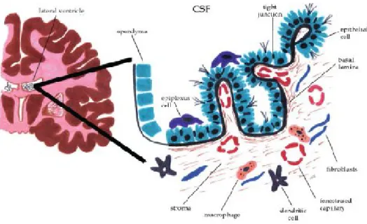
(13) II. Introduction. II. Introduction 1) Alzheimer’s Disease AD is one of the major health problems in the economically developed countries along with cardiovascular disorders and cancer (Bachurin, 2003). It is characterised by progressive deterioration of memory and higher cortical functions, that ultimately result in total degradation of intellectual and mental activities (Cocabelos, 1994; Ebly et al., 1994). AD is the most common form of dementia (Ebly et al., 1994). It affects up to 10% of people over the age of 65 and 30 to 35% or more of those over 85 years (Sykes et al., 2001). Currently, AD affects approximately 20 million people all over the world, and imposes an annual economic burden of about US $ 100 billion (Cocabelos, 1994). The key neuropathological characteristics of AD are: SP, which are associated with Aβ, NFT, and the loss of neurons in the hippocampus and nucleus basalis of Meynart (Morrison et al., 1998; Selkoe, 1991). Alterations in the morphology and in the functions of CPs have been linked to this disease. In the next chapter we explain the structure and function of this organ. Moreover, we also made a literature revision about ageing of CPs, and the cross-talk, between the CPs and AD.. 2) Choroid Plexus 2.1) Structure The CPs are located throughout the ventricles of the brain. Within the lateral ventricles, they projects from the choroidal fissure and extends from the interventricular foramen to the end of the temporal horn, and projects into the third and fourth ventricles from the ventricular roof (Emerich et al., 2005). Choroid tissues are composed of villi covered by an unistratified epithelium with a central vascular axis. Epithelial cells are cuboid with a rounded central or basal nucleus (Dohrmann, 1970). At the apical pole numerous microvilli of uniform diameter, enmeshed with each other, and a few cilia (Serot et al., 2003). Mitochondria are more numerous at the basal and apical poles, occupying 15 % of the cytoplasm in primates (Cornford et al., 1997). The Golgi apparatus contains columns of cisternae and smooth endoplasmic reticulum and clear vesicles are distributed throughout the apical cytoplasm (Emerich et al., 2005). The epithelial cells lie on an epithelial basement membrane surrounding a thin stroma with ________________________________________________________________ 3.
(14) II. Introduction numerous collagen fibbers, scarce dendritic cells, macrophages, fibroblasts and large capillaries with a fenestrated endothelium (Dohrmann, 1970). The basolateral membrane contains numerous interdigitations (Serot et al., 2003). CPs are richly innervated, receiving adrenergic, cholinergic, peptidergic and serotoninergic fibers. The distribution of nervous fibers varies widely according to species (Nilsson et al., 1990). Figure 1 represents the structure of CPs.. Figure 1: Morphological illustration of CPs within the lateral ventricle. The CPs extends from the ependymal cell layer of the ventricular wall forming a continuous strand of cuboidal epithelial cells resting upon a basal lamina and inner core of connective and highly vascularized tissue. The apical membrane of the epithelial cells faces the CSF where the cells contain numerous infoldings and scattered villi. Adjacent epithelial cells are bound together forming tight junctions and globular macrophages, dendritic cells, and fibroblasts are found throughout the stroma. Dendritic cells are also found in between the epithelial cells while epiplexus cells are located on the apical surface of the epithelial cells (modified from (Emerich et al., 2005)).. 2.2) Function The best-recognized function of the CPs is CSF production (Serot et al., 2003). CSF is produced mainly by active secretion with water entering the CSF from the blood along an osmotic gradient or by specific water channels such as aquaporins (Oshio et al., 2003). The epithelial cells replenish the CSF by moving Na2+, Cl- and HCO3- from the blood to the ventricles to create the osmotic gradient that drives the secretion of H2O (Emerich et al., 2005). The CSF is a clear, slightly viscous liquid with few cells and little protein (Rall, 1964). CSF composition is different from plasma (Segal, 2000), but similar to brain interstitial fluid (Felgenhauer, 1986). The CSF pH is slightly acid. Compared to plasma, the levels of Na+, K+, Ca2+, HCO3-, proteins and glucose are lower, but Cl- and Mg2+ levels are higher (Segal, 2000). Folate levels are 2 to 3 times ________________________________________________________________ 4.
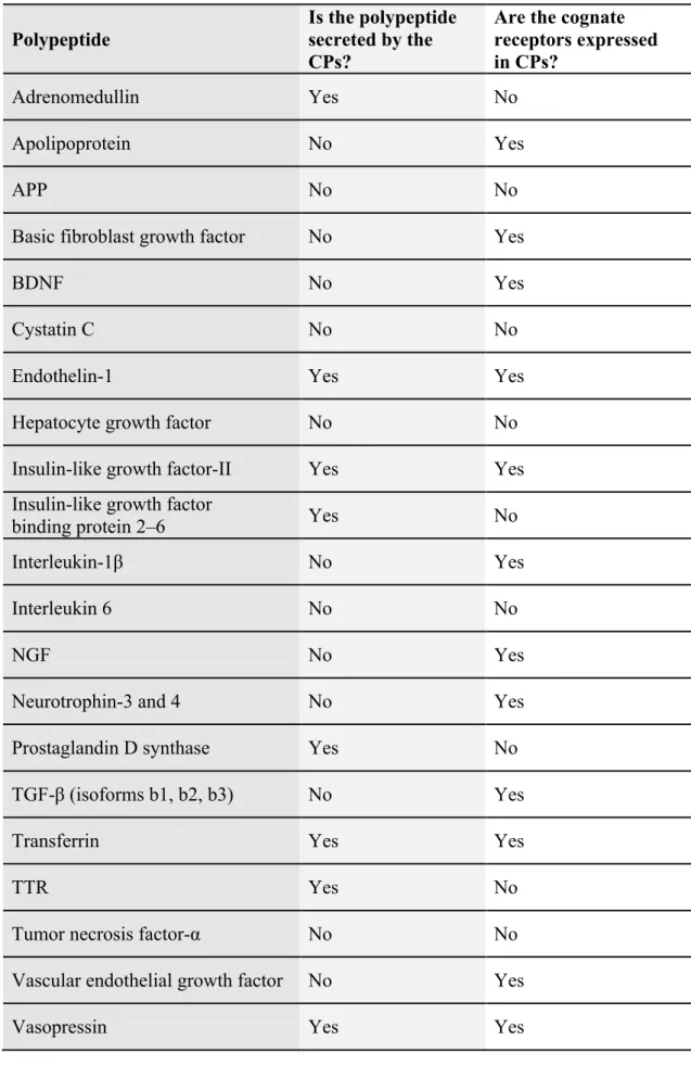
(15) II. Introduction higher in CSF than in plasma (Spector, 1977). TTR represents 25 % of proteins synthesized by CPs and 5 % of CSF proteins (Serot et al., 2003). Lying within the central ventricular system, the CPs are in an ideal position to monitor the CSF for the presence of noxious compounds, or potentially damaging cellular invasion (Emerich et al., 2005). The CPs protect the brain against acute neurotoxic insults by using a complex, multilayered detoxification system (Engelhardt et al., 2001; Gao, 2007). The CPs aid or impede the overall bio-distribution of drugs and toxic compounds by being the source of a full range of metabolizing enzymes including Phase I–III enzymes for functionalization, conjugation and transport of drugs (Emerich et al., 2005). A second example of the monitoring and modulating role of the CPs come from a recent understanding of its function within the neuroimmune system (Gao and Meier, 2001). Traditionally, the CNS has been considered an immunologically privileged site with no inherent need for immunosurveillance. The first indication that the CPs mediated interactions and/or signalling between the peripheral immune system and the brain came from demonstrations that the CPs contain inducible lymphoid cells (Emerich et al., 2005). The CPs possesses numerous specific transport systems, contains a broad array of receptors, and also serves as a major source of biologically active compounds (see Table 1). These capabilities allow the CPs to monitor and respond to the biochemistry of the brain by manipulating and maintaining baseline levels of the extracellular milieu throughout the CNS (Chodobski and Szmydynger-Chodobska, 2001; Stopa et al., 2001).. ________________________________________________________________ 5.
(16) II. Introduction Table 1: Polypeptides synthesized in CPs (Chodobski and SzmydyngerChodobska, 2001) Polypeptide. Is the polypeptide secreted by the CPs?. Are the cognate receptors expressed in CPs?. Adrenomedullin. Yes. No. Apolipoprotein. No. Yes. APP. No. No. Basic fibroblast growth factor. No. Yes. BDNF. No. Yes. Cystatin C. No. No. Endothelin-1. Yes. Yes. Hepatocyte growth factor. No. No. Insulin-like growth factor-II. Yes. Yes. Insulin-like growth factor binding protein 2–6. Yes. No. Interleukin-1β. No. Yes. Interleukin 6. No. No. NGF. No. Yes. Neurotrophin-3 and 4. No. Yes. Prostaglandin D synthase. Yes. No. TGF-β (isoforms b1, b2, b3). No. Yes. Transferrin. Yes. Yes. TTR. Yes. No. Tumor necrosis factor-α. No. No. Vascular endothelial growth factor. No. Yes. Vasopressin. Yes. Yes. ________________________________________________________________ 6.
(17) II. Introduction 2.3) Ageing of the choroid plexus Most of our knowledge about the morphology and function of human CPs during aging comes from control tissues in studies investigating changes in AD (Emerich et al., 2005). Physiological events in the ageing CPs include epithelial atrophy, weight increase (Wilson et al., 1999), and slightly different modifications according to species (Wilson et al., 1999). In humans, the height of CPs epithelial cells decreases about 10–11% during life (Serot et al., 2000), and in elderly rats epithelial cells loose 15 % of their normal height (Serot et al., 2001b). The aged epithelial cell cytoplasm becomes rich with Biondi Ring Tangles and lipofuchsin deposits (Serot et al., 2000) and the nuclei appear irregular and flattened while the basement membrane thickens (Wen et al., 1999). The stroma also thickens and contains collagen fibers, hyaline bodies, calcifications and psammoma bodies, and the infiltrating arteries become thicker and fragmented (Serot et al., 2003; Shuangshoti and Netsky, 1970). The functions of the CPs are energy-dependant, and the ageing CPs cannot maintain its normal energy output (Emerich et al., 2005). Synthesis of enzymes needed for anaerobic respiration and oxidative phosphorylation declines in aging rats (e.g. lactate dehydrogenase. and succinate-dehydrogenase. decrease 9%. and. 26%,. respectively) (Ferrante and Amenta, 1987). There are age-dependent increases in the number of epithelial cells deficient in cytochrome C oxidase, altering the respiratory mitochondrial chain with a concomitant decrease in cellular production of ATP (Miklossy et al., 1998). Reductions in Na+K+-ATPase and in the Na+K+-2Cl- cotransporter also occur (Cottrell et al., 2001). The anatomic and enzymatic modifications of CPs related to ageing are probably responsible for the drastic diminution of CSF secretion (Serot et al., 2003). In animal models, CSF secretion decreases as much as 45 % during ageing. In rats it has been evaluated that 1.2 mL/min of CSF are secreted at 3 months of age, and only 0.65 mL/min are secreted in 30 months old animals (Preston, 2001). In man, the volume of CSF secreted diminishes with age, from 0.41 mL/min at 28 years of age to 0.19 mL/min at 77 years (May et al., 1990). Due to the decreasing secretion and the simultaneously brain atrophy the CSF turnover takes longer (Preston, 2001) in elderly rats (7.9 h) than in young rats (2.2 h) (May et al., 1990). In man, the turnover of CSF is estimated to occur 6 times a day in young adults compared to 1.7 times in elderly subjects (Tanna et al., 1991).. ________________________________________________________________ 7.
(18) II. Introduction The effects of these cumulative changes on brain functioning caused by alterations in the CPs, and CSF, lead to inadequate distribution of nutritive substances, additional cellular stress, and reduced clearance of toxic compounds that may play a role in age-related cognitive decline, and in the development of specific neurological disorders. 2.4) Implication in Alzheimer’s disease Accumulating evidence supports the idea that the progressive decrease in CPs function during ageing exacerbates AD (Johanson et al., 2005). As part of a new paradigm to explain brain interstitium deterioration in age-related dementias, more attention is being given to the role of the compromised blood-CSF (Johanson et al., 2005), and blood-brain (Huber et al., 2001) barriers. Structural alterations and functional failure in CPs, as well as in brain capillary systems adversely affect fluid dynamics and composition (Rubenstein, 1998; Silverberg et al., 2001). Abnormalities similar to those observed in ageing CPs are greatly enhanced, in AD. Epithelial cell atrophy is greater, with cell height decreasing up to 22% in relation to age-matched controls. These cells also contain numerous lipofuchsin vacuoles (Serot et al., 2000). The percentage of epithelial cells containing Biondi bodies is significantly increased in AD patients (Wen et al., 1999). Epithelial basement membranes are very irregular and thickened; their thickness increases by 28% compared to controls of the same age (Serot et al., 2000). Stroma of villi is irregularly fibrotic; its thickness can reach several tenths of a µm (Jellinger, 1976; Serot et al., 2000). The CPs contains vessels with thickened walls, hyaline bodies, calcifications, and psammomas mainly at the glomus level. Immunohistology reveals many linear deposits of IgG, IgM and C1q along the epithelial basement membrane suggesting intervention of immunological processes (Serot et al., 1994). The pathological changes in CPs are resumed in Table 2. Table 2: Pathological changes of CPs in AD (in (Johanson et al., 2004)) Biondi bodies ↑ Basement membrane thickening ↑ (3-fold) Stromal fibrosis↑ Epithelial atrophy ↑ (cell size decreases by 1/3) Lipofuchsin vacuoles IgG and IgM depositions. ________________________________________________________________ 8.
(19) II. Introduction The increased atrophy of the choroidal epithelial cells in AD is associated with pronounced decreases in secretory activity and transport functions (Emerich et al., 2005). Indeed, during isotopic scinticisternographies, there is a major delay of reabsorption as well as a transitory ventricular contamination phenomena, which promotes a decrease in CSF turnover (Brusa et al., 1990; Coblentz et al., 1973) and epuration capacities. CSF production has been measured by using an intraventricular catheter in patients with AD (Silverberg et al., 2001). The mean CSF production rate decreased significantly in AD patients. Due to cerebral atrophy, and the consequent ventricular dilatation, the CSF turnover is about 36 h in AD patients compared to 14 h in age matched controls (Serot et al., 2003). The decrease of CSF production could favour the glycation of proteins and the formation of Aβ oligomers. Advanced glycation end products (AGE) are a result of a diverse class of post translational modifications, generated by the non-enzymatic reaction of a sugar ketone or aldehyde group with the free amino groups of a protein or amino acid, specifically lysine, arginine and possibly histidine. In the first step of protein glycation, a labile Schiff base is formed, which subsequently rearranges into a stable Amadori product. Finally, through a complex cascade of dehydration, fragmentation, oxidation and cyclization reactions, AGE are formed as a mixture of protein-bound nitrogenandoxygen-containing heterocyclic compounds (Harrington and Colaco, 1994). AD brain contains 3 times more advanced glycation products than controls (Vitek et al., 1994). Diffuse deposits of Aβ, SP and NFT are known to contain AGE (Smith et al., 1996). Glycation of proteins promotes their aggregation, the polymerisation of tau proteins with formation of filaments, and Aβ fibril transformation (Troncoso et al., 1993; Vitek et al., 1994). In vitro, fibril transformation is accelerated in the presence of AGE, probably through initiation of a nucleation phenomena (Munch et al., 1998). In ageing, levels of CSF proteins, glucose and Amadori bodies remain stable, the reduction in CSF turnover is insufficient to increase protein glycation. On the contrary, in AD the Amadori bodies increase significantly, while CSF protein and glucose levels remain stable (Klein et al., 2001). Aβ clearance, which is significantly reduced in elderly animals (Preston, 2001), is still unknown in AD patients (Serot et al., 2003).. In rats, clearance of. intraventricularly injected Aβ decreases from 10.4 mL/minute at 3 months of age to 0.71 mL/minute at 30 months. Consequently, the brain content of Aβ increases from 7% ________________________________________________________________ 9.
(20) II. Introduction at the end of CSF perfusion in young rats to 49% in old animals (Preston, 2001). The brain from AD patients contains 12 times as many oligomers as controls (Kuo et al., 1996). AD severity appears to be more closely correlated to Aβ oligomers levels than to SP number (Klein et al., 2001). Levels of TTR, a CPs-synthesized molecule that associates with Aβ to form complexes, are more than 10% lower in AD (Serot et al., 1997). Ascorbic acid and αtocopherol levels, the two major scavengers of free radicals of CSF, are decreased in AD likely adding to oxidative stress (Schippling et al., 2000; Tohgi et al., 1994). CSF folate and vitamin B12 (important for methylation of numerous molecules) are significantly lower (Ikeda et al., 1990; Selley et al., 2002; Serot et al., 2001a) while homocysteine, which mediates lipid peroxidation and increases the production of toxic (E)-4-hydroxy-2-nonenal, is increased in AD CSF. The impaired capacity of the CPs to clear molecules from the CSF of AD patients has potentially profound implications (Serot et al., 2003). As pointed out by Serot and colleagues (Serot et al., 2003), these changes could lead to an even greater impoverishment of the brain, conducting to methylation problems, increased oxidative stress and lipid peroxidation, decreased amyloid clearance, augmented tau protein polymerization, and amyloid peptide oligomers and fibril formation. 2.5) Neuroprotective molecules produced by CPs and regulated by androgens Recent studies reinforced the neuroprotective roles of CPs, showing that conditioned media from CPs prevents cultured embryonic neurons from death (Borlongan, 2004a; Borlongan, 2004b; Watanabe et al., 2005). CPs ependymal cell grafts promote the regeneration of damaged spinal cord (Ide, 2001), and enable the reduction of the functional and structural consequences of cerebral ischemia (Borlongan, 2004a; Borlongan, 2004b). Transcripts and/or protein for several neuroprotective molecules, such as nerve NGF, neurotrophin 3 and 4, TGF-β, BDNF, TTR, and GDNF (Dickson et al., 1986; Emerich et al., 2005; Ikeda, 1999; Koo, 2001) have been identified in CPs, as well as some of their cognate receptors. Some of these peptides involved in neuroprotection, like NGF, GDNF, BDNF and TGF, secreted by CPs, are regulated by androgens in other tissues. Studies by Katoh-Semba (1994) and his co-workers have showed that the levels of β-NGF protein decrease in the hypothalamus and hypophysis, but not in the ________________________________________________________________ 10.
(21) II. Introduction cerebellum and olfactory bulb, in male mice after castration (Katoh-Semba et al., 1994). Another study using nandrolone shows an increase of NGF levels in the hippocampus and septum and a decrease in the hypothalamus (Tirassa et al., 1997). Moreover, testosterone, but not DHT decreased hippocampal NGF protein in aged rats (BimonteNelson et al., 2003). Castration of male mice reduces the submandibular gland NGF levels to those found in control females, and androgen treatment produced an increase in the submandibular NGF mRNA (Black et al., 1992). Zhen et al, in 2003, have shown that the expression of GDNF mRNA in prostate decreased significantly after castration, in rats (Zheng et al., 2003). BDNF interacts with testosterone in the maintenance of spinal nucleus of the bulbocavernosus (dendritic arbors), in male rats (Yang et al., 2004). The castration, in rats, induced an increase of both BDNF tissue concentration and mRNA expression, in the musculature of the vesicular gland and in the fibromuscular stromal cells of both dorsal and ventral prostatic lobes (Mirabella et al., 2006). Levels of BDNF protein are increased by testosterone, in the female high vocal center of adult canary (Serinus canaria) (Rasika et al., 1999). In the case of the TGFβ, chronic DHT treatment increased TGFβ receptor binding, and also increased TGFβ induced cell proliferation in female rats fetal lung fibroblast cells (Dammann et al., 2000). In pig immature Leydig and Sertoli cells, TGFβ levels decreased after testosterone treatment (Avallet et al., 1994). Androgens negatively regulate the expression of TGFβ ligands and receptors, in the human prostate, (Chipuk et al., 2002). In ovarian cancer HEY cells treatment with DHT downregulated the expression of mRNA for TGFβ receptors I and II (Evangelou et al., 2000). Studies performed in rats have demonstrated that after castration TGF-β 1 increases in prostate, seminal vesicle and epididymis after 3 days of treatment, and decreased after 5 days (Desai and Kondaiah, 2000). We reasoned that some of these peptides could also be regulated by androgens in CPs, should AR or other androgen activated signalling pathways be present in this tissue.. ________________________________________________________________ 11.
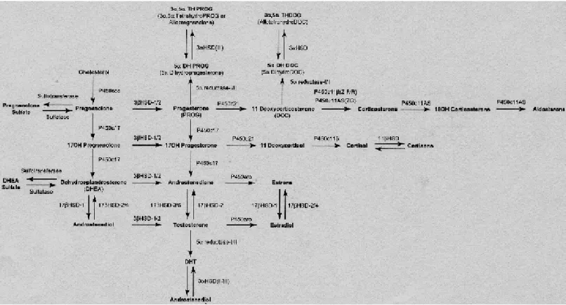
(22) II. Introduction. 3) Neurosteroids 3.1) Neurosteroids synthesis Recent evidence suggests that the brain is a steroidogenic organ, with the ability to synthesize steroid hormones from cholesterol. The steroids produced in the brain from cholesterol and other blood-born precursors, which accumulate in the nervous system at a level partially independent of traditional steroidogenic organs (adrenal glands and gonads), are termed neurosteroids (Mellon et al., 2001; Plassart-Schiess and Baulieu, 2001). Many neurosteroids were previously thought to have a passive role as precursors or metabolites of other steroids, however it has been shown, that they have effects in the nervous system, ranging from targeting gene expression to modulating neurotransmission (Bates et al., 2005). The first evidence that the brain was capable of producing steroids occurred when Corpéchot et al (1981) demonstrated the presence of pregnenolone (PREG), dehydroepiandrosterone (DHEA) and their sulfate derivates in the brains of castrated and adrenalectomized rats (Corpechot et al., 1981). Subsequent studies showed that both glial cells and neurons contain the enzymes necessary for steroid synthesis (Bates et al., 2005). The initial step in steroidogenesis is the conversion of cholesterol to PREG on the inner mitochondrial membrane by the enzyme cytochrome P450 side chain cleavage (P450scc). PREG can then be converted to progesterone (PROG) by the enzyme 3βhydroxysteroid dehydrogenase-isomerase (3β-HSD) in the endoplasmic reticulum, or to DHEA by cytochrome P450c17 (P450c17, also known as 17α-hydroxylase/c17-20lyase). The former pathway results in the synthesis of PROG and PROG metabolites, such as 20α-dihydroprogesterone and 5α-dihydroprogesterone. The latter pathway culminates in the formation of testosterone via conversion of androstenedione by 17βhydroxysteroid dehydrogenase (17β-HSD) (androstenedione is also derived from PROG). Testosterone can in turn be converted into estradiol via the enzyme P450 aromatase (Bates et al., 2005). All these pathways are represented in the figure 2.. ________________________________________________________________ 12.
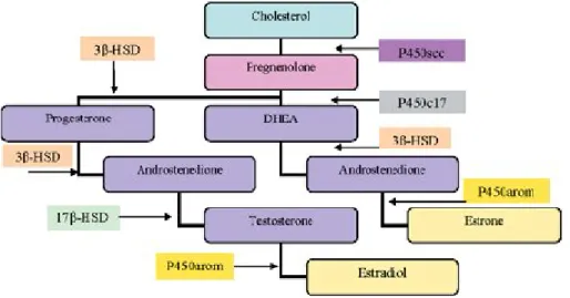
(23) II. Introduction. Figure 2: The neurosteroidogenic pathway. The names of the enzymes mediating each reaction are shown above the arrows (in (Mellon and Griffin, 2002)). The expression of the neurosteroidogenic enzymes in the CNS is cell type specific (Figure 3). In vitro analysis of messenger RNA expression and steroid production has revealed, that astrocytes are the most steroidogenic cells in the brain, expressing P450scc, P450c17, 3β-HSD, 17β-HSD, and aromatase (Zwain and Yen, 1999). Astrocytes are therefore, capable of producing PREG, PROG, DHEA, androstenedione, testosterone, estradiol and estrone. Oligodendrocytes, the myelinating cells of the CNS, express P450scc and 3β-HSD, producing PREG, PROG and androstenedione. Neurons express P450scc, P450c17, 3β-HSD and aromatase, and thus produce PREG, DHEA, androstenedione and estrogen (Bates et al., 2005). The relative production capability of these cells can be summarized as follows: astrocytes are the major producers of PROG, DHEA and androgens, oligodendrocytes are the predominant source of PREG, and neurons are the main source of estrogens (Bates et al., 2005). Furthermore, the regulation of some neurosteroidogenic enzymes is sex specific and developmentally regulated. The expression of 3α-hydroxysteroid dehydrogenase (involved in the generation of neurosteroids through ring-A reduction of hormonal precursors progesterone and corticosterone) is high on postnatal day 7, and is ________________________________________________________________ 13.
(24) II. Introduction gender specific during puberty in the rat (Mitev et al., 2003). The expression of mRNA for P450scc and 11β-hydroxylase (P450c11β, involved in the synthesis of corticosterone) in the rat is region specific. P450scc mRNA is most abundant in the cortex of both male and female adult animals, and is also found in the amygdala, hippocampus and midbrain, but absent in the cerebellum and hypothalamus (Mellon and Deschepper, 1993). P450c11β mRNA is detected mainly in the amygdala and cortex, but also in the cerebellum and hippocampus of both male and female rats (Mellon and Deschepper, 1993). Interestingly, female rats have higher expression of P450c11β in the hippocampus than male rats. Neurosteroid synthesis and metabolism is thus a complex process. Steroidogenic enzymes are differentially expressed by CNS cells, therefore adding a temporal and spatial dimension to sex steroid synthesis in the CNS (Bates et al., 2005). Neurosteroidogenesis can be envisaged as an autocrine event, with precursors produced by cells, that are required by other cell types to produce the necessary products.. Figure 3: Sex steroid synthesis in the CNS. Major products and enzymes involved are indicated. Astrocyte products are shown in purple, oligodendrocytes products in pink, and neuron products in yellow (in (Bates et al., 2005)).. 3.2) Testosterone and ageing Findings from clinical and basic science studies indicate that testosterone and its androgen metabolites have a wide range of beneficial actions in the CNS (Pike et al., 2007). Androgen actions not only influence the development of the CNS, but also help to maintain its proper function in adulthood. However, as a normal consequence of ageing in men, both circulating (Feldman et al., 2002; Gray et al., 1991) and brain ________________________________________________________________ 14.
(25) II. Introduction (Rosario et al., 2004) levels of testosterone exhibit gradual, but eventually functionally significant depletion. The decrease in testosterone production by the testes is progressive, though in men, there is no state analogous to the menopause. Total serum testosterone decreases about 30% and free testosterone by as much as 50% between 25 and 75 years of age (Morley et al., 1997; Vermeulen, 1991). Circulating testosterone not only decreases in ageing men, but also in women, as a consequence of the agedependent decline in ovarian and adrenal androgen production. The mid-cycle rise in free testosterone and androstenedione seen in younger women (19–37 years old) is consistently absent in older women (43–47 years old) (Mushayandebvu et al., 1996). The 24 h mean plasma concentrations of total and free testosterone also show a steep diminution with ageing in healthy women between the ages of 21 and 51 (Zumoff et al., 1995). This age-related androgen loss has senescent effects in androgen responsive tissues throughout the body, as demonstrated by both impaired function, and increased vulnerability to disease (Morley, 2001). As an androgen-responsive tissue, the brain is also thought to suffer deleterious consequences of age-related androgen depletion. Neural manifestations of androgen deficiency in ageing males include disturbances in mood, cognition, and libido (Gooren and Kruijver, 2002; Morley, 2001; Swerdloff and Wang, 2003). Recent evidence suggests that androgen depletion in men also increases the risk of developing age related neurodegenerative disorders including AD (Pike et al., 2007). How androgen depletion contributes to CNS dysfunction is not clear, but likely includes the diminished activation of androgen signalling pathways that affect behaviour, neuron viability, and regulation of specific pathologies. Further, the cellular and molecular mechanisms that underlie androgen mediated cell signalling pathways remain incompletely defined (Pike et al., 2007).. 3.3) Implication of testosterone in neuroprotection The non-reproductive effects of androgens in the nervous system are considerably less well characterized than those of estrogens and progestins. Testosterone influences neuroplastic changes in different nuclei of the limbic system (De Vries et al., 1994; Johnson et al., 1989; Malsbury and McKay, 1994), and also exerts neuroprotective effects, which can be mediated either directly or indirectly via its aromatization to estradiol (Balthazart and Ball, 1998). Other data suggest that testosterone may also exert neurotrophic actions. For example, Beyer et al., and Lustig ________________________________________________________________ 15.
(26) II. Introduction have observed neuronal differentiation and increase in neurite outgrowth after activation of androgen pathways in the cultured neural cells (Beyer et al., 1994; Beyer and Hutchison, 1997; Lustig, 1994). Physiological concentrations of testosterone have also been shown to protect primary cultures of human neurons against apoptosis induced by serum deprivation. The effect of testosterone was directly mediated through androgen receptors, and did not involve its aromatization to estradiol: (1) it could be mimicked by the non-aromatizable androgen mibolerone; (2) it could be blocked by the anti androgen flutamide; and (3) it was not prevented by an aromatase inhibitor (Hammond et al., 2001). Other experiments in male rodents suggest that testosterone is linked to an increase in neuron size, neuritic growth, plasticity and synaptogenesis in both motoneurons of the spinal nucleus of the bulbocavernosus (Forger et al., 1992; Matsumoto, 1997), and several populations of pelvic autonomic neurons (Keast and Saunders, 1998). Moreover, studies with motoneuron populations, including facial, spinal and pudendal motoneurons, have demonstrated that the administration of testosterone immediately after nerve injury promotes their survival and regeneration through actions mediated by the AR (Jones et al., 2001; Tanzer and Jones, 1997). Ogata et al. have reported that testosterone protected spinal cord neurons against neuronal damage induced by glutamate. The hormone reduced the extent of the spinal cord damage in vitro (Ogata et al., 1993). Testosterone also regulates the production of Aβ by neurons. Treatment of neuroblastoma cells, and of rat primary cerebrocortical neurons with testosterone increases the secretion of the non-amyloidogenic Aβ precursor protein (APP) fragment, and decreases the secretion of amyloidogenic Aβ (Gouras et al., 2000). Testosterone can prevent the hyperphosphorylation of tau, another important factor in AD pathogenesis (Papasozomenos, 1997). Androgens also play a role in myelination, and have been shown to modulate P0 gene expression in the peripherical nervous system (PNS). As Schwann cells do not express the intracellular AR, different alternative mechanisms by which androgens regulate peripheral myelin gene expression have been proposed: (1) the testosterone metabolite DHT may activate P0 expression by interacting with the progesterone receptor (PR), which is present in Schwann cells; (2) the testosterone metabolite 3α,5αandrostane-diol may interact with GABAA receptors; and (3) testosterone may ________________________________________________________________ 16.
(27) II. Introduction influence the myelination process indirectly by acting on the neurons that are myelinated (Magnaghi et al., 1999). 3.4) Testosterone and Alzheimer’s disease Progressive dysfunction and death of neurons characterize neurodegenerative diseases. There are some evidences supporting the hypothesis that testosterone may act protectively in some neurodegenerative disorders: AD, mild cognitive impairment (MCI), and depression (Bialek et al., 2004). One of the initial studies of Hogervorst et al. (2001) reported significantly reduced serum levels of testosterone in men with AD in comparison to age-matched, non-demented men (Hogervorst et al., 2001). Similar findings of low testosterone in men with AD have been reported in several studies (Almeida et al., 2004; Moffat et al., 2004; Paoletti et al., 2004; Rasmuson et al., 2002; Watanabe et al., 2004) but not all (Pennanen et al., 2004). Rosario and her team (2004) investigated the relationship between brain levels of testosterone, and of AD neuropathology, comparing the brain levels of sex steroids in men with and without AD neuropathology. An approximately 50% decrease in brain testosterone in men with AD aged 60–80 years compared to agematched men lacking any evidence of AD, or other neuropathology was observed (Rosario et al., 2004). They have not founded age-related changes in brain levels of estradiol in these men (Rosario et al., 2004). Recent findings suggest that testosterone depletion occurs prior to the development of AD, and thus may act as a contributing factor to AD pathogenesis (Pike et al., 2007). Moreover, these relationships may also be affected by apolipoprotein E ɛ4 status, which is associated with higher salivary testosterone levels in men, but lower salivary testosterone levels in women (Berteau-Pavy et al., 2007). Interestingly, low testosterone has also been linked to several other neurodegenerative diseases, including Parkinson's disease (Okun et al., 2004), vascular dementia (Watanabe et al., 2004), amyotropic lateral sclerosis (Militello et al., 2002), and Huntington's disease (Markianos et al., 2005). There are also randomized studies on various human populations that showed the effect of androgen substitution on cognition in men. Janowsky et al. (1994) have demonstrated that testosterone enhances spatial cognition of healthy older men (Janowsky et al., 1994). Some studies have reported that testosterone enanthate ________________________________________________________________ 17.
(28) II. Introduction supplementation improves spatial, verbal (Cherrier et al., 2001), and working memory (Janowsky et al., 2000), in healthy older men. One of the key events in initiating and driving AD pathogenesis is the accumulation of Aβ (Hardy and Selkoe, 2002). Androgens act as endogenous negative regulators of Aβ accumulation. Consequently, the age-related depletion of testosterone likely diminishes the ability of the brain to adequately regulate Aβ, resulting in increased of Aβ accumulation and development of AD. The mechanisms by which androgens regulate Aβ accumulation have yet to be fully determined, but apparently involve at least two pathways (Pike et al., 2007). One mechanism by which androgens may influence Aβ is via aromatization to estradiol and activation of estrogen pathways. Some studies have shown that estrogen can reduce levels of soluble Aβ (Greenfield et al., 2002; Levin-Allerhand et al., 2002; Manthey et al., 2001; Petanceska et al., 2000; Xu et al., 1998; Zheng et al., 2002) by a mechanism that involves regulation of the processing and/or trafficking of APP (Greenfield et al., 2002; Jaffe et al., 1994), the precursor protein of Aβ. APP is proteolytically cleaved at the amino- and carboxyl-termini of Aβ by βsecretase and γ-secretase, respectively, to generate the 40–42 amino acid Aβ (Sinha and Lieberburg, 1999). However, APP is alternatively processed within the Aβ sequence by α-secretase, which prevents formation of full-length Aβ. This non-amyloidogenic processing of APP results in secretion of soluble APP-α (sAPPα), which can be used as an index of APP metabolism and, indirectly, Aβ production (Pike et al., 2007). Gouras et al. (2000) evaluated the effect of testosterone on APP metabolism, and Aβ production in cultured cortical neurons, and in a neuroblastoma cell line. They found that, in parallel to the actions of estrogen, prolonged treatment of cultures with testosterone resulted in elevated levels of sAPPα and reduced levels of Aβ (Gouras et al., 2000). Their data clearly demonstrated that testosterone can regulate Aβ levels, but did not indicate whether the mechanism involves AR-dependent pathways, or aromatization to estradiol and activation of established estrogen pathways. Because aromatase is present in neurons and can effectively metabolize testosterone into estradiol (Melcangi et al., 1992; Poletti et al., 1997), an estrogen pathway is certainly reasonable. In fact, a subsequent study, in 2000, indicated that aromatization to estradiol underlies testosterone regulation of APP and, consequently, Aβ (Goodenough et al., 2000). Like Gouras (Gouras et al., 2000), these researchers found that testosterone ________________________________________________________________ 18.
(29) II. Introduction increased sAPPα levels, indicating non-amyloidogenic processing of APP (Goodenough et al., 2000). However, the testosterone-mediated increase in sAPPα was blocked by aromatase inhibition, suggesting that testosterone regulation of Aβ occurs at least in part through estrogen pathways. Consistent with these cell culture findings, there are some observations in men with prostate cancer in which treatment with anti androgen therapy reduced circulating levels of testosterone, estradiol, and increased Aβ (Almeida et al., 2004; Gandy et al., 2001). Although experimental evidence demonstrates that testosterone can regulate Aβ by an estrogen-mediated mechanism, the significance of this pathway to AD risk in men is questioned by other findings, that AD in men is associated with brain levels of testosterone, but not estradiol (Rosario et al., 2004), and that soluble Aβ levels in male rats are reduced by DHT, but not estradiol (Ramsden et al., 2003b). A second mechanism by which androgens regulate Aβ accumulation involves estrogen-independent androgen signalling pathways. To begin distinguishing between androgen versus estrogen actions of testosterone in regulation of Aβ accumulation, Lund et al, used the testosterone metabolite DHT that is not aromatized to estradiol, although it can be metabolized to form 5α-androstan-3β, 17β-diol which has some agonist effects on estrogen receptor β (ER β) (Lund et al., 2006). Pike and his team evaluated how the brain levels of soluble Aβ in adult male rats were affected by gonadectomy (GDX), and subsequent treatment with vehicle, DHT, or estradiol. If androgens negatively regulate Aβ accumulation, he has predicted that GDX-induced androgen depletion would increase Aβ levels in the brain (Pike et al., 2007). In agreement with this hypothesis, analysis of soluble Aβ in whole brain by a sensitive and specific enzyme-linked immunoadsorbent assay (ELISA) several weeks after GDX showed a significant increase in Aβ in GDX versus sham GDX male rats (Ramsden et al., 2003a). Further, this GDX-induced increase in Aβ was prevented in male rats treated with subcutaneous, slow-release hormone pellets containing DHT. Notably, GDX male rats treated with estradiol pellets did not show any reduction in Aβ (Ramsden et al., 2003a). The finding of Pike et al, that estradiol treatment did not reduce Aβ in GDX male rats (Pike et al., 2007), is in contrast to observations that estradiol can reduce Aβ in GDX female rodents (Carroll et al., 2007; Levin-Allerhand et al., 2002; Petanceska et al., 2000; Zheng et al., 2002).. ________________________________________________________________ 19.
(30) II. Introduction Together, these findings suggest that brain levels of Aβ in males are regulated by androgens, and that the underlying mechanism involves, at least in part, androgen signalling pathways that are independent of estrogen (Pike et al., 2007). If androgens are in fact endogenous regulators of Aβ, might expect that changes in androgen levels more subtle than GDX-induced depletion may affect Aβ levels. To study this possibility, Pike et al, examined whether inherent differences in circulating DHT levels in gonadally intact, adult male rats predicted brain levels of soluble Aβ. They observed that male rats with relatively high DHT levels showed lower levels of the two primary forms of Aβ protein, Aβ1-40 and Aβ1-42, than male rats with relatively low DHT levels (Pike et al., 2007). This finding predicts that, even the gradual age-related loss of testosterone associated with normal male aging may promote Aβ accumulation (Pike et al., 2007). Moreover, Pike et al, also have found that in men lacking AD, but characterized by mild neuropathological changes, brain levels of testosterone are inversely correlated with brain levels of soluble Aβ. Further, in male brown Norway rats, they have found that age-related decreases in testosterone and DHT are associated with increased brain levels of Aβ (Pike et al., 2007). One interesting aspect of the data obtained in aging rats is that the observed changes in androgens and Aβ occurred prior to significant increases in the levels of luteinizing hormone, a variable linked to testosterone loss which some have argued that also contributes to regulation of Aβ (Casadesus et al., 2005). Considering the testosterone and Aβ literature, one obvious and clinically important prediction is that low testosterone may promote the development of AD neuropathology, Rosario et al, evaluated how androgen status affects the development of AD-like neuropathology in the 3xTg-AD triple transgenic mouse model of AD. They observed that depletion of endogenous androgens by GDX in male 3xTg-AD mice at 3 months of age resulted in a significant increase in accumulation of Aβ in subiculum, hippocampus CA1, and amygdala at 6 months of age (Rosario et al., 2006). This GDXinduced increase in Aβ was associated with a significant worsening in performance in spontaneous alternation behaviour, a hippocampal dependent task of working memory. However, continuous treatment with DHT beginning at the time of GDX prevented the increase in Aβ accumulation, and worsening in behavioural performance (Rosario et al., 2006). These data confirm in another context that androgen pathways regulate Aβ accumulation, and predict a protective role against AD. Although research findings have established that, androgens can regulate Aβ accumulation through estrogen________________________________________________________________ 20.
(31) II. Introduction independent, androgen pathways, the signalling mechanism(s) underlying this action remain to be elucidated (Pike et al., 2007). Tau protein, a microtubular binding protein, predominantly axonal, which stabilizes the neuronal cytoskeleton, is another neuropathological hallmark of AD (Bialek et al., 2004). Papasozomenos et al, has demonstrated that heat shock-induced hyperphosphorylation of tau protein in the brain of orchidectomized male rats can be reduced by testosterone (Papasozomenos, 1997). Correlations between two markers of AD and level of neurosteroids have been founded. Aβ peptide levels were negatively correlated with PREGS levels in the striatum and cerebellum. Phosphorylated tau protein levels were negatively correlated with DHEAS concentrations in the hypothalamus. Other data demonstrated that administration of progestins reversed the age-dependent myelin abnormalities whereas administration of androgens was without effect (Azcoitia et al., 2003). Ibanez et al, have revealed that PROG administration slows remyelination of axons by oligodendrocytes after toxin-induced demyelination in old male rats (Ibanez et al., 2004). Morales et al, have reported that DHEA increases physical and psychological well-being in woman and men, increases lean body mass and muscle strength in men (Militello et al., 2002; Morales et al., 1994). However, most human trials demonstrating DHEA effect on cognitive performance have failed (Wolkowitz et al., 1995). Some of the numerous effects of androgens, in neuroprotection, and in AD, previously described are mediated by the androgen receptor. The structure, function and its relation with AD are described below.. 4) Androgen receptor 4.1) Gene and protein structure AR is a member of the steroid and nuclear receptor superfamily (Freedman, 1998), which is composed of over 100 members, and continues to grow. Among this large family of proteins, only five vertebrate steroid receptors, estrogen, progesterone, androgen, glucocorticoid, and mineralocorticoid receptors are known. Like other steroid receptors, AR is a soluble protein, that functions as an intracellular transcriptional factor. AR function is regulated by the binding of androgens, which initiates sequential conformational changes of the receptor that affect receptor protein interactions and. ________________________________________________________________ 21.
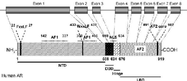
(32) II. Introduction receptor-DNA interactions (Gao et al., 2005). AR-regulated gene expression is responsible for male sexual differentiation and male pubertal changes (Gao et al., 2005). In 1981, Migeon et al, first localized the AR gene to the human X chromosome (Migeon et al., 1981). In 1998, Lubahn et al, cloned human AR genomic DNA from a human X chromosome library using a consensus nucleotide sequence from the DNAbinding domain of the nuclear receptor family (Lubahn et al., 1988). In the same year, several groups cloned human AR cDNAs (Chang et al., 1988; Colvard et al., 1989; Lubahn et al., 1988; Trapman et al., 1988). To date, only one AR gene has been identified in humans. The AR gene is more than 90 kb long, and codes for a protein of 919 amino acids, that has three major functional domains, as illustrated in figure 4, (Gao et al., 2005).. Figure 4: Structural organization of the human AR gene and protein. (from (Gao et al., 2005)). The N-terminal domain (NTD), which serves a modulatory function, is encoded by exon 1 (1586 bp). The DNA-binding domain (DBD) is encoded by exons 2 and 3 (152 and 117 bp, respectively) (McEwan, 2004). The ligand binding domain (LBD) is encoded by five exons, which vary from 131 to 288 bp in size. There is also a small hinge region between the DNA-binding domain and ligand-binding domain. Two transactivation functions have been identified. The N-terminal activation function 1 (AF1) is constitutively active in the truncated receptor, that does not contain the ligandbinding domain, and its sequence is not conserved compared to other steroid receptors, whereas the C-terminal activation function 2 (AF2) functions in a ligand-dependent ________________________________________________________________ 22.
(33) II. Introduction manner, and is relatively more conserved compared to other steroid hormone receptors, particularly in the charged-clamp residues (Chawnshang, 2002). A nuclear localization signal (NLS) spans the region between the DNA-binding domain and the hinge region. The human and rat AR amino acid sequence is very similar with identical sequences in the DNA- and ligand-binding domains, and an overall sequence identity of 85% (Lubahn et al., 1988). 4.2) Function and physiologic roles AR is mainly expressed in androgen target tissues, such as the prostate, skeletal muscle, liver, and CNS. In the brain from monkeys, AR mRNA was found in the medial basal hypothalamus, the bed nucleus of the stria terminalis, the medial preoptic area, anterior hypothalamus, and in the lateral dorsomedial hypothalamus, septum and amygdale, in the hippocampus, cingulate cortex, parietal cortex, cerebellum, anterior pituitary (Abdelgadir et al., 1999), with the highest expression level observed in the prostate, adrenal gland, and epididymis as determined by real-time polymerase chain reaction (PCR) (Keller et al., 1996). AR can be activated by the binding of endogenous androgens, including testosterone and DHT (Gao et al., 2005). Physiologically, functional AR is responsible for male sexual differentiation in utero, and for male pubertal changes. In adult males, androgens are mainly responsible for maintaining libido, spermatogenesis, muscle mass and strength, bone mineral density, and erythropoisis (Goodman, 2001; Johansen, 2004). The actions of androgen in the reproductive tissues, including prostate, seminal vesicle, testis, and accessory structures, are known as the androgenic effects, while the nitrogen retaining effects of androgen in muscle and bone, are known as the anabolic effects (Gao et al., 2005). Numerous and varied site mutations in AR have been identified (The Androgen Receptor Gene Mutations Database World Wide Web Server, http://www.androgendb.mcgill.ca/) (Gao et al., 2005). The majority of these mutations are associated with diseases (Gao et al., 2005). Besides the site mutations documented, AR gene polymorphism, have also been identified, particularly, the poly-Q (CAG)n at exon I. The polymorphic (CAG)10-35 triplet repeat sequence, starting from codon 58, codes for polyglutamine. The length of the repeat is inversely correlated with the transactivation activity of AR (Oettel, 2003).. ________________________________________________________________ 23.
(34) II. Introduction 4.3) Implication in neurodisease / Alzheimer’s disease The AR is present in the brain, including regions that can be severely affected in AD such as the cerebral cortex and hippocampus (Simerly et al., 1990). AR are bound to heat shock protein 90 (Hsp90) in the cytosol, stabilising the apoAR in a high affinity ligand binding conformation, and protecting them from degradation (Solit et al., 2003). AR change in structure and dimerize upon binding to androgens, following which, the androgen–receptor complexes enter the nucleus and bind to DNA modulating gene transcription (Fuller et al., 2007). Downstream effects are still being investigated, however the antioxidant enzyme catalase (Ahlbom et al., 2001), and hsp70 (Magrane et al., 2004; Zhang et al., 2004) appear to be induced. Increases in Hsp70 levels may be particularly relevant to AD, as this protein has been shown to be part of a neuroprotective response, against Aβ-induced toxicity (Magrane et al., 2004; Zhang et al., 2004). AR-dependent activation of a mitogen activated protein kinase (MAPK)/extracellular signal regulated kinase (ERK) pathway, eventually inactivating the pro-apoptotic protein Bad, has also been shown to promote neuroprotection in hippocampal cultures (Nguyen et al., 2005). AR may affect nongenotropic changes as well, for example androgen receptor activation can promote a Src/Shc/ERK signalling pathway, attenuating apoptosis in cell culture models (Kousteni et al., 2001). In other tissues, AR-driven transcription controls muscle growth, bone growth spermatogenesis, and the development of secondary sexual characteristics (Mooradian et al., 1987). The AR gene contains a polymorphic trinucleotide CAG-repeat in exon 1, which encodes a functional polyglutamine tract of variable length (Fuller et al., 2007). The normal CAGrepeat length is within the range 5–35; several studies have attempted to establish an association between variation in AR CAG-repeat length and serum steroid levels, with limited success (for a review see (Kaufman and Vermeulen, 2005)). However, one study has found that short AR CAG-repeat lengths combined with lower than average testosterone levels appears to increase the risk of AD in men (Leder et al., 2004). Few studies of age-related changes in brain expression of AR have been conducted. One study has shown that AR synthesis declines with age in both male and female mice (Tan and Pu, 2001), although AR phosphorylation, thought to be necessary to make the AR transcriptionally active (Wang et al., 1999), has been found to be higher in old mice of either sex compared to younger adult mice. In this study, testosterone supplementation caused a remarkable increase in AR phosphorylation (Tan and Pu, 2001), whereas in ________________________________________________________________ 24.
(35) II. Introduction gonadectomised mice, testosterone supplementation caused a drop in AR synthesis. High levels of AR mRNA have been found in the human hippocampus CA1 region (Beyenburg et al., 2000), and hippocampal AR mRNA has been shown to decrease significantly with age (Tohgi et al., 1995). In another study of post-mortem human brains, the vertical limb of the diagonal band of Broca and the nucleus basalis of Meynert, major cholinergic nuclei of the basal forebrain, nuclear AR expression was also found to decrease with aging (Ishunina et al., 2002). In contrast, a study in rat hippocampus showed that AR mRNA levels were significantly higher in old (22 month) than in young adult (5 month) male rats (Kerr et al., 1995). Discrepancies in these results may be due to differences in post-mortem delay, as well as species differences, and further studies are needed to clarify this issue. In the brain, methylation of the AR promotor in mouse brain cortex is induced following testosterone treatment in gonadectomised mice, and results in a decrease in AR mRNA expression (Kumar, 2004). AR, responds to circulating androgen levels to alter AR expression (Kerr et al., 1995). Studies of AR CAG-repeats have shown that shorter repeat lengths are associated with higher expression of AR (Krithivas et al., 1999), and as mentioned above, shorter repeat lengths have also been associated with AD risk (Lehmann et al., 2004), particularly in subjects with low testosterone levels. Alteration of testosterone levels appears to have a significant effect on AR levels, AR phosphorylation and AR promotor methylation, suggesting a complex feedback mechanism that will need much further research before we can predict the outcomes of testosterone supplementation, or other hormonal treatment in putative AD-preventive treatment of elderly males (Patchev et al., 2004). Individual variations in AR CAG-repeats may also cause individual variations in responses to androgen treatments, providing another area that requires further research (Fuller et al., 2007). Nevertheless, AR-targeting therapies have been suggested for AD, including non-steroidal small molecule compounds (selective androgen receptor modulators - SARMS) that have specifically high affinity for brain androgen receptors (Chen et al., 2002). Such compounds could potentially signal through brain receptors and activate only essential pathways, e.g. anabolic pathways in the brain. This may have therapeutic advantages in AD and related disorders in men (Kumar et al., 1999).. ________________________________________________________________ 25.
(36) III. Objectives. III. Objectives The presence of AR in CPs has never been investigated. Otherwise, the information provided by this literature revision, gives us enough information to raise the hypothesis that should CPs be an androgen responsive tissue, this property may have implications in AD onset and progression. Therefore the present study proposes the following objectives: - investigate and characterised AR distribution and expression in male and female rats CPs; - analysed the response of AR to DHT, in castrated male and female mice, subjected to DHT replacement.. ________________________________________________________________ 26.
(37) IV. Material and methods. IV. Material and methods 1) Animals All animals were handled in compliance with the NIH guidelines and the National and European Union rules for the care and handling of laboratory animals (Directive 86/609/EEC). Male (n=5) and female (n=5) Wistar rats (3 months old), and mice (see DHT stimulation experiment for details) were housed in appropriate cages at constant temperature in a 12h light /12h dark photoperiod and given standard laboratory chow, and water ad libitum. Tissue sampling was carried out in animals euthanized under anaesthesia (Clorketam 1000, Vétoquinol, Lure, France). CPs (dissected from the lateral and fourth ventricles of brains), liver, and prostate were collected from rats and frozen at -80ºC for protein, or RNA extraction. Brains including CPs, were fixed in 4% paraformaldehyde (PFA) in phosphate buffered saline (PBS) for in situ hybridization (ISH), and immunohistochemistry (IHC). Rat CPs samples were used for Western blotting, ISH, and IHC as larger amounts of tissue were required for these experiments.. 2) DHT stimulation experiment An experiment aiming to determine the response of AR to DHT treatment in CPs was carried out in mice. Female and male mice (129S1/Sv strain, 5 months±2 weeks) were either ovariectomised (n=17) or orchidectomised (n=20) under anaesthesia (Clorketam1000), following standard procedures. Five weeks after surgery, castrated animals were implanted with Alzet mini-osmotic pumps (Charles River Laboratories, Model 1007D, DurectTM, Barcelona, Spain) delivering 419μg/Kg/day of DHT (Fluka, Seelze,Germany) (7 females and 10 males), or vehicle only (placebos, 10 females and 10 males) (0.5% ethanol:99.5% polypropyleneglicol, Aldrich, Saint Louis, USA). Implants were placed in the subscapular region under anaesthesia. Sham operated animals, not implanted (5 females and 5 males), were also included in the experiment. After one week, mice were euthanized under anaesthesia, and CPs were dissected, and frozen at -80ºC until RNA extraction.. ________________________________________________________________ 27.
(38) IV. Material and methods. 3) AR mRNA detection 3.1) RNA isolation Total RNA was extracted from CPs dissected from rat and mice brains, upon homogenization in TRI Reagent (Ambion, Applied Biosystems, Austin USA) according to the manufacturers’ instructions. RNA was quantified by UV spectrophotometry at 260nm (Pharmacia Biotech, Ultrospec 3000, Denmark), and its quality was assessed by agarose gel electrophoresis containing ethidium bromide (Sigma, St. Louis, USA).. 3.2) RT- PCR Total RNA (60ng) from rat and mice CPs, was reverse transcribed for 1h at 37ºC in a 20μL reaction containing First Strand-5X buffer (50mM Tris-HCl, 75mM KCl, 3mM MgCl2) (Invitrogen, Karlsruhe LMA, Germany), 10mM DTT, 0.5mM of each dNTP (dATP, dCTP, dGTP, dTTP) (Amersham, Uppsala, Sweden), 20U of RNAse Out (Invitrogen), 25ρmol of random hexamer primers (Invitrogen), and 200U of M-MLV reverse transcriptase (Invitrogen). PCRs were carried out using 1μL of rat or mice CPs cDNA, in a 25μL reaction containing 20ρmol of forward and reverse primers, 0.2mM of dNTPs, 1.5mM MgCl2 (Promega, Madinson, USA), 1Xbuffer (10mM Tris-HCl, 50mM KCl and 0.1%Triton® X-100) (Promega), and 1.25U of Taq DNA polymerase (Promega). Amplifications were carried out over 35 cycles of 1min denaturation at 94ºC, 1min annealing at 57ºC, and 30s extension at 72ºC. Primer sequences for amplification of rat and mice AR were the following: sense- 5´ GCC AGT GCG TGA GGA TGA 3´ and anti-sense- 5´ GGT GAG CTG GTA GAA GCG C 3´( rat ); and sense-5´ GGC GGT CCT TCA CTA ATG TCA CTC 3´ and anti-sense -5´GAG ACT TGT GCA TGC GGT ACT CAT 3´ (Waters et al., 2001). Aliquots of PCR products were resolved by agarose gel electrophoresis containing ethidium bromide (Sigma), and visualized with the Molecular Imager FX Pro Plus MultiImager system (Biorad, Hercules, USA). PCR products were cloned in pGEM-T easy vector (Promega) and sequenced (Stabvida, Oeiras, Portugal) to confirm the identity of the amplicons.. ________________________________________________________________ 28.
(39) IV. Material and methods 3.3) ISH Digoxigenin-labeled AR (sense and anti-sense) cRNAs were generated by in vitro transcription using the DIG RNA Labeling Kit, according to the manufacturers’ instructions (Roche, Basel, Switzerland), from a pGEM-T easy vector (Promega) containing a 237bp fragment of rat AR cDNA, which had been obtained by RT-PCR as described above. Paraffin embedded male and female rat CPs sections (3μm) were hydrated, permeabilised with RNAse free proteinase K (20μg/mL) in TE buffer (100mM Tris, 50mM EDTA, pH 8.0) for 30min at 37ºC, post-fixed in 4% PFA for 10 min, and pre-hybridized for 2h at 45ºC in a buffer containing 50% formamide, 5X saline sodium citrate (SSC), and 40μg/mL of denatured fish sperm DNA (Sigma). Hybridization was carried out at 45ºC for 18h in pre-hybridization buffer containing the synthesized riboprobe (~2.5 μg/mL). Slides were washed at room temperature for 10min in 2XSSC, twice at 65ºC for 15min in 1XSSC, twice at 65ºC for 15min in 0,5XSSC, and blocked with Tris buffered saline (TBS) containing 0.1%Triton X-100, and 2% normal sheep serum, for 30min at room temperature. Sections were then incubated with alkaline phosphatise coupled anti-digoxigenin antibody (Roche) for 4h at room temperature, and subsequently stained using 4-nitro blue tetrazolium and 5-bromo-4chloro-3-indolylphosphate as substrate (NBT/BCIP, Roche), for 16h at room temperature. As negative controls, sections were hybridised with the sense probe, or without any probe, and reactions allowed proceeding during the same time, as with the antisense probe. For further details see point 1 in the annexes.. 4) AR protein analysis 4.1) Antibodies Anti-AR antibodies raised in rabbit against the AR C-terminal (C-19:SC-815), and the AR N-terminal (N20:SC-816), and the corresponding blocking peptides (SC815P and SC-816P) were purchased from Santa Cruz Biotechnology (Santa Cruz). Preabsorption of the antibodies was carried out by incubation with a five fold (by weight) excess of the corresponding blocking peptide in PBS, overnight at 4ºC.. ________________________________________________________________ 29.
Imagem
+6
Documentos relacionados