Printed version ISSN 0001-3765 / Online version ISSN 1678-2690 http://dx.doi.org/10.1590/0001-3765201820180199
www.scielo.br/aabc | www.fb.com/aabcjournal
New technique of intragastric sleeve: viability and survival in a pig model
MARIANA S. RIBEIRO1
, RICARDO P.A.S. ZORRON2,3
, SAULO JOSÉ Q. SILVA1
, SILVIA M.R. CADENA1
, FERNANDA ANTUNES1
, MARCELO B. DOS SANTOS JUNIOR1
, LUCIANA M. MELLO1
, HAROLDO JOSÉ S. IGREJA JUNIOR1
, VILSON L. BATISTA1
, JUSSARA P. SCHEFFER1
and ANDRÉ L.A. OLIVEIRA1
1
Laboratory of Animal Medicine and Surgery, State University of Northern Rio de Janeiro, Av. Alberto Lamego, 2000, Parque Califórnia, 28035-200 Campos dos Goitacazes, RJ, Brazil
2
Department of Surgery, Division of Innovative Surgery, Klinikum Bremerhaven Reinkenheide, Postbrookstraße 103, 27574, Bremerhaven, Germany 3
Department of Surgery, Hospital Municipal Miguel Couto, R. Mario Ribeiro, 117, Leblon, 22430-160 Rio de Janeiro, RJ, Brazil
Manuscript received on March 3, 2018; accepted for publication on May 21, 2018
ABSTRACT
Developing a less invasive, practical and cost-effective operative technique for obesity treatment represents a pressing need for our society. In this way, intragastric single port sleeve by endoplication was tested in six pigs during 18 weeks. Celiotomy was performed with animal placed in dorsal decubitus position. Single port gastrostomy was performed and double tobacco pouch sutures were made in fundic region, making a gastric sleeve. At the end, stomach layers and skin were closed in a conventional manner. Means and the standard deviations of surgical time were calculated. The procedure was simple and all animals survived; there were no significant blood loss and no intra and postoperative complications. The procedure was fast (67.4 minutes). The technique has the advantage of not requiring the use of mechanical sutures, making it less costly. The innovation of this procedure was the use of a single port gastrostomy device to perform an intraluminal sleeve. What made this technique less invasive were the use of a single port, nonmanipulation of the stomach intra-abdominally, ease of execution and no need of pneumoperitoneum. The new technique is acceptable and has reproducible viability, had a short procedure time without intra and postoperative complications.
Key words: bariatric, intragastric plicature, invasiviness, single-port.
Correspondence to: André Lacerda de Abreu Oliveira E-mail: [email protected]
INTRODUCTION
Although its satisfactory results, bariatric surgery already represents low number of procedures, in contrast to the number of existing cases. This is due to its high cost, limited access, patient’s choice and associated risks. Against that background, intragastric surgical procedures to treat obesity are
promising and less invasive than others bariatric surgeries and can result in good weight loss (Dayyeh et al. 2013). A new technique, a variant of gastric sleeve that aims to minimize the reported problems, can have great value for solving the
difficulties of access to this technique, as well as in the good results that it offers.
represents a need of our society. In order to make the surgical procedure less invasive, practical and with lower cost, a new laparoscopic technique called intragastric single port sleeve by endoplication was developed (Muller et al. 2017).
Morbid obesity comorbidities reached epidemic proportions. As a result, obesity therapy is a complexity intervention and weight loss maintenance is one of its challenges (Haddock et al. 2002, Familiari et al. 2011). Lifestyle interventions (diet, exercise and behavioral changes) result in a moderate weight loss (5 to 10% of initial body weight) and high percentage of patients regain weight after 1 or 2 years. In addition, pharmacological treatments does not provide a
definitive result, since its long-term effectiveness is
limited (Haddock et al. 2002, Li et al. 2005). Despite all impediments, bariatric surgery is nowadays the most effective treatment, since it provides massive weight reduction associated with the improvement or remission of comorbidities, generating an improvement in the quality of life (Moreira et al. 2010).
Within the complications most commonly associated with bariatric surgery are fistula and dehiscence in procedures with gastric section; obstruction, erosion and herniation in those that use
rings or bands; inflammatory reaction associated
with foreign body, in case of introduction of an intragastric balloon (Mathus-Vliegen and Tytgat 2002, Niville et al. 2005, Maggard et al. 2005, Shikora et al. 2005, Gagner et al. 2008).
A preliminary research using post mortem swine stomachs have already proved that intragastric sleeve by endoplication produces a significant reduction of gastric volume, restricting its area of distension without causing gastric ischemia, besides the impossibility of the occurrence of leaks, gastric necrosis or bleeding, since the technique does not induce gastric ischemia (Muller et al. 2017).
In view of the great technical and scientific
relevance of the subject and with the objective of
offering a technique that is easy to execute and has
low cost, we developed this technique, which for
the first time is performed by endoplication using a
single port in an experimental study in pig, and also we evaluate post-surgical viability and survival.
MATERIALS AND METHODS
ANIMALS
The project was approved by the Ethics Committee on Animals Use of State University of Northern Rio de Janeiro (765311). The experiment was developed at the Animal Experimental Unity with 75-days-old healthy pigs (n=6), weighting 33-45 kg. Animals were confined during 18 weeks to evaluate survival time.
SURGICAL TECHNIQUE
The pigs were kept fasting from food and water during 16 and 12 hours, respectively. Sedation was done using an intramuscular association of acepromazine 2 mg.kg-1 (Acepran® 1%) and
midazolam 0.1 mg.kg-1 (Dormire®). Subsequently, marginal vein of ear was cannulated and fluid therapy started. The intravenous anesthetic induction was done with sodium thiopental 1 mg.kg-1 (Thiopentax® sódico) and propofol 10 mg.mL-1 (Provive® 1%). After endotracheal intubation, the anesthesia was maintained with
isoflurane (Isoforine®) and oxygen.
Surgical procedures were performed by the same surgeon in all animals, who had experience in laparoscopic surgery, but who had not yet performed this technique.
Animal was positioned in supine position and a left pre-umbilical paramedian celiotomy of 2 cm was performed between cranial and caudal mammary glands, using a monopolar electronic scalpel (Deltronix Precision TC4®). Then, Farabeuf retractors were positioned and the body
of stomach was identified, which was fixed to the
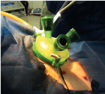
After the percutaneous gastrostomy, an unique gel portal system device (GelPoint, Applied Med. RS Margarita, CA, EUA) with three trocars (5-10 mm) was introduced (Fig. 2).
Stomach was insufflated with CO2 (12 mm Hg)
and a 30º (10mm) rigid endoscope was introduced
transgastrically. Fundic region was identified and
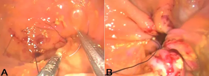
endoplicature was performed using double tobacco pouch sutures with 2-0 polypropylene (Fig. 3). The purpose of this suture was to separate the major curvature by making a gastric sleeve.
After the end of the endoplication, the surgical instruments and single-port device were removed, and stomach was closed with 2-0 nylon in 2 layers.
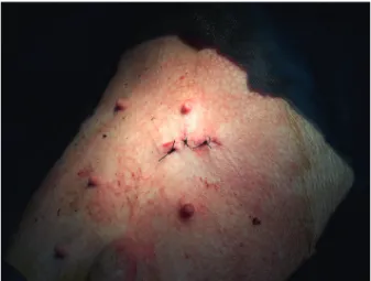
The fixation points were removed and skin was
closed in a conventional manner with 0 nylon.
OPERATORY TIME
The operatory time was timed (minutes) and
subdivided in: Celiotomy (CEL), Identification of
the Body Region (IDBODY), Fixation of the Body Region (FIXBODY), Portal Introduction (PORT),
Insuflation (INSLU), Suture (SUT), Portal removal
(PORTR), Stomach Closure (STOCLO), Skin Closure (SKICLO) and Procedure Time (TOTAL).
POST-OPERATORY
A healing spray (Bactrovet Prata®) was used during 12 days. Animals were medicated with meloxicam 0,4 mg.kg-1 (Maxicam® 1%) during five days and sulfadiazine/trimethoprim 15 mg.kg-1 (Borgal®) during 10 days. The amount and consistency of feed were gradually increased so that the pigs
were fed with liquid feed for the first seven days
followed by slurry feed for the next seven days.
Pigs were sacrificed with an anesthetic overdose 18
weeks after surgery.
Data of operatory time were saved in software (Microsoft Excel), in which means and standard deviation of each variable were subsequently calculated.
Figure 1 - Single-port Intragastric sleeve in pigs - Fixation of the stomach to the skin through four interrupted sutures with 0 nylon.
Figure 2 - Single-port Intragastric Sleeve in pigs - Positioning the single portal system (GelPoint, Applied Med. RS Margarita, CA, EUA).
RESULTS
Means and standard deviations were presented in Table I. The maximum values were related to the
first animal to be operated and the minimum to the
last. The total time of the surgical procedure ranged
from 46 (the last animal) to 82 minutes (the first
animal), mean of 67.40 minutes.
The operative procedure was simple, all
animals survived with no significant blood loss and
complications were observed and all animals were already feeding normally after anesthesia. No pig reported diarrhea, prostration and signs of pain in its evolution.
On the seventh postoperative day, all patients had a surgical scar with no signs of infection. The pigs recovered uneventfully and remained healthy for 18 weeks of follow-up.
DISCUSSION
The technique of intragastric single port sleeve by endoplication was relatively faster than other similar procedures recently developed, such as the endoscopic sleeve.
The first report of the endoscopic sleeve technique in humans initially lasted 3.48 h, with a reduction of 57.47% at the end (Kumar et al. 2015). Lopez-Nava et al. (2016) performed this technique in humans with an average time of 80 min (50-120 min). The OverStitch endoscopic suture device was tested by Galvão Neto et al. (2016) and the procedure was finished in 50 minutes. Another internal suture system called EndoCinch was clinically tested by Fogel et al. (2005) in 10 overweight volunteers, in which an endoscopic gastric plication was performed along the shorter curvature of the stomach. The time required for the procedure ranged from 60 to 90 min and there were no intraprocedural complications.
However, none of the reported techniques were similar to intragastric sleeve, because those used mechanical sutures that increase the cost of the procedure, although they are very reliable and with satisfactory results. The cost of those procedures using mechanical sutures has been a limiting factor of their wider use. Thus, the proposed technique Figure 3 - Single-port Intragastric Sleeve in pigs. a) Needle passing through the mucosa and submucosa of stomach. b) Traction
of surgical thread for making the suture in tobacco pouch (final appearance).
TABLE I
Operative times (minutes) to perform the single-port intragastric sleeve by endoplication technique in pigs.
Minutes
Times Mean DP(±) Maximum Minimum
CEL 4.80 3.49 11 3
IDBODY 2.40 1.14 4 1
FIXBODY 2.20 0.45 3 2
PORT 3.60 1.67 6 2
INSUFLA 1.40 0.55 2 1
SUT 39.20 14.29 53 23
PORTR 1.00 0 1 1
STOCLO 4.00 1.58 6 2
SKICLO 4.40 1.95 6 1
TOTAL 67.40 15.37 82 46
demonstrated results compatible with those described, with the advantage that there is no need of mechanical sutures.
Boza et al. (2012) demonstrated a comparison between two currently used bariatric surgical procedures, Roux-en-Y Laparoscopic Gastric Bypass (LRYGB) and Laparoscopic Sleeve Gastrectomy (LSG), and observed that the means of operative time were 106.2 minutes in the LRYGB group and 76.6 minutes in the LSG group. Lee et
al. (2012) submitted five pigs to the technique of
single-port vertical gastrectomy and had an average of 140 minutes of operative time.
In contrast, the new technique of the present
study lasted on average 67.4 min, with the first
animal being operated in 1 h and 22 minutes, with a reduction of 43.9% in operative time in
the last animal after refinement of the technique.
The reduction of more than 40% of the surgical time of a new technique performed in few animals demonstrates the ease of execution and learning of this technique.
The time to perform the double tobacco pouch suture made the procedure more time-consuming,
increasing surgeon’s learning curve and influenced
in approximately 58% of the total mean time. We expected that if there was an improvement of this
technique, the final time of the procedure might
have a significant time reduction, increasing surgical safety and decreasing cases of morbidity.
An important innovation of this technique is the introduction of a single port in a gastrostomy access. In this way, the sleeve was performed intraluminally, without the need of viscera/organs’ traction. Conversely, Morales-Conde et al. (2013) reported that one of the problems related to the single-port laparoscopic approach is the lack of triangulation and traction capacity of the stomach and liver during dissection, requiring the addition of one more trocar. It is important to note that in that article, the access was intraperitoneal, and the sleeve was not performed by intragastric sutures. In our view, that could characterize a disadvantage,
because it makes the procedure technically difficult.
When comparing the intragastric sleeve technique with restrictive bariatric procedures,
the first is less invasive. What make this technique
less invasive were the use of a single-port, no manipulation of the stomach intrabdominally (which could cause bleeding, pancreatitis and gastrointestinal perforation), and ease of execution, even though it is performed with a single port, but in an intragastric form.
Another relevant point to emphasize is that we didn’t performed pneumoperitoneum, which could be an additional risk factor. We know that pneumoperitoneum and consequent increase in intra-abdominal pressure during laparoscopic procedures produce hemodynamic changes, as the reduction of splanchnic irrigation and portal venous flow, which promote venous thrombotic events in spleen (Ishizaki et al. 1993, Jakimowicz et al. 1998, Sternberg et al. 1998). Salinas et al. (2014) reported a case of portmesenteric vein thrombosis after sleeve gastrectomy, which is mostly observed after bariatric surgery.
The single-port intragastric sleeve by endoplication technique is safe, with acceptable technical and reproducible viability, with short Figure 4 - Skin closure after Single-port Intragastric Sleeve
procedure time, without intra and postoperative complications.
ACKNOWLEDGMENTS
We thank to Coordenação de Aperfeiçoamento do Pessoal de Ensino Superior (CAPES) and the team of the Animal Experimental Unity, department of surgery and anesthesiology, for the intellectual and technical contribution.
REFERENCES
BOZA C, GAMBOA C, SALINAS J, ACHURRA P, VEGA A AND PÉREZ G. 2012. Laparoscopic Roux-en-Y gastric bypass versus laparoscopic sleeve gastrectomy: a case-control study and 3 years of follow-up. Surg Obes Relat Dis 8(3): 243-249.
DAYYEH BKA, RAJAN E AND GOSTOUT CJ. 2013. Endoscopic sleeve gastroplasty: a potential endoscopic alternative to surgical sleeve gastrectomy for treatment of obesity. Gastrointest Endosc 78(3): 530-535.
FAMILIARI P ET AL. 2011. Transoral gastroplasty for morbid obesity: a multicenter trial with a 1-year outcome. Gastrointest Endosc 74: 1248-1258.
FOGEL R, DE LA FUENTE R AND BONILLA, Y. 2005. Endoscopic vertical gastroplasty: a novel technique for treatment of obesity: a preliminary report. Gastrointest Endosc 61: AB106.
GAGNER M, MILONE L, YUNG E, BROSEUS A AND GUMBS AA. 2008. Causes of early mortality after laparoscopic adjustable gastric banding. J Am Coll Surgeons 206(4): 664-669.
GALVÃO-NETO MD, GRECCO E, SOUZA TF, QUADROS LG SILVA LB AND CAMPOS JM. 2016. Endoscopic sleeve gastroplasty - minimally invasive therapy for primary obesity treatment. Arq Bras Cir Dig 29(Suppl 1): 95-97.
HADDOCK CK, POSTON WSC, DILL PL, FOREYT JP AND ERICSSON M. 2002. Pharmacotherapy for obesity: a quantitative analysis of four decades of published randomized clinical trials. Int J Obesity 26(2): 262-273. ISHIZAKI Y, BANDAI Y, SHIMOMURA K, ABE H,
OHTOMO Y AND IDEZUKI Y. 1993. Changes in splanchnic blood flow and cardiovascular effects following peritoneal insufflation of carbon dioxide. Surg Endosc 7(5): 420-423.
JAKIMOWICZ J, STULTIENS G AND SMULDERS F. 1998. Laparoscopic insufflation of the abdomen reduces portal venous flow. Surg Endosc 12(2): 129-132.
KUMAR N ET AL. 2015. 934 Endoscopic sleeve gastroplasty: multicenter weight loss results. Gastroenterology 148(4): S-179.
LEE JH, LEE MS, KIM HH, PARK DJ, LEE HJ, YANG HK AND PARK KU. 2011. Comparison of single-incision laparoscopic distal gastrectomy and laparoscopic distal gastrectomy for gastric cancer in a porcine model. J Laparoendosc Adv S 21(10): 935-940.
LI Z ET AL. 2005. Meta-analysis: pharmacologic treatment of obesity. Ann Intern Med 142(7): 532-546.
LOPEZ-NAVA G, GALVAO M, BAUTISTA-CASTANO I, FERNANDEZ-CORBELLE JP AND TRELL M. 2016. Endoscopic sleeve gastroplasty with 1-year follow-up: factors predictive of success. Endosc Int Open 4(2): E222-E227.
MAGGARD MA ET AL. 2005. Meta-analysis: surgical treatment of obesity. Ann Intern Med 142(7): 547-559. MATHUS-VLIEGEN EMH AND TYTGAT GNJ. 2002.
Gastro-oesophageal reflux in obese subjects: influence of overweight, weight loss and chronic gastric balloon distension. Scand J Gastroentero 37(11): 1246-1252. MORALES-CONDE S, DOMINGUEZ G, GOMEZ JC,
SOCAS M, BARRANCO A, MORENO JG AND PADILLO FJ. 2013. Magnetic-assisted singleport sleeve gastrectomy. Surg Innov 20(4): NP9-NP11.
MOREIRA MA, SILVA AS, ARAÚJO CMS AND NASCIMENTO CCC. 2010. Avaliação clínico-nutricional de obesos submetidos ao bypass gástrico em Y de Roux. Acta Gastroen Latinoam 40(3): 244-250.
MULLER V, FIKATAS P, GÜL S, NOESSER M, FUEHRER KT, SAUER L, PRATSCHKE J AND ZORRON R. 2017. New Technique For Obesity Surgery: Internal Gastric Plication Technique Using Intragastric Single-Port (Igs-Igp) In Experimental Model. Arq Bras Cir Dig (São Paulo) 30(1): 60-64.
NIVILLE E, DAMS A, VAN DER SPEETEN K AND VERHELST H. 2005. Results of lap rebanding procedures after Lap-Band® removal for band erosion–a mid-term evaluation. Obes Surg 15(5): 630-633.
SALINAS J ET AL. 2014. Portomesenteric vein thrombosis after laparoscopic sleeve gastrectomy. Surg Endosc 28(4): 1083-1089.
SHIKORA SA, KIM JJ, TARNOFF ME, RASKIN E AND SHORE R. 2005. Laparoscopic Roux-en-Y gastric bypass: results and learning curve of a high-volume academic program. Arch Surg-Chicago 140(4): 362-367.