R E S E A R C H
Open Access
Deep sequencing analysis of toad
Rhinella
schneideri
skin glands and partial
biochemical characterization of its
cutaneous secretion
Priscila Yumi Tanaka Shibao
1, Camila Takeno Cologna
1, Romualdo Morandi-Filho
2, Gisele Adriano Wiezel
1,
Patricia Tiemi Fujimura
2, Carlos Ueira-Vieira
2and Eliane Candiani Arantes
2,3*Abstract
Background:Animal poisons and venoms are sources of biomolecules naturally selected.Rhinella schneideritoads are widespread in the whole Brazilian territory and they have poison glands and mucous gland. Recently, protein from toads’secretion has gaining attention. Frog skin is widely known to present great number of host defense peptides and we hypothesize toads present them as well. In this study, we used a RNA-seq analysis fromR. schneideriskin and biochemical tests with the gland secretion to unravel its protein molecules.
Methods:Total RNA from the toad skin was extracted using TRizol reagent, sequenced in duplicate using Illumina Hiseq2500 in paired end analysis. The raw reads were trimmed and de novo assembled using Trinity. The resulting sequences were submitted to functional annotation against non-redundant NCBI database and Database of Anuran Defense Peptide. Furthermore, we performed caseinolytic activity test to assess the presence of serine and metalloproteases in skin secretion and it was fractionated by fast liquid protein chromatography using a reverse-phase column. The fractions were partially sequenced by Edman’s degradation.
Results:We were able to identify several classes of antimicrobial peptides, such as buforins, peroniins and brevinins, as well as PLA2, lectins and galectins, combining protein sequencing and RNA-seq analysis for the first time. In addition, we could isolate a PLA2from the skin secretion and infer the presence of serine proteases in cutaneous secretion.
Conclusions:We identified novel toxins and proteins fromR. schneiderimucous glands. Besides, this is a pioneer study that presented the in depth characterization of protein molecules richness from this toad secretion. The results obtained herein showed evidence of novel AMP and enzymes that need to be further explored.
Keywords:RNA-seq,Rhinella schneideri, Toad secretion, Transcriptome, Illumina, Cutaneous secretion, Skin secretion, Toad protein
* Correspondence:[email protected] 2Laboratory of genetics
–LABGEN, Institute of Genetics and Biochemistry, Campus Umuarama, Federal University of Uberlândia, Avenida Pará, Uberlândia, MG 1720, Brazil
3
Department of Physics and Chemistry, University of São Paulo, School of Pharmaceutical Sciences of Ribeirão Preto, Av. do Café s/n°, Monte Alegre, Ribeirão Preto, SP 14040-903, Brazil
Full list of author information is available at the end of the article
Background
Animal’s and microorganisms’secretions, as well as plant extracts, have been used as folk medicine since the dawn of the humanity [1].Therefore, molecules found in poisons and venoms are interesting, once they were selected by evolution to act in their molecular targets with high speci-ficity [1,2]. Such molecules can be used for feeding (pre-dation), defense or even to dispose advantage in inter and intra-specific competition [3,4]. As an example, the glan-dular secretory product from toads Bufo melanostictus Schneider and Bufo Bufo gargarizans cantor, known as Chan Su, is used as medicine in treatment for several physiological disturbances [5].
Rhinella schneideri toads are widely found in South America territory: Paraguay, Bolivia, Argentina, Uruguay and Brazil. Regarding the Brazilian territory, they are found especially in cerrado. These toads have shown re-markable adaptations skills and live in urban areas as well as rural [6].
These toads present two types of glands: granulous or parotoid and mucous glands. The first one is re-sponsible for the animal protection against predators and are located in the postorbital region of the animal’s body; they can look bigger when the animal feels en-dangered due to body’s inflation, and acts as airbags against predator bites [7, 8]. The secretions are com-posed mainly by biogenic amines and steroids, as bufo-dienolides and bufotoxines, but they also produce proteins and glicoconjugate molecules [9, 10]. Al-though it was previously believed that this poison pre-sented only few or even no protein, lately it was revealed this secretion possess up to 30% of weight in proteins, but there is a lack of data to full assess them [11, 12]. Rhinella schneideriparotoid gland poison has shown activity against different human cancer cells proliferation [13], activate human complement system [14] and inhibit chymotrypsin [15]. Protein compo-nents present anti-inflammatory, anti-nociceptive and toxic activities in mice [16] Similarly, protein from Bufo bufo parotoid secretions are likely to play a role in cardiotoxic effects [17] and B. bufo, B. verrucosissi-mus and Bufotes variabilis both parotoid and skin se-cretion protein are capable of inhibiting different gram-negative bacteria and cytotoxic effects on differ-ent types of cells [18].
Anuran cutaneous gland secretions are widely known to contain several classes of antimicrobial peptides (AMP) and function as the first barrier against micro-organisms. Although frogs’ mucous gland secretions are broadly studied and there are more than 40 classes of AMP reported up to date [19], there is no survey re-gardingR. schneideriputative protein and AMPs in the cutaneous secretions. The “omics” technologies are powerful tools to overcome this issue.
Transcriptomics, one of the “omics techniques”, is one powerful approach to unravel peptides and protein in a holistic manner. Currently, RNA-seq is the state-of-art technique used to predict all protein mole-cules that can be produced by a specific issue with the greatest outcome of information, thus making possible the discovery of minor toxins that could not be de-tected through traditional techniques due to their low abundance in the final secretion [20]. This approach was used to unravel frogs AMP and adaptations [21,
22] and immune system [23]. However, there is no transcriptomic information regarding toads’ skin pep-tides and proteins.
In order to overcome this lack of information, we constructed a RNA-seq transcriptome from skin of an individual R. schneideri toad. The transcriptome was sequenced in duplicate using Illumina HiSeq 2500, the reads were treated and the contigs were de novo as-sembled with the aid of Trinity. The results were anno-tated against non-redundant (nr) NCBI database and enriched with database of anuran defense peptides (DADP). Thus, the cutaneous secretion from the same toad, milked prior its death, was used to carry out bio-chemical analysis, as assessing its protein profile by SDS-PAGE, RP-FPLC fractionation in C18 column, peptide and protein sequencing by Edman’s degrad-ation and activity upon casein to better investigate this secretion. To the best of our knowledge, this is the first study to unravel the potential of R. schneideri cutane-ous gland secretions.
Methods
Ethics statement
Animal’s experiments were designed according to the Nor-mative Resolution N. 13, from Brazilian Minister of Sci-ence, Technology and Innovation. The experiments were reviewed and approved by the Animals usage Ethic Com-mittee from School of Pharmaceuticals Science of Ribeirão Preto–University of São Paulo (N°: 15.1.341.60.2).
Sample collection and RNA extraction
The total RNA was extracted using liquid nitrogen and TRIzol® reagent (Life technologies) following the manufac-turer’s instructions. RNA integrity was assessed with 1% agarose gel and quantified with a Qubit® RNA assay Kit with a Qubit® 2.0 Fluorometer (Life technologies). Thus, the RNA integrity was attested using 2100 Bioanalyzer (Agilent, USA) analysis.
Toad’s cutaneous secretion (CS) was stored at −20 ° C until the moment of usage for RP-FPLC and bio-chemical analysis.
Transcriptome construction and sequencing
The transcriptome was constructed using the TruSeq Stranded mRNA library kit (Illumina, USA) accord-ing to the manufacturer’s instructions. The library containing 100 bp fragments was paired-end se-quenced in duplicate in the Illumina HiSeq 2500 platform (Illumina).
De novo assembling and functional annotation
Raw reads were trimmed using FastQC (Q < 20) [24] and the adaptor sequences were discarded. The quality control was confirmed using the FastQC tool and the reads with good quality were submitted to the de novo assembling using the Trinity software with K-mer = 31. The reads were mapped against the constructed tran-scriptome using the Tophat tool to identify splice junc-tions between exons. Transcripts per million (TPM) was calculated using Salmon tool. The contigs were as-sembled against National Center for Biotechnology In-formation (NCBI) non-redundant (NR) database, with the aid of FunctionAnnotator website available at
http://163.25.92.60/index.php [25], and specific anuran antimicrobial peptides (AMP) database DADP [26] using the blastx algorithm, to obtain the functional an-notation. The resulting annotated sequences were the ones with cut-off value of significance lower than 1 × 10⁻⁵, coverage higher than 70% and protein identity (pident) higher than 60.
Fractionation of cutaneous secretion (CS) by RP-FPLC, SDS-PAGE and N-terminal sequencing
CS was lyophilized and the dried secretion (25 mg) was dispersed in deionized water (5 mL). The insoluble part was separated after centrifugation (10.000 x g, 5 min, room temperature) and the supernatant was filtered in 0.22μm polyvinylidene fluoride (PVDF) membrane. CS
solution (1,5 mL) was submitted to fast protein liquid chromatography (FPLC) in a C18 column (5μm, 250 ×
10.0 mm, 300 Å, Jupiter, Phenomenex) using Äkta Pure system (GE Healthcare) as described by Shibao et al. [15]. C18 column was firstly equilibrated with solution A (TFA 0, 1%) and the fractions were eluted with seg-mented gradient of acetonitrile until 100% of solution
B (acetonitrile 60% in TFA 0, 1%) under 5 mL/min flow rate and 214 nm monitoring. The resulting fractions were collated and storage at−20 °C until the moment of usage. The chromatographic profile was generated using the software Unicorn 5.20 (GE Healthcare).
An aliquot of 100μL of each fraction was dried and
dis-persed in 50% acetonitrile (ACN) solution. Each fraction was submitted to sodium dodecyl-sulphate-polyacrylamide gel electrophoresis (SDS-PAGE), according to Schagger and Von Jagow [27]. In addition, different volumes (5, 10 and 20μL) of the crude secretion used to RP-FPLC was
also submitted to SDS-PAGE. Bench marker Amersham low molecular weight calibration kit for SDS electrophor-esis (GE Healthcare) was also used to estimate protein mo-lecular weight. The gel was submitted to 90 V, 40 mA and 15 W for 4 h and stained with the PlusOne Silver Staining Kit (GE Healthcare).
Protein fractions identified in the SDS-PAGE were submitted to amino terminal sequencing through Edman degradation [28] by the automatic protein sequenator model PPSQ-334 (Shimadzu).
Peptides and protein alignment
Primary peptides and proteins sequences were deduced from the cDNAs sequences from the transcriptomes with Expasy translator tool. The deduced sequences and the sequences determined by N-terminal sequen-cing were aligned using Multalin algorithm [29]. The alignments were formatted using Espript 3.0 [30].
Caseinolytic activity
A chromogenic proteolytic assay with the CS was per-formed in the presence and absence of ethylenediamine tetraacetic acid (EDTA) and phenylmethylsulfonyl fluor-ide (PMSF). The assay was conducted following the method described by Wang [31]. For this assay, we used 90μL azocasein (10 mg/mL) in 50 mM Tris-HCl buffer
with 0.15 M NaCl and 0.15 M CaCl2 (pH 8.0), different volumes (10μl, 20μl and 30μl) of CS (5 mg of dried
se-cretion in 1 mL of deionized water), 100 mM EDTA or 100 mM PMSF and Tris-HCl buffer solution (100 mM) to complete the reactions to 120μL. Positive control was
performed using 10μL Trypsin (100 mM) and negative
control was carried out using the same volume of buffer. The reactions were incubated at 37 °C for 90 min and stopped by adding 120μL of 0.5 M trichloroacetic acid.
All the tubes were centrifuged at 1000 x g for 5 min, 150μL of the supernatant was mixed with the same
Results
Transcriptome sequencing, de novo assembly and functional annotation
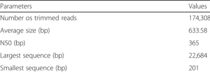
The same transcriptome was sequenced in duplicate resulting in 129,467,414 and 131,652,320 raw reads (considering forward and reverse reads) for each dupli-cate. The data obtained from de novo assembling is summarized in Table 1. The contigs were analyzed ac-cording to their functional annotation regarding Gene Ontology (Additional file 1), hits with deposited nu-cleotide and protein sequences from nr NCBI database and DADP, being the latter very important for results enrichment, once there is not much information re-garding toads in NCBI database.
AMP assessment
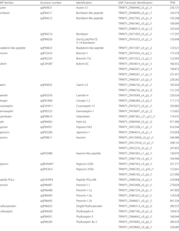
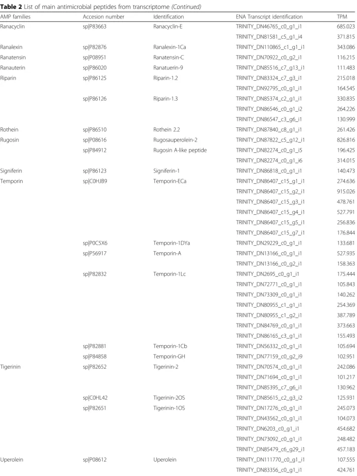
The functional analysis of the transcriptome data and the AMP Database showed the presence of 43 different peptides and protein classes. Table 2 summarizes the more abundant contigs (TPM > 100) and are clustered in 33 classes of AMP. The five major classes of AMP, considering the TPM values, are kassinin, temporin, peroniin, rugosauperolein and buforin.
Other proteins of interest
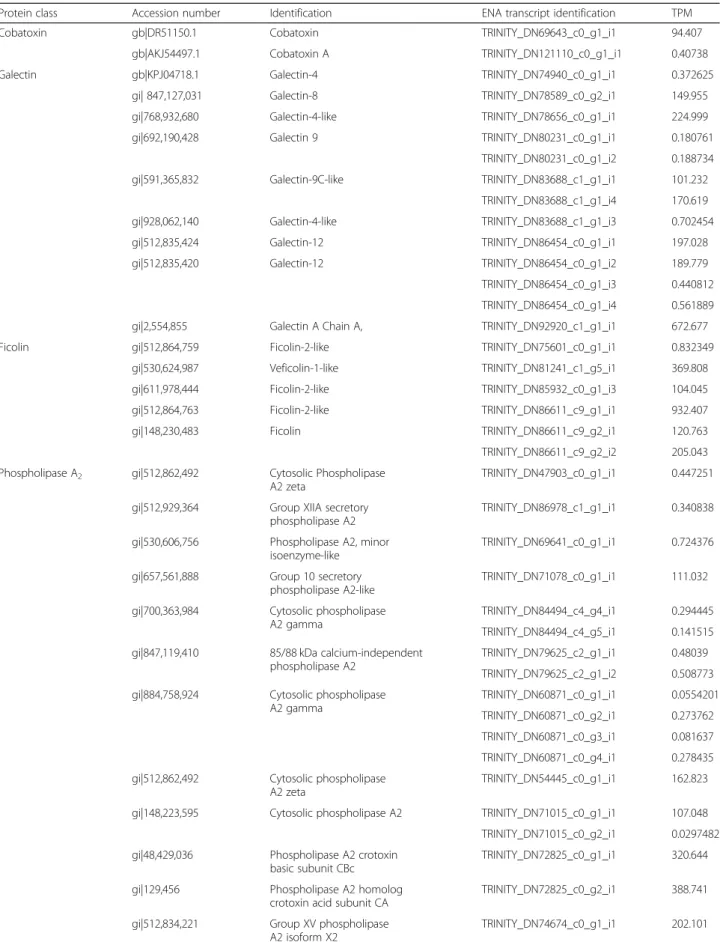
The main protein of interest that are not considered AMP are listed in Table 3. Two contigs related to cobatoxin were found in this study. The first one, iden-tified as TRINITY_DN69643_c0_g1_i, is identical to cobatoxin fromHelicoverpa armigera,identified by ac-cess number ADR51150.1 (gi|313,247,974). The second one, identified as TRINITY_DN121110_c0_g1_i1, has
matched cobatoxin A from Spodoptera exigua
(gi|827,029,657).
In the present transcriptome we found 19 full-length se-quences with high homology to PLA2. Interestingly, two contigs are similar to snake PLA2. Contig TRINI-TY_DN72825_c0_g1_i1 encodes a PLA2highly similar to Crotalus durissus terrificus PLA2 crotoxin basic subunit (gi 48,429,036) (Fig. 1a). This PLA2was also identified in the raw skin secretion in the fractions 24A, 25 and 26 (Fig.2) and confirmed by Edman degradation sequencing of the fractions. Contig TRINITY_DN72825_c0_g2_i1 is
also related to PLA2from C. d. terrificus(Fig.1b), but to the acid subunit (gi|129,456).
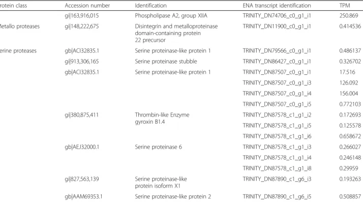
This study identified one full-length contig related to metalloproteases and 14 contigs related to serine pro-teases. Contig TRINITY_DN11900_c0_g1_i1 is highly homologous to a metallo-disintegrin from Xenopus laevis.Fourteen full-length sequences related to serine proteases were obtained. Six of them showed high similarity to Crotalus ssp snakes, being three (TRINI-TY_DN87578_c1_g1_i2, TRINITY_DN87578_c1_g1_i5, TRINITY_DN87578_c1_g1_i6,) containing the same coding sequence (herein named RsSVSP) highly related to gyroxin (Fig.3). Contigs TRINITY_DN87578_c1_g1 _i3, TRINITY_DN87578_c1_g1_i4 and TRINITY_DN8 7578_c1_g1_i8 encode a protein (RsSVSP2) very similar to serine protease 6 from C. adamanteus (gi|338,855,342).
Thirteen complete open reading frames (ORFs) related to galectins and 6 related to ficolins were found in the transcriptome. From those, 12 are related to predicted galectin from different genomes and transcriptomes. Con-tig TRINITY_DN92920_c1_g1_i1 is similar to galectin fromRhinella arenarumovary. Four complete ORFs were found matching different galectins fromXenopusgenome assessment. All contigs related to ficolins were annotated against model organisms’ genome (Xenopus spp. and Monodelphis domestica).
Fractionation of CS by RP-FPLC, SDS-PAGE and N-terminal sequencing
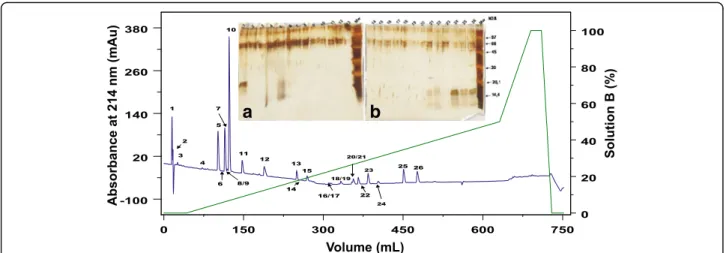
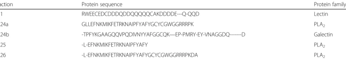
Crude secretion SDS-PAGE profile is shown in Additional file 2. CS was separated in 26 fractions, named CS1 to CS26 (Fig.2). The fractions were further submitted to SDS-PAGE, gel was stained with silver and the fractions named CS1, CS24, CS25 and CS26 (Fig.2, in-sert) were identified containing protein compounds. In addition, probably fractions CS5, CS21 and CS22 also con-tain protein molecules, but due to its low concentration, they were not investigated in this study.
Both gels show some interference on their top, prob-ably caused by the silver staining. It is possible to see bands with approximately 16 kDa in the fractions CS24, CS25 and CS26, which showed similar diffusion profiles. Therefore, these fractions were submitted to N-terminal sequencing by Edmans’degradation, but it was possible to obtain only CS1 and CS24–CS26 par-tial sequences (Table4).
Caseinolytic activity
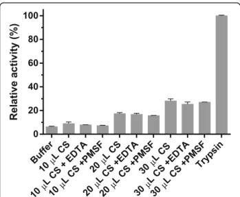
The transcriptome functional annotation showed some sequences that can be related to serine and metallo-proteases. In order to investigate if the sequences could really deduce these enzymes, we performed a proteolytic test using azocasein as substrate (Fig. 4). Table 1Statistical analysis of the transcriptome sequencing and
de novo assembling with Trinity
Parameters Values
Number os trimmed reads 174,308
Average size (bp) 633.58
N50 (bp) 365
Largest sequence (bp) 22,684
Table 2List of main antimicrobial peptides from transcriptome
AMP families Accesion number Identification ENA Transcript identification TPM
Aurein sp|P69021 Aurein-3.1 TRINITY_DN64440_c0_g1_i1 256.721
Bombesin sp|P84211 Bombesin-like peptide TRINITY_DN48099_c0_g1_i1 164.379
sp|P84212 Bombesin-like peptide TRINITY_DN27392_c0_g1_i1 192.298
TRINITY_DN61842_c0_g2_i1 106.099
TRINITY_DN80913_c0_g1_i2 102.443
sp|P84214 Bombesin TRINITY_DN71876_c0_g1_i1 117.297
sp|P86026 [Asn3,Lys6,Phe13]
3–14-bombesin
TRINITY_DN76333_c0_g1_i1 135.698
Bradykinin-like peptide sp|P84823 Bradykinin-like peptide TRINITY_DN71047_c0_g1_i2 123.521
Brevinin sp|P32423 Brevinin-1 TRINITY_DN70354_c0_g3_i1 175.329
sp|P82233 Brevinin-1Ta TRINITY_DN73322_c2_g2_i1 122.993
Buforin sp|C0HJB7 Buforin-EC TRINITY_DN54614_c0_g1_i1 766.952
TRINITY_DN60267_c0_g1_i1 190.875
TRINITY_DN60267_c1_g1_i1 231.457
TRINITY_DN60267_c0_g2_i1 228.282
Caerin sp|P69032 Caerin-2.3 TRINITY_DN66726_c0_g1_i1 182.654
TRINITY_DN66726_c0_g1_i2 121.242
Caeridin sp|P82076 Caeridin-4 TRINITY_DN79589_c4_g3_i1 226.024
Citropin sp|P81840 Citropin-1.2 TRINITY_DN86384_c3_g3_i1 111.715
Cruzioseptin sp|C0HK11 Cruzioseptin-15 TRINITY_DN70275_c0_g1_i1 293.883
Dermaseptin sp|P85523 Dermaseptin-1 TRINITY_DN76697_c0_g1_i2 115.602
Hylambatin sp|P08614 Hylambatin TRINITY_DN87362_c21_g12_i1 174.475
Hylin sp|P84003 Hylin-b2 TRINITY_DN84946_c0_g1_i2 971.000
Hyposin sp|P84957 Hyposin-HA3 TRINITY_DN72338_c1_g1_i1 616.344
Japonicin sp|P83306 Japonicin-1 TRINITY_DN64014_c0_g1_i1 310.058
Kassinin sp|P08611 Kassinin TRINITY_DN120600_c0_g1_i1 346.986
TRINITY_DN127618_c2_g1_i1 5081.92
TRINITY_DN52216_c0_g1_i1 247.853
sp|P42988 Kassinin-like peptide TRINITY_DN81065_c1_g3_i1 130.979
TRINITY_DN87139_c3_g3_i1 164.946
Nigrocin sp|B3A0M7 Nigrocin-2JDb TRINITY_DN87453_c9_g4_i1 327.271
sp|P0C8U1 Nigrocin-2HSb TRINITY_DN85182_c2_g10_i1 122.641
TRINITY_DN85182_c2_g3_i1 221.000
Peptide PGLa sp|C0HKP4 Peptide PGLa-R6 TRINITY_DN80328_c3_g2_i1 229.068
Peroniin sp|P86487 Peroniin-1.1 TRINITY_DN53498_c0_g1_i1 279.654
sp|P86488 Peroniin-1.1a TRINITY_DN67234_c0_g1_i1 347.803
sp|P86493 Peroniin-1.3a TRINITY_DN81622_c0_g1_i1 129.528
sp|P86495 Peroniin-1.1b TRINITY_DN46821_c0_g1_i1 841.554
Phyllocaerulein sp|P86625 [Arg4]-Phyllocaerulein TRINITY_DN85513_c6_g2_i2 280.537
Phylloseptin sp|P84569 Phylloseptin-4 TRINITY_DN87185_c0_g1_i2 109.815
sp|P84931 Phylloseptin-3 TRINITY_DN64642_c0_g2_i1 168.944
sp|P86283 Phylloseptin Bu-2 TRINITY_DN78083_c0_g4_i1 389.329
TRINITY_DN78083_c0_g6_i1 334.985
Table 2List of main antimicrobial peptides from transcriptome(Continued)
AMP families Accesion number Identification ENA Transcript identification TPM
Ranacyclin sp|P83663 Ranacyclin-E TRINITY_DN46765_c0_g1_i1 685.023
TRINITY_DN81581_c5_g1_i4 371.815
Ranalexin sp|P82876 Ranalexin-1Ca TRINITY_DN110865_c1_g1_i1 343.086
Ranatensin sp|P08951 Ranatensin-C TRINITY_DN70922_c0_g2_i1 116.215
Ranauterin sp|P86020 Ranatuerin-9 TRINITY_DN85516_c7_g13_i1 111.483
Riparin sp|P86125 Riparin-1.2 TRINITY_DN83324_c7_g3_i1 215.018
TRINITY_DN92795_c0_g1_i1 164.545
sp|P86126 Riparin-1.3 TRINITY_DN85374_c2_g1_i1 330.835
TRINITY_DN86546_c0_g1_i2 264.226
TRINITY_DN86547_c3_g6_i1 130.999
Rothein sp|P86510 Rothein 2.2 TRINITY_DN87840_c8_g1_i1 261.426
Rugosin sp|P08616 Rugosauperolein-2 TRINITY_DN87822_c5_g12_i1 826.816
sp|P84912 Rugosin A-like peptide TRINITY_DN82274_c0_g1_i5 196.425
TRINITY_DN82274_c0_g1_i6 314.015
Signiferin sp|P86123 Signiferin-1 TRINITY_DN86818_c0_g1_i1 140.473
Temporin sp|C0HJB9 Temporin-ECa TRINITY_DN86407_c15_g1_i1 274.636
TRINITY_DN86407_c15_g2_i1 915.026
TRINITY_DN86407_c15_g3_i1 478.761
TRINITY_DN86407_c15_g4_i1 527.791
TRINITY_DN86407_c15_g5_i1 256.836
TRINITY_DN86407_c15_g7_i1 176.844
sp|P0C5X6 Temporin-1DYa TRINITY_DN29229_c0_g1_i1 133.681
sp|P56917 Temporin-A TRINITY_DN13166_c0_g1_i1 527.935
TRINITY_DN13166_c0_g2_i1 158.363
sp|P82832 Temporin-1Lc TRINITY_DN2695_c0_g1_i1 175.444
TRINITY_DN72771_c0_g1_i1 105.843
TRINITY_DN73309_c0_g1_i1 140.262
TRINITY_DN80955_c1_g1_i1 254.369
TRINITY_DN80955_c1_g2_i1 387.789
TRINITY_DN84769_c0_g1_i1 373.663
TRINITY_DN86165_c3_g1_i1 155.493
sp|P82881 Temporin-1Cb TRINITY_DN56332_c0_g1_i1 105.694
sp|P84858 Temporin-GH TRINITY_DN77159_c0_g2_i9 102.951
Tigerinin sp|P82652 Tigerinin-2 TRINITY_DN70574_c0_g1_i1 242.086
TRINITY_DN71694_c0_g1_i1 101.217
TRINITY_DN85395_c7_g6_i1 130.962
sp|C0HL42 Tigerinin-2OS TRINITY_DN85615_c2_g3_i2 125.931
sp|P82651 Tigerinin-1OS TRINITY_DN17276_c0_g1_i1 245.073
TRINITY_DN43562_c0_g1_i1 104.073
TRINITY_DN6203_c0_g1_i1 454.682
TRINITY_DN73092_c0_g1_i1 248.482
TRINITY_DN85479_c6_g29_i1 457.183
Uperolein sp|P08612 Uperolein TRINITY_DN111770_c0_g1_i1 107.555
CS was capable of degrading azocasein. The tests in presence of EDTA and PMSF did not show differences.
Discussion
Although toads are widely spread in Brazilian and Latin America territory, its cutaneous peptides and proteins have come to the spotlight only recently and the scarce in-formation hinders its assessment [11,16,17].
The present study reveals the abundance of the Rhi-nella schneideri mucous gland defense peptides and protein through the RNA-seq transcriptome and some peptides and proteins present in its secretions. We used the cutaneous secretions from one specimen that was milked prior the gland extraction to obtain a more accurate result comparing the biochemical tests with the transcriptome.
Transcriptome survey is snapshots of mRNA obtained in a specific time. Therefore, the first step of this study was milking the toad’s skin secretions to maximize the transcripts production. We also used the secretion to carry out biochemical analysis subsequently. Using the se-cretions from the same toad to perform the experiments carried out in this paper allows us to discard any variation that may occur and obtain a more accurate analysis. RNA-seq analysis revealed the presence of distinct pro-teins and peptides. Most of the transcripts are related to housekeeping function, as expected, but we found novel proteins in the skin. As frog defense peptides are better characterized, we enriched the functional annotation using DADP database. Using this approach, there were several classes of AMP that are summarized in Table2. The con-tigs that presented the highest values of TPM belong to tachykynins, temporin, peroniin and buforin classes.Bufo bufo, B. verrucossismus and B. variabilis skin secretions act as antimicrobial agent against Escherichia fecalis and E. faeciumwith lower minimum inhibitory concentration than ampicillin [18].
The molecular classes of AMPs found in this tran-scriptome are discussed below.
Kassinin and Rugosauperolein
Kassinin and rugasoperolein AMPS belong to the tachyki-nin family of neuropeptides. Tachykitachyki-nins are well present in amphibian secretions and higher organisms, acting as neurotransmitters and neuroprotective agents in the latter. They present a C-terminal conserved region Phe-X-Gly-Leu-Met, that is known for this family’s activity [32]. Re-garding amphibians secretions, kassinin, a dodecapeptide, was the first tachykinin described fromKassina senegalen-sis methanol skin extract, which shows similar activity as substance P, physalaemin and eldosein, being able to stimulated salivary secretion, act as hypotensive agent and stimulate smooth muscle [33, 34]. Contig TRINI-TY_DN127618_c2_g1_i1 is very similar to the first kassi-nin ever identified.
Rugosauperolein was discovered in Uperoleia rugosa skin methanol extracts and was named after the tachyki-nin uperolein [35].
Tachykinins are widely studied mainly in higher organ-isms due their interesting neuroprotective activity. Des-pite it is known that they are present in amphibian skin, their role as AMP is poorly studied. As their role as neu-rotransmitters is well established, here we hypothesize they act not as a AMP, but as a host defense molecule that prevents the toad from predation along with micro molecules that are already known to play this role [36]. We use the same hypotheses to the presence of peroniin. Although we cannot discard the possibility of peroniin present any antimicrobial activity, we believe it is pro-duced to prevent R. schneideri to be swallowed, once tachykinins are known to show neuroactivity being able to cause gut tissue contraction [37].
Temporin
Temporins were discovered in a cDNA library from Rana temporaria skin and later obtained in R. tempor-alis’skin secretions. They are effective against gram posi-tive Bacillus megaterium and Escherichia coli with different sensitivities. This AMP family comprises
Table 2List of main antimicrobial peptides from transcriptome(Continued)
AMP families Accesion number Identification ENA Transcript identification TPM
TRINITY_DN83356_c0_g1_i2 339.468
TRINITY_DN83356_c0_g1_i3 101.171
TRINITY_DN81958_c0_g1_i1 277.617
TRINITY_DN81958_c0_g1_i2 216.205
Uperin sp|P82036 Uperin-5.1 TRINITY_DN87247_c0_g1_i1 332.281
TRINITY_DN87247_c1_g1_i1 137.665
TRINITY_DN87247_c0_g1_i3 185.119
TRINITY_DN77327_c0_g2_i1 238.182
TRINITY_DN87210_c0_g1_i1 105.306
Table 3List of other peptides and protein of interest from the transcriptome
Protein class Accession number Identification ENA transcript identification TPM
Cobatoxin gb|DR51150.1 Cobatoxin TRINITY_DN69643_c0_g1_i1 94.407
gb|AKJ54497.1 Cobatoxin A TRINITY_DN121110_c0_g1_i1 0.40738
Galectin gb|KPJ04718.1 Galectin-4 TRINITY_DN74940_c0_g1_i1 0.372625
gi| 847,127,031 Galectin-8 TRINITY_DN78589_c0_g2_i1 149.955
gi|768,932,680 Galectin-4-like TRINITY_DN78656_c0_g1_i1 224.999
gi|692,190,428 Galectin 9 TRINITY_DN80231_c0_g1_i1 0.180761
TRINITY_DN80231_c0_g1_i2 0.188734
gi|591,365,832 Galectin-9C-like TRINITY_DN83688_c1_g1_i1 101.232
TRINITY_DN83688_c1_g1_i4 170.619
gi|928,062,140 Galectin-4-like TRINITY_DN83688_c1_g1_i3 0.702454
gi|512,835,424 Galectin-12 TRINITY_DN86454_c0_g1_i1 197.028
gi|512,835,420 Galectin-12 TRINITY_DN86454_c0_g1_i2 189.779
TRINITY_DN86454_c0_g1_i3 0.440812
TRINITY_DN86454_c0_g1_i4 0.561889
gi|2,554,855 Galectin A Chain A, TRINITY_DN92920_c1_g1_i1 672.677
Ficolin gi|512,864,759 Ficolin-2-like TRINITY_DN75601_c0_g1_i1 0.832349
gi|530,624,987 Veficolin-1-like TRINITY_DN81241_c1_g5_i1 369.808
gi|611,978,444 Ficolin-2-like TRINITY_DN85932_c0_g1_i3 104.045
gi|512,864,763 Ficolin-2-like TRINITY_DN86611_c9_g1_i1 932.407
gi|148,230,483 Ficolin TRINITY_DN86611_c9_g2_i1 120.763
TRINITY_DN86611_c9_g2_i2 205.043
Phospholipase A2 gi|512,862,492 Cytosolic Phospholipase
A2 zeta
TRINITY_DN47903_c0_g1_i1 0.447251
gi|512,929,364 Group XIIA secretory phospholipase A2
TRINITY_DN86978_c1_g1_i1 0.340838
gi|530,606,756 Phospholipase A2, minor isoenzyme-like
TRINITY_DN69641_c0_g1_i1 0.724376
gi|657,561,888 Group 10 secretory
phospholipase A2-like
TRINITY_DN71078_c0_g1_i1 111.032
gi|700,363,984 Cytosolic phospholipase A2 gamma
TRINITY_DN84494_c4_g4_i1 0.294445
TRINITY_DN84494_c4_g5_i1 0.141515
gi|847,119,410 85/88 kDa calcium-independent phospholipase A2
TRINITY_DN79625_c2_g1_i1 0.48039
TRINITY_DN79625_c2_g1_i2 0.508773
gi|884,758,924 Cytosolic phospholipase A2 gamma
TRINITY_DN60871_c0_g1_i1 0.0554201
TRINITY_DN60871_c0_g2_i1 0.273762
TRINITY_DN60871_c0_g3_i1 0.081637
TRINITY_DN60871_c0_g4_i1 0.278435
gi|512,862,492 Cytosolic phospholipase A2 zeta
TRINITY_DN54445_c0_g1_i1 162.823
gi|148,223,595 Cytosolic phospholipase A2 TRINITY_DN71015_c0_g1_i1 107.048
TRINITY_DN71015_c0_g2_i1 0.0297482
gi|48,429,036 Phospholipase A2 crotoxin basic subunit CBc
TRINITY_DN72825_c0_g1_i1 320.644
gi|129,456 Phospholipase A2 homolog
crotoxin acid subunit CA
TRINITY_DN72825_c0_g2_i1 388.741
gi|512,834,221 Group XV phospholipase
A2 isoform X2
anionic, hydrophobic, small peptides (8–17 amino acids) that are folded in alpha-helices and which potency is re-lated to the final net charge [38, 39]. Following its first discovery, temporins were also found in secretions of different species of the genus Euphycits, Limnonectes, Hypsiboas, Amolops, Hylarana and Lithobates [39]. Re-garding their biotechnological applications, there are
more than 20 deposited patents related to them, which varies from their pharmacological use as anti-HIV to obtaining of transgenic plant resistant to pathogens [39].
Deposited contig TRINITY_DN86407_c15_g2_i1 is similar to temporin-ECa, from Euphlyctis cyanophlyctis, the skittering frog. As other temporins, it shows activity against gram-positive bacteria E. coli, K. pneumonia,
Table 3List of other peptides and protein of interest from the transcriptome(Continued)
Protein class Accession number Identification ENA transcript identification TPM
gi|163,916,015 Phospholipase A2, group XIIA TRINITY_DN74706_c0_g1_i1 250.869
Metallo proteases gi|148,222,675 Disintegrin and metalloproteinase domain-containing protein 22 precursor
TRINITY_DN11900_c0_g1_i1 0.414536
Serine proteases gb|ACI32835.1 Serine proteinase-like protein 1 TRINITY_DN79566_c0_g1_i1 0.486137
gi|913,306,165 Serine proteinase stubble TRINITY_DN86427_c0_g1_i1 0.326702
gb|ACI32835.1 Serine proteinase-like protein 1 TRINITY_DN87507_c0_g1_i1 17.516
TRINITY_DN87507_c0_g1_i3 126.092
TRINITY_DN87507_c0_g1_i4 156.004
TRINITY_DN87507_c0_g1_i5 0.772103
gi|380,875,411 Thrombin-like Enzyme
gyroxin B1.4
TRINITY_DN87578_c1_g1_i2 0.172693
TRINITY_DN87578_c1_g1_i5 0.125578
TRINITY_DN87578_c1_g1_i6 0.658672
gb|AEJ32000.1 Serine proteinase 6 TRINITY_DN87578_c1_g1_i3 0.266027
TRINITY_DN87578_c1_g1_i4 0.246148
TRINITY_DN87578_c1_g1_i8 0.29959
gi|827,563,139 Serine proteinase-like protein isoform X1
TRINITY_DN87890_c1_g6_i3 0.193263
gb|AAM69353.1 Serine proteinase-like protein 2 TRINITY_DN87890_c1_g6_i5 0.508857
a
b
Fig. 1Sequence alignment of phospholipases A2discovered in this transcriptome (RsPLA2) and those available in databases.aAlignment of
protein codified by contig TRINITY_DN72825_c0_g1_i2, named RsPLA2 and different basic crotoxins subunits fromCrotalus durissus terrificusCdtPLA2
(PA2B6_CRODO),Crotalus durissus collilineatusCdcPLA2(PA2B6_CRODO) andCrotalus durissus ruruima(PA2BA_CRODR).bAlignment of protein
Micrococcus luteus and Staphylococcus aureus, and low hemolytic activity [40] .
Peroniin
Peroniins discovery was made through mass spectrom-etry analysis ofLitoria peroniiskin secretions in winter and summer. Albeit they are considered AMP, there is no report of their test against neither gram positive nor gram negative bacteria. In fact, peronnins are the major component ofL. peroniisecretions both summer and winter and they possess activity over smooth muscle causing its contraction. There is only one re-port of peroniin up to date [41].
Buforin
This AMP family was first discovered inBufo Bufo gar-garizans stomach. This family comprises small pep-tides (approximately 6.5 kDa) and are effective against several gram positive and negative bacteria [42]. They are derived from histone H2A and belong to the toads’ innate immune system. While buforin I is secreted in the stomach protecting the toad against pathogen ingested microorganisms and further binding to the mucosa biofilm enhancing its protection, buforin II does not have its mechanism of action fully elucidated, but it is hypothesized they bind to microorganism’s nu-cleic acid destroying it [43]. In this study, contig TRI-NITY_DN54614_c0_g1_i1 is related to buforin-EC, isolated from skin secretions of frog Euphlyctis cya-nophlyctis, which has shown activity against Staphyloc-coccus aureus and Escherichia coli [40]. We believe temporins and buforins, along with other AMPs, act as protection against microorganisms, due to the well-established activity of these molecules against gram
positive and negative bacteria. The diversity of AMP found in the skin proves that glandular secretions work as a biochemical enriched barrier for the toads’ protection.
In addition to the AMPs many other protein compo-nents, which probably have relevant roles for frog defense, have been identified in the transcriptome, among them cobatoxins, PLA2, proteases, ficolin and galectins. These molecular classes are discussed below. Interestingly, ficolin was one of the major contigs found in the transcriptome survey.
Cobatoxin
Cobatoxins were firstly reported in theCentruroides nox-iusscorpion poison as potassium channel-blocking toxins, belonging to theα-K-toxins, subfamily 9. They are
moder-ate affinity blockers of K+ voltage-dependent channels Shaker and Kv1.1 [44]. Cobatoxin fromHelicoverpa armi-gerais mostly connected to the insect defense. Its level of expression has significantly raised after infection with both gram positive and negative bacteria [45]. Regarding Spodo-petra exiguacobatoxin, gene expression analysis from the insect midgut after the exposition of the insect toB. thur-ingiensistoxins revealed an increase of the mRNA coding for this protein, indicating that it plays a fundamental role in the insect defense [46]. Furthermore, an analysis of Gal-leria mellonellachallenged againstMicrococcus luteushas shown that cobatoxin is likely to maximize the potential of other innate AMPs from the insect [47].
Here, we hypothesize that cobatoxin is also part of the immune system of the toad.
We also found other proteins of interest that are not catalogued as AMP with the aid of non-redundant NCBI database annotation:
a
b
Phospholipase A2
Phospholipase A2 (PLA2) catalyze the hydrolysis of phospholipids in the sn2 position releasing arachidonic acid and lysophosphatidic acid, which are precursors of signaling molecules in immune response, inflamma-tion, pain and cell regulatory processes [48–50]. They
can be found in different tissues and organelles and are often small proteins (14–18 kDa), and their stability varies according to the number of disulfide bonds. Se-creted PLA2s are one of the major components of Ela-pidae and Viperidae snake venoms [51]. In fact, the crotoxin was the first toxin isolated almost 100 years
a
b
Fig. 3Sequence alignment of serine protease discovered in this transcriptome (RsSP) and those from different snake venoms (SVSP) available in datadases.aAlignment of protein codified by contigs TRINITY_DN87578_c1_g1_i3, TRINITY_DN87578_c1_g1_i4 and TRINITY_DN87578_c1_g1_i8, named RsSP1 and gyroxin serine proteases from snake venoms (SVSPs) fromCrotalus durissus terrificus(CdtSVSP3, VSP13_CRODU), CdtSVSP 2 (VSP21_CRODU) CdtSVSP4 (VSP14_CRODU).bAlignment of serine protease codified by contigs TRINITY_DN87578_c1_g1_i2, TRINITY_DN87578_c1_g1_i5 and TRINITY_DN87578_c1_g1_i6, named RsSP2 and gyroxin SVSP fromCrotalus adamanteus1, CaSVSP1 (VSP1_CROAD), CaSVSP2 (VSP2_CROAD), CaSVSP5 (VSPE_CROAD) and CaSVSP6 (A0A1W7RB84_CROAD). Signal peptide is indicated above the sequences and the arrows beside the alignment indicates the amino acids that are important to the catalytic activity. Alignment was generated with the aid of Multalin and formatted using Espript 3.0
Table 4N-terminal sequences from the poison fractions by Edman’s degradation technique
Fraction Protein sequence Protein family
CS1 RWEECEDCDDDQDDQQQQQCAKDDDDE---Q-QQD Lectin
CS24a GLLEFNKMIKFETRKNAIPFYAFYGCYCGWGGRRRPK PLA2
CS24b -TPFYKGAAGQQVPQDIVNYYAFGGCQK—EP-PMRY-EY-VNAGGDQ---D Galectin
CS25 -L-EFNKMIKFETRKNAIPFYAFY PLA2
ago and its sequence has been determined for more than 30 years now, and the cloning was successfully obtained in the 80’s [52,53].
InCrotalussnake venoms, crotoxin is composed of two non-covalently bound subunits (one acidic and one basic). The basic component (CB) is a catalytic active PLA2 whereas the acidic component (CA) is a PLA2catalytically inactive responsible to direct CB towards specific sites that lead to neurotoxic actions [54–58].
In humans, secreted PLA2group IIA can be found in tears and it is most likely to play a defensive role in eyes defense against gram-positive bacteria, but no re-sponse to gram-negative bacteria [59]. These mole-cules are also found in dromedary tears and showed activity against both gram positive and negative bac-teria [60]. Thus, a PLA2isolated from Daboia Russelli venom was able to strongly inhibit gram negative bac-teria and also showed activity against gram positive bacteria isolated from human [61] . Therefore, we also assume the protective action in toad skin.
Serine and metalloproteases
The contig TRINITY_DN11900_c0_g1_i1 is highly homologous to a metallo-disintegrin protease that has been related to Xenopus laevis reproduction [62] and neural crest development [63]. Although it was pos-sible to obtain a full-length transcript in neural cells, the metallo-disintegrin did not have the catalytic do-main, which indicates this protein acts as a transmem-brane receptor [63].
Several full-length sequences were related to serine proteases. Three of them containing the same coding se-quence (herein named RsSVSP) highly related to gyroxin, a non-lethal serine protease with neurotoxic effects that causes a neurological syndrome in mice known for the animal’s movements as rotation to a barrel roll [64]. The contigs TRINITY_DN87578_c1_g1_i3, TRINITY_DN87578_c1_g1_i 4 and TRINITY_DN87578_c1_g1_i8 encode a protein, named RsSVSP2, very similar to serine protease 6 from C. adamanteus (gi|338,855,342), also found in a tran-scriptoma survey [65].
Ficolin and galectin
Ficolins are a group of oligomeric lectin that present fibrinogen-like and collagen-like domains and possess a carbohydrate binding domain (CRD), being N-acetyl-glucosamine (GlcNAc) the carbohydrate that presents the major number of galectins specificity [66,67]. They are capable of activating complement system via lectin pathway, and aggregate some bacteria enhancing phagocytosis showing their relevant role in organisms’ defense [68].
Galectins are lectins which major ligand isβ
-galacto-se-containing glycoconjugates and show their CRD conserved. They may bind to cell-surface and matrix glycans, being able to control intracellular signaling, and protein interactions dependent pathways [69]. Due to its action over neurological system, we believe it also plays a possible role as host defense peptides (HDP) and may provide protections against animal predation adding or even enhancing the symptoms of micro molecules present in parotoid glands that show neurological effects [10].
The contig TRINITY_DN92920_c1_g1_i1 is similar to galectin, an S-type lectin, from Rhinella arenarumovary, probably playing a developmental regulatory role [70]. In contrast,Xenopusgalectins from the animal’s skin are be-lieved to act as HDP [71]. Recently, a galectin was found in parotoid secretion fromR. schneideriusing mass spec-trometry de novo sequencing [11].
Crude secretion has shown a different profile from parotoid poison presented before [11,12]. In the cuta-neous secretion (Additional file 2) it is possible state there are a richness of protein content which molecu-lar weight varies from high molecumolecu-lar mass (around 100 kDa) to low molecular mass (less than 14 kDa), but there is a strong band with approximately 60 kDa that is common to the profile presented in male and female parotoid secretions from Piaui, Brazil [11]. After ana-lyzing the variation of mass, crude secretion was sub-mitted to RP-FPLC to further investigation.
The RP-FPLC of the secretion resulted in 26 frac-tions, which were further analyzed by SDS-PAGE. The fractions CS1, CS24-CS26 were submitted to Edman’s
µ µ µ
µ µ
µ µ
µ µ
degradation sequencing, because they showed protein bands on SDS-PAGE. Other fractions might contain mi-cro molecules from poison and this method of chroma-tography was already used for isolation of those molecules fromR. schneideriparotoid poison before [15].
Fractions CS24, CS25 and CS26 showed sequences regarding a PLA2 from snake venom [72]. Fractions CS24a, CS25 and CS26 contain PLA2isoforms similar to a basic PLA2isolated fromCrotalus durissus terrifi-cus (PA2BF_CRODU). As expected, we were able to predict the protein sequence from the contig TRINI-TY_DN72825_c0_g1_i1. We also retrieved contigs re-lated to the acidic PLA2. In crotoxin, one of the major components of Crotalus spp. venom, both units (basic and acidic PLA2) are bonded by a non-covalent bound. The acid subunit (also named crotapotin) is devoid of activity, acting as a stabilizer, and the basic subunit is catalytically active and toxic [52]. Despite its role in the snake venom, we hypothesize this PLA2 is related to toad’s immune system, similarly to secreted PLA2in human [59] and dromedary tears [60], especially facing the probability of the toad infection by amoeba.
CS24b sequencing also presents a protein that showed similarity to a galectin-1 from Rhinella are-narum (gi|255a855; sp.|P5621). It is a beta-galactosyl-binding lectin discovered in toad ovary and similar to the mammalian one that binds to different carbohy-drates intra and extra-cellular, probably regulating de-velopmental process in toads oocytes [70]. There are three contigs related to this protein, but none of them is complete. Xenopus laevis is known to secrete a lec-tin, along with other defense peptides, which plays a role not only in defense against pathogenic microor-ganisms, but also against predators and another un-known structural role [71].
CS1 sequenced revealed this fraction contains more than one protein; the first putative protein is similar to a C-type lectin isolated from Helicoverpa armigera (gi|385,202,653) that is up regulated in the presence of Escherichia coliinjection, indicating it role as a defense protein [45], but it was not found in the transcriptome. CS1 is also similar to the lectin fromNaegleria gliberia (gi|290,983,012), an amoeba that can be found in moist habitats and freshwater, that was unrevealed in its gen-ome [73]. We could not retrieve any contigs related to this protein, which indicates a possible contamination of the toad skin by this microorganism. This contamin-ation may explain the high levels of defense peptides and proteins found in the transcriptome and other fractions. It is known that toads might produce toxins that are specific against pathogens [74].
Beyond the AMP assessment and the discovery of other peptides and proteins of defense, we also re-trieved some proteases. As the transcriptome showed
some evidence of serine and metalloproteases, we de-cided to carry out an exploratory experiment to survey the presence of these proteins in the poison. Azocasein degradation assay is a classic test to determine the presence of either of these enzymes since proteolytic activity is evaluated. Raw secretion can result in dose dependent azocasein degradation. We used EDTA, which is a chelator agent that inhibits metallopro-teases, and PMSF that inhibits serine proteases to in-vestigate which class is most likely to be present in the raw secretion and cause the casein degradation. Des-pite our best efforts, it was not possible to determine which class of protease was more active, once the con-trols with PMSF and EDTA did not show any statistical relevance. However, we believe serine protease are the main responsible for this result, since there are more full-length contigs with higher TPM in the transcrip-tome and only one low expressed (TPM < 1) full-length contig coding for a metalloprotease. In addition, a serine protease was found in Bufo bufoparotoid se-cretion through a proteomic analysis [17], indicating it may be produced in this toad secretion too, but further characterization is needed.
Here we presented the first transcriptome survey from R. schneideri skin. As the results showed, this study paves the way for discovering new molecules be-sides characterizing an important secretion and the glands where they are produced.
Conclusions
Although Rhinella schneideri toads are known to pos-sess bioactive molecules in its secretion, it is still poorly studied when compared to other venomous and poisonous animals. Most of the studies comprises the parotoid micro molecules secretions in detriment of cutaneous secretion. This is the first study to use a high throughput RNA-seq technology to investigate R. schneideri cutaneous secretions and the first one to focus on defense peptides and proteins. Furthermore, using the milked secretion and skin from the same toad allowed an accurate analysis of protein expres-sion, once individual variation was dismissed. The re-sults obtained herein showed evidence of novel AMP and enzymes that need to be further explored.
Additional files
Additional file 1:Gene ontology of Rhinella schneideri skin transcriptome. The Gene Ontology is divided in biological proccess, molecular function and cellular component. (DOCX 13 kb)
Additional file 2:Eletrophoretic profile of Rhinella schneideri’s cutaneous secretion. Different volumes (5, 10 and 20 µL) of CS were analyzed by 12,5 % SDS-PAGE and stained with Coomasie Blue PhastGel
β-mercaptoethanol and boiled for 10 minutes befora application in the SDS-PAGE. (PPTX 189 kb)
Abbreviations
ACN:Acetonitrile; AMP: Antimicrobial peptides; CS: Cutaneous secretion; DADP: Anuran antimicrobial peptides database; DEPC: Diethylpyrocarbonate; EDTA: Ethylenediamine tetraacetic acid; ENA: European Nucleotide Archive; GO: Gene ontology; HDP: Host defense peptides; NCBI: National Center for Biotechnology Information; pident: Protein identity; PLA2: Phospholipase A2;
PMSF: Phenylmethylsulfonyl fluoride; PVDF: Polyvinylidene fluoride; RNA-seq: RNA sequencing; RP-FPLC: Reversed phase fast protein liquid chromatography; SDS-PAGE: Denaturing polyacrylamide electrophoresis gel; SVSP: Snake venom serine protease; TCA: Trichloroacetic acid; TFA: Acid trifluoracetic; TPM: Transcripts Per Kilobase Million
Acknowledgements
The authors are thankful for Karla de Castro Figueiredo Bordon, PhD, for the N-terminal sequencing, Iara Aimê Cardoso for technical support and biologist Luiz Henrique Anzaloni Pedrosa.
Funding
The São Paulo Research Foundation (FAPESP; grant n. 2015/18432–0 and scholarships to PYTS n.2014/15644–3, CTC n. 2013/26200–6 and GAW n. 201/ 06170–8) supported this work. This work was supported in part by the Coordination for the Improvement of Higher Education Personnel (CAPES) through Programa Editoração CAPES (edital n. 13/2016, auxílio n. 0722/2017, processo n. 88881.142062/2017–01) and by the National Council for Scientific and Technological Development (CNPq) through Programa Editorial CNPq/ CAPES (process n. 440954/2017–7).
Availability of data and materials
The datasets generated and/or analyzed during the current study are available in the European Nucleotide Archive- ENA repository,http://www.ebi.ac.uk/ena.. The accession number for the transcriptome is: PRJEB27393 and for the coding sequences mentioned in this paper is: ERZ693103.
Authors’contributions
PYTS was responsible for carrying out the transcriptome data analysis, toad’s poison biochemical analysis and was a major contributor in writing the manuscript. CTC also was responsible for data analysis, design experiment and writing the manuscript. RMF performed the de novo assembling and FPM calculations. GAW performed part of the functional annotation and deposited the data. CUV surpervisioned the in silico analysis and assisted in the project design. ECA coordinated the whole team, assisted in the experiments design and advised PYTS. All authors read and approved the final manuscript.
Ethics approval and consent to participate
Not applicable.
Consent for publication
Not applicable.
Competing interests
The authors declare there are no competing interests.
Publisher’s Note
Springer Nature remains neutral with regard to jurisdictional claims in published maps and institutional affiliations.
Author details
1
Laboratory of Animal Toxins, School of Pharmaceutical Scienes of Ribeirão Preto, University of São Paulo, Avenida do Café s/n, Ribeirão Preto, SP, Brazil.
2
Laboratory of genetics–LABGEN, Institute of Genetics and Biochemistry, Campus Umuarama, Federal University of Uberlândia, Avenida Pará, Uberlândia, MG 1720, Brazil.3Department of Physics and Chemistry, University of São Paulo, School of Pharmaceutical Sciences of Ribeirão Preto, Av. do Café s/n°, Monte Alegre, Ribeirão Preto, SP 14040-903, Brazil.
Received: 4 July 2018 Accepted: 7 November 2018
References
1. Koehn FE, Carter GT. The evolving role of natural products in drug discovery. Nat Rev Drug Discov. 2005;4(3):206–20.
2. Newman DJ, Cragg GM, Snader KM. The influence of natural products upon drug discovery. Nat Prod Rep. 2000;17(3):215–34.
3. Casewell NR, Wüster W, Vonk FJ, Harrison RA, Fry BG. Complex cocktails: the evolutionary novelty of venoms. Trends Ecol Evol. 2012;28(4):219–29. 4. Fry BG, Roelants K, Champagne DE, Scheib H, Tyndall JD, King GF, et al. The
toxicogenomic multiverse: convergent recruitment of proteins into animal venoms. Annu Rev Genomics Hum Genet. 2009;10:483–511.
5. Chan WY, Ng TB, Yeung HW. Examination for toxicity of a Chinese drug, the toad glandular secretory product chan su, in pregnant mice and embryos. Biol Neonate. 1995;67(5):376–80.
6. Frost DR. Amphibian Species of the World: an Online Reference. vol. 6.0; 2018.http://research.amnh.org/vz/herpetology/amphibia/
7. Regis-Alves E, Jared SGS, Maurício B, Mailho-Fontana PL, Antoniazzi MM, Fleury-Curado MC, et al. Structural cutaneous adaptations for defense in toad (Rhinella icterica) parotoid macroglands. Toxicon. 2017;137:128–34. 8. Jared C, Antoniazzi MM, Jordao AE, Silva JR, Greven H, Rodrigues MT.
Parotoid macroglands in toad (Rhinella jimi): their structure and functioning in passive defence. Toxicon. 2009;54(3):197–207.
9. Clarke BT. The natural history of amphibian skin secretions, their normal functioning and potential medical applications. Biol Rev Camb Philos Soc. 1997;72(3):365–79.
10. Daly JW, Spande TF, Garraffo HM. Alkaloids from amphibian skin: a tabulation of over eight-hundred compounds. J Nat Prod. 2005;68(10):1556–75. 11. Sousa-Filho LM, Freitas CD, Lobo MD, Monteiro-Moreira AC, Silva RO,
Santana LA, et al. Biochemical profile, biological activities, and toxic effects of proteins in theRhinella schneideriparotoid gland secretion. J Exp Zool A Ecol Genet Physiol. 2016;325(8):511–23.
12. Sciani JM, Angeli CB, Antoniazzi MM, Jared C, Pimenta DC. Differences and similarities among parotoid macrogland secretions in South American toads: a preliminary biochemical delineation. Sci World J. 2013;2013:ID 937407.
13. Schmeda-Hirschmann G, Quispe C, Theoduloz C, de Sousa PT Jr, Parizotto C. Antiproliferative activity and new argininyl bufadienolide esters from the
“cururu”toadRhinella(Bufo)schneideri. J Ethnopharmacol. 2014;155(2):1076–85. 14. Anjolette FA, Leite FP, Bordon KC, Azzolini AE, Pereira JC, Pereira-Crott LS, et al. Biological characterization of compounds fromRhinella schneideripoison that act on the complement system. J Venom Anim Toxins incl Trop Dis. 2015;21:25.https://doi.org/10.1186/s40409-015-0024-9.
15. Shibao PY, Anjolette FA, Lopes NP, Arantes EC. First serine protease inhibitor isolated fromRhinella schneideripoison. J Venom Anim Toxins incl Trop Dis. 2015;21:30.https://doi.org/10.1186/s40409-015-0029-4.
16. Freitas CDT, Sousa-Filho LM, Lima MSCS, Pereira ACTC, Ferreira GP, Lopes FES, et al. Proteins from theRhinella schneideriparotoid gland secretion exhibit anti-nociceptive effect against nociception induced by inflammation. Biomed Pharmacother. 2017;93:705–8.
17. Kowalski K, Marciniak P, Rosinski G, Rychlik L. Toxic activity and protein identification from the parotoid gland secretion of the common toadBufo bufo. Comp Biochem Physiol C Toxicol Pharmacol. 2018;205:43–52. 18. Nalbantsoy A, Karis M, Yalcin HT, Göcmen B. Biological activities of skin and
parotoid gland secretions of bufonid toads (Bufo bufo,Bufo verrucosissimus
andBufotes variabilis) from Turkey. Biomed Pharmacother. 2016;80:298–303. 19. Konig E, Bininda-Emonds OR, Shaw C. The diversity and evolution of anuran
skin peptides. Peptides. 2014;63:96–117.
20. Boldrini-Franca J, Cologna CT, Pucca MB, Bordon KC, Amorim FG, Anjolette FA, et al. Minor snake venom proteins: structure, function and potential applications. Biochim Biophys Acta Gen Subj. 2016;1861(4):824–38. 21. Huang L, Li J, Anboukaria H, Luo Z, Zhao M, Wu H. Comparative
transcriptome analyses of seven anurans reveal functions and adaptations of amphibian skin. Sci Rep. 2016;6:24069.
22. Yang W, Qi Y, Fu J. Genetic signals of high-altitude adaptation in amphibians: a comparative transcriptome analysis. BMC Genet. 2016;17:134. 23. Zhao F, Yan C, Wang X, Yang Y, Wang G, Lee W, et al. Comprehensive
24. Andrews S. FastQC: a quality control tool for high throughput sequence data; 2010.
25. Chen TW, Gan RC, Fang YK, Chien KY, Liao WC, Chen CC, et al. FunctionAnnotator, a versatile and efficient web tool for non-model organism annotation. Sci Rep. 2017;7:10430.
26. NovkovićM, SimunićJ, BojovićV, Tossi A, JuretićD. DADP: the database of anuran defense peptides. Bioinformatics. 2012;28(10):1406–7.
27. Schägger H, von Jagow G. Tricine-sodium dodecyl sulfate-polyacrylamide gel electrophoresis for the separation of proteins in the range from 1 to 100 kDa. Anal Biochem. 1987;166(2):368–79.
28. Edman P, Begg G. A protein sequenator. Eur J Biochem. 1967;1(1):80–91. 29. Corpet F. Multiple sequence alignment with hierarchical clustering. Nucleic
Acids Res. 1988;16(22):10881–90.
30. Robert X, Gouet P. Deciphering key features in protein structures with the new ENDscript server. Nucleic Acids Res. 2014;42:W320–4.
31. Wang WJ, Shih CH, Huang TF. A novel P-I class metalloproteinase with broad substrate-cleaving activity, agkislysin, fromAgkistrodon acutusvenom. Biochem Biophys Res Commun. 2004;324(1):224–30.
32. Severini C, Improta G, Falconieri-Erspamer G, Salvadori S, Erspamer V. The tachykinin peptide family. Pharmacol Rev. 2002;54(2):285–322. 33. Anastasi A, Montecucchi P, Erspamer V, Visser J. Amino acid composition
and sequence of kassinin, a tachykinin dodecapeptide from the skin of the African frog Kassina senegalensis. Experientia. 1977;33(7):857–8.
34. Erspamer GF, Erspamer V, Piccinelli D. Parallel bioassay of physalaemin and kassinin, a tachykinin dodecapeptide from the skin of the African frogKassina senegalensis. Naunyn Schmiedeberg's Arch Pharmacol. 1980;311(1):61–5. 35. Nakajima T, Yasuhara T, Erspamer V, Erspamer GF, Negri L, Endean R.
Physalaemin- and bombesin-like peptides in the skin of the Australian leptodactylid frogUperoleia rugosa. Chem Pharm Bull (Tokyo). 1980;28(3): 689–95.
36. Sakate M, Oliveira PCL. Toad envenoming in dogs: effects and treatment. J Venom Anim Toxins. 2000;6(1):52–62www.scielo.br/scielo.php?script=sci_ arttext&pid=S0104-79302000000100003.
37. Carter MS, Krause JE. Structure, expression, and some regulatory mechanisms of the rat preprotachykinin gene encoding substance P, neurokinin a, neuropeptide K, and neuropeptide gamma. J Neurosci. 1990; 10(7):2203–14.
38. Simmaco M, Mignogna G, Canofeni S, Miele R, Mangoni ML, Barra D. Temporins, antimicrobial peptides from the European red frogRana temporaria. Eur J Biochem. 1996;242(3):788–92.
39. Ladram A, Nicolas P. Antimicrobial peptides from frog skin: biodiversity and therapeutic promises. Front Biosci (Landmark Ed). 2016;21:1341–71. 40. Asoodeh A, Azam AG, Chamani J. Identification and characterization of
novel antibacterial peptides from skin secretions ofEuphlyctis cyanophlyctis. Int J Pept Res Ther. 2012;18(2):107–15.
41. Bilusich D, Jackway RJ, Musgrave IF, Tyler MJ, Bowie JH. The host-defence skin peptide profiles of Peron's tree frogLitoria peroniiin winter and summer. Sequence determination by electrospray mass spectrometry and activities of the peptides. Rapid Commun Mass Spectrom. 2009;23(17):2628–36. 42. Park CB, Kim MS, Kim SC. A novel antimicrobial peptide fromBufo bufo
gargarizans. Biochem Biophys Res Commun. 1996;218(1):408–13. 43. Cho JH, Sung BH, Kim SC. Buforins: histone H2A-derived antimicrobial
peptides from toad stomach. Biochim Biophys Acta. 2008;1788(8):1564–9. 44. Selisko B, Garcia C, Becerril B, Gomez-Lagunas F, Garay C, Possani LD.
Cobatoxins 1 and 2 fromCentruroides noxiusHoffmann constitute a subfamily of potassium-channel-blocking scorpion toxins. Eur J Biochem. 1998;254(3):468–79.
45. Wang Q, Liu Y, He HJ, Zhao XF, Wang JX. Immune responses ofHelicoverpa armigerato different kinds of pathogens. BMC Immunol. 2010;11:9. 46. Crava CM, Jakubowska AK, Escriche B, Herrero S, Bel Y. Dissimilar regulation
of antimicrobial proteins in the midgut ofSpodoptera exigualarvae challenged withBacillus thuringiensistoxins or baculovirus. PLoS One. 2015; 10(5):e0125991.
47. Bolouri Moghaddam MR, Tonk M, Schreiber C, Salzig D, Czermak P, Vilcinskas A, et al. The potential of theGalleria mellonellainnate immune system is maximized by the co-presentation of diverse antimicrobial peptides. Biol Chem. 2016;397(9):939–45.
48. Narumiya S, Sugimoto Y, Ushikubi F. Prostanoid receptors: structures, properties, and functions. Physiol Rev. 1999;79(4):1193–226.
49. Lin ME, Herr DR, Chun J. Lysophosphatidic acid (LPA) receptors: signaling properties and disease relevance. Prostaglandins Other Lipid Mediat. 2010;91(3–4):130–8.
50. Murakami M, Kudo I. Phospholipase A2. J Biochem. 2002;131(3):285–92. 51. Zambelli VO, Picolo G, Fernandes CAH, Fontes MRM, Cury Y. Secreted
phospholipases A2 from animal venoms in pain and analgesia. Toxins (Basel). 2017;9(12):406.
52. Aird SD, Kaiser II, Lewis RV, Kruggel WG. A complete amino acid sequence for the basic subunit of crotoxin. Arch Biochem Biophys. 1986;249(2):296–300.
53. Bouchier C, Ducancel F, Guignery-Frelat G, Bon C, Boulain JC, Menez A. Cloning and sequencing of cDNAs encoding the two subunits of Crotoxin. Nucleic Acids Res. 1988;16(18):9050.
54. Bon C. Synergism of the two subunits of crotoxin. Toxicon. 1982;20(1):105–9. 55. Bon C, Changeux JP, Jeng TW, Fraenkel-Conrat H. Postsynaptic effects of
crotoxin and of its isolated subunits. Eur J Biochem. 1979;99(3):471–81. 56. Canziani G, Seki C, Vidal JC. The mechanism of inhibition of phospholipase
activity of crotoxin B by crotoxin A. Toxicon. 1983;21(5):663–74. 57. Habermann E, Walsch P, Breithaupt H. Biochemistry and pharmacology of
the cortoxin complex. II. Possible interrelationships between toxicity and organ distribution of phospholipase a, crotapotin and their combination. Naunyn Schmiedebergs Arch Pharmacol. 1972;273(4):313–30.
58. Rübsamen K, Breithaupt H, Habermann E. Biochemistry and pharmacology of the crotoxin complex. I. Subfractionation and recombination of the crotoxin complex. Naunyn Schmiedebergs Arch Pharmakol. 1971;270(3):274–88. 59. Qu XD, Lehrer RI. Secretory phospholipase A2 is the principal bactericide for
staphylococci and other gram-positive bacteria in human tears. Infect Immun. 1998;66(6):2791–7.
60. Ben Bacha A, Abid I. Secretory phospholipase A2 in dromedary tears: a host defense against staphylococci and other gram-positive bacteria. Appl Biochem Biotechnol. 2013;169(6):1858–69.
61. Sudharshan S, Dhananjaya BL. Antibacterial potential of a basic
phospholipase A2 (VRV-PL-VIIIa) fromDaboia russelii pulchella(Russell’s viper) venom. J Venom Anim Toxins incl Trop Dis.2015;21:17.https://doi.org/10. 1186/s40409-015-0014-y.
62. Shilling FM, Krätzschmar J, Cai H, Weskamp G, Gayko U, Leibow J, et al. Identification of metalloprotease/disintegrins inXenopus laevistestis with a potential role in fertilization. Dev Biol. 1997;186(2):155–64.
63. Cai H, Krätzschmar J, Alfandari D, Hunnicutt G, Blobel CP. Neural crest-specific and general expression of distinct metalloprotease-disintegrins in earlyXenopus laevisdevelopment. Dev Biol. 1999;204(2):508–24.
64. Alexander G, Grothusen J, Zepeda H, Schwartzman RJ. Gyroxin, a toxin from the venom ofCrotalus durissus terrificus, is a thrombin-like enzyme. Toxicon. 1988;26(10):953–60.
65. Rokyta DR, Wray KP, Lemmon AR, Lemmon EM, Caudle SB. A high-throughput venom-gland transcriptome for the eastern diamondback rattlesnake (Crotalus adamanteus) and evidence for pervasive positive selection across toxin classes. Toxicon. 2011;57(5):657–71.
66. Matsushita M. Ficolins in complement activation. Mol Immunol. 2013;55(1):22–6. 67. Stryer JMB, John LT, Stryer L. Lectins are specific carbohydrate-binding
proteins; 2002.
68. Matsushita M. Ficolins: complement-activating lectins involved in innate immunity. J Innate Immun. 2010;2(1):24–32.
69. Yang RY, Rabinovich GA, Liu FT. Galectins: structure, function and therapeutic potential. Expert Rev Mol Med. 2008;10:e17.
70. Bianchet MA, Ahmed H, Vasta GR, Amzel LM. Soluble beta-galactosyl-binding lectin (galectin) from toad ovary: crystallographic studies of two protein-sugar complexes. Proteins. 2000;40(3):378–88.
71. Marschal P, Herrmann J, Leffler H, Barondes SH, Cooper DN. Sequence and specificity of a soluble lactose-binding lectin fromXenopus laevisskin. J Biol Chem. 1992;267(18):12942–9.
72. Hernandez-Oliveira S, Toyama MH, Toyama DO, Marangoni S, Hyslop S, Rodrigues-Simioni L. Biochemical, pharmacological and structural characterization of a new PLA2 fromCrotalus durissus terrificus(south American rattlesnake) venom. Protein J. 2005;24(4):233–42.
73. Fritz-Laylin LK, Prochnik SE, Ginger ML, Dacks JB, Carpenter ML, Field MC, et al. The genome ofNaegleria gruberiilluminates early eukaryotic versatility. Cell. 2010;140(5):631–42.