Prophylactic application of laser light restores L-FABP
expression in the livers of rats submitted to partial
ischemia
Kelvin Henrique Vilalva,IRebeca Lopes Figueira,IIMarina Silveira,ICatarina Graf,I Frances Lanhellas Gonc¸alves,
II Lourenc
¸o Sbragia,
IIMaria Cecı´lia Gomes,IFabrı´cia Mumic,IJose´ Dirceu Vollet-Filho,III
Vanderlei Salvador Bagnato,IIILuiz Augusto Carneiro D
’Albuquerque,IVOrlando Castro-e-SilvaI,IV,*
IDivisao de Transplante de Figado, Departamento de Cirurgia e Anatomia, Faculdade de Medicina de Ribeirao Preto, Universidade de Sao Paulo, Ribeirao
Preto, SP, BR.IILaboratorio de Cirurgia Fetal Experimental, Divisao de Cirurgia Pediatrica, Departamento de Cirurgia e Anatomia, Faculdade de Medicina
de Ribeirao Preto, Universidade de Sao Paulo, Ribeirao Preto, SP, BR.IIIDepartamento de Fisica e Ciencia dos Materiais, Instituto de Fisica de Sao Carlos,
Universidade de Sao Paulo, Sao Carlos, SP, BR.IVDepartamento de Gastroenterologia, Faculdade de Medicina FMUSP, Universidade de Sao Paulo, Sao
Paulo, SP, BR.
Vilalva KH, Figueira RL, Silveira M, Graf C, Gonc¸alves FL, Sbragia L, et al. Prophylactic application of laser light restores L-FABP expression in the livers of
rats submitted to partial ischemia. Clinics. 2018;73:e113
*Corresponding author. E-mail: [email protected]
OBJECTIVES: The objective of the present study was to evaluate the protective effect of pre-conditioning treatment with laser light on hepatic injury in rats submitted to partial ischemia using mitochondrial function and liver fatty acid binding protein as markers.
METHODS:Rats were divided into four groups (n=5): 1) Control, 2) Control + Laser, 3) Partial Ischemia and 4) Partial Ischemia+Laser. Ischemia was induced by clamping the hepatic pedicle of the left and middle lobes of the liver for 60 minutes. Laser light at 660 nm was applied to the liver immediately prior to the induction of ischemia at 22.5 J/cm2, with 30 seconds of illumination at five individual points. The animals were sacrificed after 30 minutes of reperfusion. Blood and liver tissues were collected for analysis of mitochondrial function, determination of malondialdehyde and analysis of fatty acid binding protein expression by Western blot.
RESULTS: Mitochondrial function decreased in the Partial Ischemia group, especially during adenosine diphosphate-activated respiration (state 3), and the expression of fatty acid binding protein was also reduced. The application of laser light prevented bioenergetic changes and restored the expression of fatty acid binding protein.
CONCLUSION:Prophylactic application of laser light to the livers of rats submitted to partial ischemia was found to have a protective effect in the liver, with normalization of both mitochondrial function and fatty acid binding protein tissue expression.
KEYWORDS: Ischemia; Reperfusion Injury; Laser Therapy; L-FABP.
’ INTRODUCTION
The hepatic ischemia/reperfusion (HIR) process has serious local and systemic repercussions, resulting in high morbidity and mortality rates, mainly among surgical patients (1).
Laser light therapy, or photobiostimulation, has been used for decades in the treatment of lower limb ulcers, vascular neoformation, fibroblast proliferation, nerve regeneration and even tumors. Its efficacy in hepatic treatment has been
demonstrated in several experimental studies on rats sub-mitted to partial resection or liver transplantation (2).
Laser light is assumed to cause conformational changes in the cytochromes of hepatocytes, leading to a probable increase in adenosine triphosphate (ATP) production, which results in an increase in the energy metabolism of the liver.
In recent years, investment in studies on pre-conditioning treatments has increased. In pathological conditions or surgi-cal procedures, where tissue stress is already expected, sti-mulating the tissue with small cycles of stress results in less pain, inflammation and edema (3). Several modalities, such as ischemic pre-conditioning, hyperbaric chambers and hypo-thermia, are already well-known and have been extensively studied in the literature. However, little information is avai-lable regarding pre-conditioning with laser light, and even less information is available regarding its preventive action on hepatic ischemic damage (4-6).
Liver fatty acid binding protein (L-FABP) is a constitutive cytoplasmic protein responsible for the transport of long-chain DOI:10.6061/clinics/2018/e113
Copyright&2018CLINICS–This is an Open Access article distributed under the terms of the Creative Commons License (http://creativecommons.org/licenses/by/ 4.0/) which permits unrestricted use, distribution, and reproduction in any medium or format, provided the original work is properly cited.
No potential conflict of interest was reported.
fatty acids that is found in the non-injured liver. Recently, L-FABP has been reported to be an effective marker for the detection of liver damage and has been investigated as a potential candidate for the detection of hepatocyte death or rupture. Because it is a low molecular-weight protein (15 kDa), L-FABP is easily detected in the plasma and urine after tissue damage (7). The use of L-FABP in the early diagnosis of various diseases of the digestive system or in surgical procedures is considered an effective method for preventing further organ damage. The main investigations available in the literature refer to inflammatory and infectious diseases, such as necrotizing enterocolitis, renal dysfunction and kidney transplantation (8-11).
Therefore, our objective was to evaluate the protective effect of pre-conditioning treatment with laser light on hepatic injury in rats submitted to partial ischemia using L-FABP as a marker.
’ MATERIALS AND METHODS
Animals
The project was approved by the Ethics and Animal Expe-rimentation Committee of the Ribeirão Preto Medical School, University of São Paulo. Adult male Wistar rats were main-tained under controlled conditions of light (12 hours of light/ 12 hours of dark), temperature (average 23o
C) and relative humidity (close to 55%) and had free access to water and rations for rodents. All procedures involving animals were approved by the Ethics Committee on Animal Experimenta-tion of the Ribeirão Preto Medical School (CETEA–
FMRP-USP), protocol number 014/2006.
Formation of Experimental Groups
The rats were divided into four groups (n=5 per group): 1) Control (C), 2) Control+Laser (CL), 3) Partial Ischemia (PI) and 4) Partial Ischemia+Laser (PIL).
Anesthesia, Induction of Ischemia and Sample Collection
All animals were submitted to general anesthesia. The drugs used were xilazin, 20 mg/ml (Dopasers
, Gepec, Brazil), and ketamine, 50 mg/ml (Ketalars
Pfizer, Brazil). The dose was 15 units/100 g of weight via intraperitoneal administration.
The control group was submitted to the same surgical procedure of median laparotomy as the experimental groups with liver exposure, but not the ischemia procedure. After 90 minutes, the liver and blood were harvested.
For the induction of partial ischemia (PI), a median lapa-rotomy with liver exposure was performed, followed by hepatic pedicle clamping of the left and middle lobes of the liver for 60 minutes. Laser light was applied to the liver prior to the induction of PI at 22.5 J/cm2. The animals were sacrificed after 30 minutes of reperfusion, for a total of 90 procedural minutes. Blood and liver tissues were collected for biochemical and molecular biology analyses.
Laser Application
This study used a BW 660, class III b LASER apparatus with continuous emission of visible light on low power, with an active semiconductor medium and a transducer of fused optical fibers of 80 mm in length and 7 mm in diameter, produced by M.M. Optics LTDA (São Carlos, SP, Brazil). The wavelength of choice was 660 nm for high penetrability through biological tissue, regulated at a power output of 30 mW
for a 0.04-cm2irradiation probe (for a total of 750 mW/cm2 irradiance), with an irradiation dose of 22.5 J/cm2for 30 sec-onds at 5 points, resulting in a total dose of 112.5 J/cm2. The five points of application were: two points in the right lobe (subdivided: 1 in the anterior lobule and 1 in the posterior lobule), one point in the median lobe, one point in the left lobe and one point in the caudate lobe.
Mitochondrial Function
The parameters studied were respiration and membrane permeability of mitochondria isolated from hepatic tissue. For mitochondrial preparation, a Potter-Elvehjem-type homo-genizer was used in medium containing 250 mM sucrose, 1 mM EGTA and 10 mM Hepes-KOH with a pH of 7.2. Mitochondria were isolated by differential centrifugation at 4o
C (12), and mitochondrial proteins were determined by the Coomassie method (Coomassie plus - Bradford Assayt kit) (Pierce Biotechnology, Road Rockford, IL-USA) (13). Oxygen consumption was measured polarographically at 30o
C using an Oxytherm (Hansatech Instruments, Norfolk, United Kingdom) in respiration medium containing 125 mM sucrose, 65 mM KCl, 10 mM Hepes, 1 mM MgCl2, 2 mM KH2PO and 0.1 mM EGTA with a pH of 7.4, and 2.0 mg/ml of mitochondrial protein energized with 5 mM potassium succinate. State 3 corresponds to oxygen consumption coupled to ATP synthesis by the addition of 200 nmol of adenosine diphosphate (ADP) and is therefore called activated respiration. On the other hand, state 4 represents oxygen consumption after consumption of the added ADP, which is a basal measure of mitochondrial bio-energetic metabolism. The measurements of states 3 and 4 are expressed asnoxygen atoms/min/mg of mitochondrial pro-tein. The respiratory control ratio (RCR) was calculated as the ratio between the rate of oxygen consumption in state 3 and the rate of oxygen consumption in state 4. Alteration of the RCR indicates the degree of coupling between oxygen consump-tion and the synthesis of ATP by mitochondria (14).
Determination of Mitochondrial Osmotic Swelling (Swelling)
The transition of internal mitochondrial membrane perme-ability induced by 20mM calcium chloride and 1 mM potas-sium phosphate was determined at 540 nm in a Beckman DU-640B spectrophotometer (GMI - Ramsey, MN-USA) by reducing the optical density (DDO). The results are expressed as absorbance variation (DAbs) (15).
Determination of Malondialdehyde (MDA)
Malondialdehyde is one of the final products derived from the peroxidation of fatty acids, and its determination represents a convenient measure of lipid peroxidation. Colo-rimetric determination of MDA was performed in the liver. A reaction with thiobarbituric acid was carried out at 532 nm on a Versamax (Molecular Devices-LLC, San Jose, CA- USA) microplate reader using 1,1,3,3-tetramethoxypropane (0 to 100mM) as the standard, and the results are expressed as
mM/mg of protein (16).
Serum Aminotransferases
Western Blotting Analysis of L-FABP
After homogenization of the liver, aliquots containing 20 mg of protein from each group were added to 12% polyacrylamide gel containing 0.1% sodium lauryl sulfate (SDS-PAGE) and run buffer. Electrophoretic separation of the proteins was performed at a constant current inten-sity (100 V) for approximately 2 hours. Subsequently, the protein bands were electrophoretically transferred to a nitrocellulose membrane in 120-V transfer buffer at 4o
C for 90 minutes. Nonspecific binding sites of the primary anti-body to the membrane were blocked by incubating the membrane in 5% skim milk solution in 0.01 M PBS buffer at a pH of 7.4 under constant stirring for one hour. The membranes were then incubated for 12 hours at 4o
C with rabbit anti-FABP-L primary antibody (sc-50380, Santa Cruz Biotechnology, CA, USA) diluted 1:1000 in PBS/3% BSA. After incubation, the membrane was washed with 0.01 M PBS buffer at a pH of 7.4 and incubated with anti-rabbit secondary antibody (sc-2768, Santa Cruz Biotechnology, CA, USA) diluted 1:2000 in PBS/3% BSA for two hours. The membrane was then visualized by chemiluminescence for 5 minutes. Membranes were developed using the Chemi-Doct XRS+System Photodocumentation System (Bio-Rad Laboratories, Alfred Nobel Drive, Hercules, CA, USA), and bands were analyzed and measured with Image Labt (Media Cybernetics Inc., Rockville, MD, USA).
Statistical Analysis
Analysis of Variance (ANOVA) followed by the Tukey-Kramer post-test was used for the data analysis, with the level of significance set atpo0.05. The results were expressed
as the mean ± standard deviation. Calculations were
per-formed using GraphPad Prism 5.0 software (GraphPad Software Inc., La Jolla, CA, USA).
’ RESULTS
Mitochondrial Function
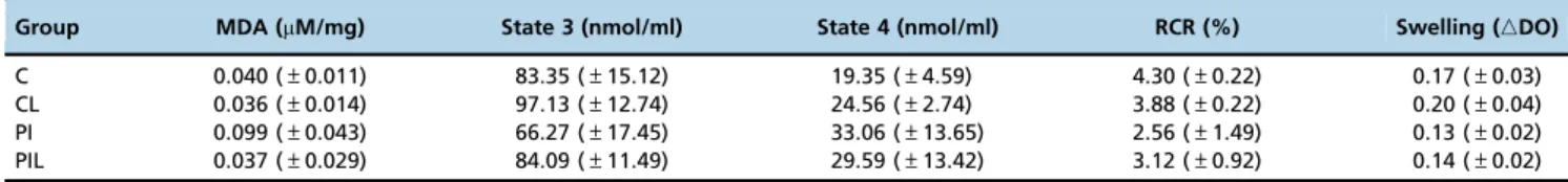
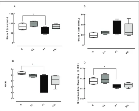
A decrease in state 3 (the rate of oxygen consumption in the activated state) was observed in the PI group (Partial Ischemia) compared to that in the C group (Control) [PIvs.C (po0.05)]. No statistically significant difference was
identi-fied between the C and Partial Ischemia+Laser (PIL) groups [PILvs.C (p40.05)]. For state 4 (basal mitochondrial respira-tion), no statistically significant difference was identified between the groups studied. The RCR was similar to state 3: PIvs.C (po0.05) (Table 1) (Figure 1).
Mitochondrial Swelling
Significant differences were observed between the PI and C groups (po0.05) and the PIL and C groups (po0.05)
(Table 1) (Figure 1).
MDA (Malondialdehyde)
A statistically significant difference in MDA was found between the Partial Ischemia group (PI) and the other groups (po0.05) (Table 1) (Figure 2).
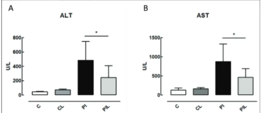
Serum Aminotransferases
The ALT and AST enzyme measurements revealed signif-icant differences between the PIL and PI groups (po0.05),
with the PIL group showing decreased levels of these enzymes. The levels in the C and CL groups were reduced compared with those in the PI and PIL groups (**po0.005).
In Figure 3, we show the differences between the PI and PIL groups.
L-FABP Analysis by Western Blot
The evaluation of L-FABP expression by Western blot revealed decreased expression in the PI group compared to that in groups C, CL and PIL (po0.005) (Figure 4). Optical
den-sity values, in arbitrary units, were (n=5): C=15.04 (±2.25);
CL=12.58 (±0.15); PI: 8.92 (±0.83); and PLI: 14.12 (±1.44).
Comparisons between groups: CxCL: [15.04 (±2.25)vs.12.58
(±0.15) (NS)]; CxPI: [15.04 (±2.25)vs.8.92 (±0.83) (po0.005)];
CxPLI: [15.04 (±2.25) vs. 14.12 (±1.44) (NS)]; CLxPI: [12.58
(±0.15)vs. 8.92 (±0.83) (po0.05)]; CLxPLI: [12.58 (±0.15)vs.
14.12 (±1.44) (NS)]; PIxPLI: [8.92 (±0.83) vs. 14.12 (±1.44)
(po0.005)].
’ DISCUSSION
Ischemia/reperfusion (IR) is a phenomenon characterized by cellular damage after a period of hypoxia followed by reperfusion (18). Hepatic ischemia/reperfusion (HIR) is a complex process that can gravely impair liver function and represents a systemic process that affects multiple tissues, thus promoting a cascade of multiple organ dysfunction (19). Several mechanisms are linked to HIR, including reactive oxygen species (ROS) generation, alteration of calcium homeostasis, lipid peroxidation, mitochondrial dysfunction, activation of liver Kupffer cells (KCs) and cytokine produc-tion (20). The funcproduc-tional integrity of the liver after injury can be determined indirectly by the mitochondrial function of the tissue, i.e., by the ability of the previously injured liver to synthesize energy (21). The present study evaluated mito-chondrial respiration based on the oxygen consumption rate in state 4 (basal respiration), the oxygen consumption rate in state 3 (respiration activated by ADP) and the RCR, which indicates the degree of coupling between oxygen uptake and ADP phosphorylation. Furthermore, inner mitochondrial membrane permeability, an important marker of the struc-tural and functional integrity of this organelle, was evaluated by osmotic mitochondrial swelling.
The main purpose of this project was to evaluate the potential protective role of 660-nm laser light application to
Table 1-Malondialdehyde (MDA) and mitochondrial evaluation (State 3, State 4, RCR: Respiratory Control Ratio, Mitochondrial Swelling). The results are expressed as the mean±standard deviation. C: Control; CL: Control+Laser; PI: Partial Ischemia; PIL: PI+Laser.
Group MDA (mM/mg) State 3 (nmol/ml) State 4 (nmol/ml) RCR (%) Swelling (nDO)
liver tissue by analyzing hepatic energy capacity based on previous studies from our research group concerning hepatic dysfunction, mitochondrial function and tissue levels of L-FABP. The liver consists of a complex organization of atoms that form molecules, structures, tissues and organs. Light has dual behaviors as a particle and a wave. The energy carried by light can be understood as an oscillation of electric and magnetic fields with no mass. These fields, when interacting with the charges of matter, cause matter to move by oscillation, especially electrons, which are lighter than atom nuclei, and create other electric and magnetic fields produ-cing different effects, including electromagnetic waves (22). Some of these effects are more relevant to comprehension of the processes that occur when matter is illuminated, i.e., the
energy transfer from light to molecules (absorption) and vice-versa (emission), and the changes in the course of light propagation (scattering). These three phenomena are very important to understanding the interactions between light and biological structures.
A fundamental concept in phototherapy is called biologi-cal optibiologi-cal window or therapeutic optibiologi-cal window, which represents the range of wavelengths that are most suitable for phototherapy applications when the abovementioned interactions are considered in biological tissues (23).
The results of the present study showed that prophylactic application of 660-nm laser light was able to minimize the harmful effects of ischemia and reperfusion, thus improving hepatic energy capacity, as observed by comparing the levels of oxygen consumption by hepatic mitochondria after the addition of ADP to the groups analyzed. Compared with the C group, a reduction in state 3 was observed in the PI group; however, no difference was found between the C and PIL groups, indicating that prophylactic laser application pre-served mitochondrial energy functions.
This observation is supported by effects reported for other cells. Wong-Riley et al. (24) showed beneficial effects on neuron functionality after laser therapy at similar wavelengths. Eels et al. (25) observed similar effects for retinal injury, with pre-served and stimulated mitochondrial function.
Changes in mitochondrial permeability include loss of inner mitochondrial membrane integrity due to gaps in nonspecific pores that release a flux of small molecules (o1500 Da) and protons. Consequently, significant
mito-chondrial swelling, loss of membrane potential, outer mem-brane rupture and apoptotic or necrotic death occur. Formation of these pores can occur in response to many stimuli, including Ca+2
overload and oxidative stress (15).
Studies of calcium- and phosphate-induced mitochon-drial swelling have shown significant changes in internal mito-chondrial membrane permeability in ischemic (PI) conditions,
Figure 1 - Mitochondrial bioenergetic evaluation. C: Control; CL: Control+Laser; PI: Partial ischemia; PIL: Partial ischemia+Laser.
Panel A) ADP: Activated Oxygen Consumption Velocity (state 3); Panel B) Basal Oxygen Consumption Rate (state 4); Panel C) RCR: Respiratory Control Ratio; Panel D) Mitochondrial Swelling. *po0.05.
Figure 2 - Determination of malondialdehyde (MDA).C: Control;
with no evidence of these changes in laser-treated ischemic groups (PLI). These results show that the IR process induced mitochondrial swelling with a change in mem-brane permeability, probably through interactions with ROS. However, this alteration was not sufficient to com-pletely dissipate the electrochemical proton gradient since oxygen consumption velocity was maintained by the mito-chondria in state 4.
Peng and Jou (26) showed how the interference of light in the blue-green range (488 nm) modifies ion balance, producing reactive oxygen species that open ion pores and changing the balance towards mitochondrial swelling. How-ever, Shi et al. (27) showed that for IR wavelengths, laser irradiation can positively contributes to instead interfere with fragmented mitochondria recovery, including in liver cells, although laser-induced mitochondrial reactive oxygen species do not damage mitochondria through mitochondrial permeability transition pores. The transient opening of these pores may help release these species into the cytosol to protect the mitochondria against damage. This observation seems to corroborate the results obtained here.
The prophylactic use of laser light prevented mitochon-drial swelling as evaluated using absorbance, reflecting an indirect measure of the degree of membrane damage, as shown in Figure 1, panel D. Studies have demonstrated that L-FABP is an effective marker of liver injury in various diseases, such as necrotizing enterocolitis, non-alcoholic fatty liver disease and liver tumors, and in liver transplantation (8,28,29). Due to its low molecular weight and high solu-bility, L-FABP can rapidly reach the bloodstream and be expressed in serum after the organ of origin has been dam-aged. L-FABP is found only in the cytoplasm of hepatocytes and, because these cells are in direct contact with the blood without an interstitial barrier, damage can cause its rapid release into the circulation (28). In the liver, inflammatory diseases that induce cytokine and bacterial factor release by Kupffer cells are known to decrease the expression of L-FABP in tissues. However, independent of these factors, simple rupture of the cellular barrier may be the cause of its rapid release (28). Akbal et al. (28) evaluated the serum levels of L-FABP in a clinical study of non-fatty alcoholic liver disease and found that it was an effective marker for early diagnosis of the disease. In an experimental study, Monbaliu et al. (7) demonstrated an increase in serum L-FABP in proportion to the time of exposure to hot ischemia in transplants perfor-med in pigs. Other clinical studies have also demonstrated the efficacy of L-FABP as a biomarker of liver injury (30-32). Our results were similar to those reported by Monbaliu et al. (7). We can hypothesize that L-FABP increased in serum as it decreased in tissues, although we did not measure L-FABP levels in plasma. A decrease in L-FABP was obser-ved in hepatic tissues from the group submitted to PI, and a higher tissue level was observed in the group pre-conditioned with laser light (PLI); the results of the treated group and the control group were similar. The present results for tissue L-FABP expression are similar to those of Mitidiero et al. (8) who evaluated the tissue expression of L-FABP in necrotizing enterocolitis patients and found lower hepatic expression in this group. In the study, an inverse relationship was observed between intestinal fatty acid binding protein (I-FABP), a biomarker of intestinal tissue damage, and hepatic injury marked by L-FABP.
The effect of radiation in the far-red wavelengths of the spectrum on biological molecules has been reported in the literature. Cytochrome c oxidase (CcO) is reported to be a light absorber in mammalian cells, where it accelerates electron transfer reactions and increases ATP synthesis, ion antiporters and bomb activity. Light also dissociates nitric
Figure 3 - Serum aminotransferases.Panel A) ALT: alanine aminotransferase. Panel B) AST: aspartate aminotransferase. C: Control;
CL: Control+Laser; PI: Partial Ischemia and PIL: Partial Ischemia+Laser. *po0.05.
Figure 4 -L-FABP expression, showing protein detection at 15 kDa
oxide (NO) bonds to CcO, increasing the respiratory rate and preventing NO-based cell death (32). Renno et al. (33) also showed that 660-nm light has regenerative effects, including stimulation of gene expression factors and ATP reactions. Although light can reportedly stimulate regeneration and the respiratory chain and prevent NO-based cell death, this stimulation has been reported to occur in tissues under regular conditions of temperature and blood flow. Ischemia and reperfusion are responsible for several sources of damage promotion in tissues, which are mediated by biochemical processes in which light can interfere. Therefore, these pro-cesses may interrupt the chain of biochemical responses to light stimulation. On the other hand, irradiation prior to ischemia may induce early activation of the biochemical chains that upregulate cell metabolism and activate rege-nerative processes in tissues. Thus, when damage actually occurs, these processes have already been initiated, which is probably the reason for the protective effect indicated by the increase in L-FABP expression.
Zhu et al. (34) showed that the same wavelength could be used to protect rat heart cells from damage similarly to liver cells, revealing that either pre- or post-ischemia irradiation can enhance tissue preservation. Later, Carnevalli et al. showed that IR laser therapy, in addition to establishing positive biomodulation, contributes to the reestablishment of cellular homeostasis in cells under nutritional stress and prevented apoptosis in animal ovarian (CHO K-1) cells (35). In the present study, we induced ischemia under normother-mic conditions and found positive results for IR laser irra-diation, demonstrating evidence of the favorable effect of light in the maintenance of tissues after damage-induced stress.
Therefore, the application of 660-nm laser light as a pre-conditioning treatment to the livers of rats submitted to partial ischemia was shown to be effective in protecting the liver, with L-FABP levels similar to those of controls.
Finally, redox-sensitive cellular targets have roles in cellular bioenergetics associated with changes of environ-mental stimuli, such as hormones, nutrients and oxygen tension, providing rapid and sensitive responses to changes in metabolism and fluxes in ROS. Conversely, ROS also have roles in cellular processes through redox-dependent sig-naling. In the present study, as expected, an increase in lipoperoxidation was observed with HIR (PI group), and the application of laser light was able to reverse or prevent ischemia-induced cellular changes, as shown by MDA levels, mitochondrial function parameters and L-FABP levels (36).
This project has limitations, including the lack of liver histology and immunohistochemistry analyses. Further stu-dies are required to investigate L-FABP levels through these techniques and to study alternative pathways in liver injury to confirm the protective effects of laser light in hepatic ischemia.
’ ACKNOWLEDGMENTS
The authors wish to thank the São Paulo Research Foundation, FAPESP and CNPq (Higher Education Consortia Program - Brazilian Ministry of
Education) forfinancial support.
’ AUTHOR CONTRIBUTIONS
Vilalva KH performed the technical procedures, collected data and analyzed and interpreted the data in collaboration with Silveira M, Graf C
and Mumic F. Figueira RL, Gonc¸alves FL, Bagnato VS, D’Albuquerque
LA, Sbragia L and Vollet-Filho JD were responsible for the intellectual and
scientific content of the study. Gomes MC contributed to the bioenergetics
evaluations. Castro e Silva O served as the mentor and tutor for the project and designed the protocol. Vilalva KH, Castro e Silva O, Figueira RL and Sbragia L were responsible for manuscript writing.
’ REFERENCES
1. Losada DM, Jordani ME, Jordani MC, Piccinato MA, Fina CF, Feres O, et al. Should preconditioning hyperbaric oxygenation protect the liver against ischemia-reperfusion injury? An experimental study in a rat model. Transplant Proc. 2014;46(1):56-62, http://dx.doi.org/10.1016/ j.transproceed.2013.10.044.
2. Castro e Silva Júnior O, ilveira MR. The liver and light. A precise com-bination, from mythology to medicine. Revis Med. 2013;46(3):318-21. 3. Agrawal T, Gupta GK, Rai V, Carroll JD, Hamblin MR. Pre-conditioning
with low-level laser (light) therapy: light before the storm. Dose Response. 2014;12(4):619-49, http://dx.doi.org/10.2203/dose-response.14-032.Agrawal.
4. Bailey TG, Birk GK, Cable NT, Atkinson G, Green DJ, Jones H, et al. Remote ischemic preconditioning prevents reduction in brachial artery flow-mediated dilation after strenuous exercise. Am J Physiol Heart Circ Physiol. 2012;303(5):H533-8, http://dx.doi.org/10.1152/ajpheart. 00272.2012.
5. Lu PG, Feng H, Yuan SJ, Zhang RW, Li M, Hu R, et al. Effect of pre-conditioning with hyperbaric oxygen on neural cell apoptosis after spinal cord injury in rats. J Neurosurg Sci. 2013;57(3):253-8.
6. Katz LM, Frank JE, Dvorak A, Finch A, Szymanowski A, Gordon CJ. Independence of brain and trunk temperature during hypothermic pre-conditioning in rats. J Neurosci Methods. 2009;179(2):179-83, http://dx. doi.org/10.1016/j.jneumeth.2009.01.025.
7. Monbaliu D, de Vries B, Crabbé T, van Heurn E, Verwaest C, Roskams T, et al. Liver fatty acid-binding protein: an early and sensitive plasma marker of hepatocellular damage and a reliable predictor of graft viability after liver transplantation from non-heart-beating donors. Transplant Proc. 2005;37(1):413-6, http://dx.doi.org/10.1016/j.transproceed.2004. 12.103.
8. Mitidiero LF, Simões AL, Gonc¸alves FL, Figueira RR, Castro e Silva O, Sbragia L. L-FABP and I-FABP expression in newborn rats changes inversely in the model of necrotizing enterocolitis. Acta Cir Bras. 2014; 29 Suppl 2:43-9, http://dx.doi.org/10.1590/S0102-8650201400140009. 9. Benkoe TM, Mechtler TP, Weninger M, Pones M, Rebhandl W, Kasper DC.
Serum levels of interleukin-8 and gut-associated biomarkers in diagnos-ing necrotizdiagnos-ing enterocolitis in preterm infants. J Pediatr Surg. 2014;49(10): 1446-51, http://dx.doi.org/10.1016/j.jpedsurg.2014.03.012.
10. Okazaki Y, Furuhashi M, Tanaka M, Mita T, Fuseya T, Ishimura S, et al. Urinary excretion of fatty acid-binding protein 4 is associated with albu-minuria and renal dysfunction. PLoS One. 2014;9(12):e115429, http://dx. doi.org/10.1371/journal.pone.0115429.
11. Iguchi N, Uchiyama A, Ueta K, Sawa Y, Fujino Y. Neutrophil gelatinase-associated lipocalin and liver-type fatty acid-binding protein as bio-markers for acute kidney injury after organ transplantation. J Anesth. 2014;29(2):249-55, http://dx.doi.org/10.1007/s00540-014-1909-4. 12. Jordani MC, Santos AC, Prado IM, Uyemura SA, Curti C. Flufenamic acid
as an inducer of mitochondrial permeability transition. Mol Cell Biochem. 2000;210(1-2):153-8, http://dx.doi.org/10.1023/A:1007185825101. 13. López JM, Imperial S, Valderrama R, Navarro S. An improved Bradford
protein assay for collagen proteins. Clin Chim Acta. 1993;220(1):91-100, http://dx.doi.org/10.1016/0009-8981(93)90009-S.
14. Mingatto FE, Santos AC, Uyemura SA, Jordani MC, Curti C.In vitro
interaction of nonsteroidal anti-inflammatory drugs on oxidative phos-phorylation of rat kidney mitochondria: respiration and ATP synthesis. Arch Biochem Biophys. 1996;334(2):303-8, http://dx.doi.org/10.1006/ abbi.1996.0459.
15. Zoratti M, Szabò I. The mitochondrial permeability transition. Biochem Biophys Acta. 1995;1241(2):139-76.
16. Lapenna D, Ciofani G, Pierdomenico SD, Giamberardino MA, Cuccurullo F. Reaction conditions affecting the relationship between thiobarbituric acid reactivity and lipid peroxides in human plasma. Free Radic Biol Med. 2001;31(3):331-5, http://dx.doi.org/10.1016/S0891-5849(01)00584-6. 17. Henry JR, Chiamori N, Golub OJ, Berkman S. Revised
spectrophoto-metric methods for the determination of glutamic-oxalacetic transami-nase, glutamic-pyruvic transamitransami-nase, and lactic acid dehydrogenase. Am J Clin Pathol. 1960;34:381-98, http://dx.doi.org/10.1093/ajcp/ 34.4_ts.381.
18. Teoh NC, Farrell GC. Hepatic ischemia reperfusion injury: pathogenic mechanisms and basis for hepatoprotection. J Gastroenterol Hepatol. 2003;18(8):891–902, http://dx.doi.org/10.1046/j.1440-1746.2003.03056.x.
19. Duarte S, Baber J, Fujii T, Coito AJ. Matrix metalloproteinases in liver injury, repair and fibrosis. Matrix Biol. 2015;44-46:147-56, http://dx.doi. org/10.1016/j.matbio.2015.01.004.
21. Tolentino EC, Castro e Silva O, Zucoloto S, Souza ME, Gomes MC, San-karankutty AK, et al. Effect of hyperbaric oxygen on liver regeneration in a rat model. Transplant Proc. 2006;38(6):1947-52, http://dx.doi.org/ 10.1016/j.transproceed.2006.06.066.
22. Bagnato VS, Kurachi C, Ferreira J, Sankarankutty AK, Zucoloto S, de Castro e Silva O. New photonic technologies for the treatment and diag-nosis of hepatic diseases: an overview of the experimental work per-formed in collaboration, between Physics Institute of São Carlos and Ribeirão Preto Faculty of Medicine of the University of São Paulo. Acta Cir Bras. 2006;21Suppl 1: 3-11, http://dx.doi.org/10.1590/S0102-8650 2006000700002.
23. Bagnato VS, Kurachi C, Castro e Silva O. New perspectives for optical techniques in diagnostic and treatment of hepatic diseases. Acta Cir Bras. 2010;25(2):214-6, http://dx.doi.org/10.1590/S0102-865020100002 00016.
24. Wong-Riley MT, Liang HL, Eells JT, Chance B, Henry MM, Buchmann E, et al. Photobiomodulation directly benefits primary neurons functionally inactivated by toxins: role of cytochrome c oxidase. J Biol Chem. 2005; 280(6):4761-71, http://dx.doi.org/10.1074/jbc.M409650200.
25. Eells JT, Wong-Riley MT, VerHoeve J, Henry M, Buchman EV, Kane MP, et al. Mitochondrial signal transduction in accelerated wound and retinal healing by near-infrared light therapy. Mitochondrion. 2004;4(5-6):559-67, http://dx.doi.org/10.1016/j.mito.2004.07.033.
26. Peng TI, Jou MJ. Mitochondrial swelling and generation of reactive oxygen species induced by photoirradiation are heterogeneously dis-tributed. Ann N Y Acad Sci. 2004;1011:112-22, http://dx.doi.org/10.1196/ annals.1293.012.
27. Shi F, He H, Wang Y, Liu D, Hu M, Wang C. Mitochondrial swel-ling and restorable fragmentation stimulated by femtosecond laser. Biomed Opt Express. 2015;6(11):4539–45, http://dx.doi.org/10.1364/
BOE.6.004539.
28. Akbal E, Koc¸ak E, Akyürek O, Köklü S, Batgi H, Senes M. Liver fatty acid-binding protein as a diagnostic marker for non-alcoholic fatty liver dis-ease. Wien Klin Wochenschr. 2014;128(1-2):48-52.
29. Inoue M, Takahashi Y, Fujii T, Kitagawa M, Fukusato T. Significance of downregulation of liver fatty acid-binding protein in hepatocellular car-cinoma. World J Gastroenterol. 2014;20(46):17541-51, http://dx.doi.org/ 10.3748/wjg.v20.i46.17541.
30. Halpern MD, Holubec H, Dominguez JA, Meza YG, Williams CS, Ruth MC, et al. Hepatic inflammatory mediators contribute to intestinal damage in necrotizing enterocolitis. Am J Physiol Gastrointest Liver Physiol. 2003;284(4):G695-702, http://dx.doi.org/10.1152/ajpgi.00353.2002. 31. Ishimitsu T, Ohta S, Saito M, Teranishi M, Inada H, Yoshii M, et al.
Urinary excretion of liver fatty acid-binding protein in health-check par-ticipants. Clin Exp Nephrol. 2005;9(1):34-9, http://dx.doi.org/10.1007/ s10157-004-0331-x.
32. Farivar S, Malekshahabi T, Shiari R. Biological effects of low level laser therapy. J Lasers Med Sci. 2014;5(2):58-62.
33. Renno AC, Iwama AM, Shima P, Fernandes KR, Carvalho JG, De Oliveira P, et al. Effect of low-level laser therapy (660 nm) on the healing of second-degree skin burns in rats. J Cosmet Laser Ther. 2011;13(5):237-42, http:// dx.doi.org/10.3109/14764172.2011.606466.
34. Zhu Q, Yu W, Yang X, Hicks GL, Lanzafame RJ, Wang T. Photo-irradiation improved functional preservation of the isolated rat heart. Lasers Surg Med. 1997;20(3):332-9, http://dx.doi.org/10.1002/(SICI)1096-9101(1997) 20:3o332::AID-LSM1243.0.CO;2-F.
35. Carnevalli CM, Soares CP, Zangaro RA, Pinheiro AL, Silva NS. Laser light prevents apoptosis on Cho K-1 cell line. J Clin Laser Med Surg. 2003;21(4): 193-6, http://dx.doi.org/10.1089/104454703768247756.
36. Rasbach KA, Schnellmann RG. Signaling of mitochondrial biogenesis following oxidant injury. J Biol Chem. 2007;282(4):2355–62, http://dx.doi.