Calcium is essential for a number of vital processes, from bone formation to the modulation of permeability and excitability of membranes. As small changes in [Ca2+] have a marked effect on neural, cardiovascular and muscular function, maintenance of circulating [Ca2+] within narrow limits is a fundamental requirement, and a constant supply of calcium must be ensured to the body. The ability of adult teleosts to maintain constant plasma [Ca2+] in diverse calcium environments is well documented (Flik et al., 1995; Pang et al., 1980). Calcium uptake becomes of increasing importance during early life stages of vertebrates, including fish, as the skeleton and other bony parts are undergoing rapid development. In the gilthead sea bream (Sparus auratus), a marine teleost fish widely cultivated in southern Europe, the development of the skeleton initiates with the formation of a cartilaginous template, followed by gradual mineralization (Faustino and Power, 1998, 1999, 2001). The onset of this process takes place a few days after hatching, and ossification of the vertebral column (Faustino and Power, 1998), fins (Faustino and Power, 1999) and cranial complex (Faustino and Power, 2001) continues during the first 70–90·days post-hatching (DPH). Scales, developing in the hitherto ‘naked’ fish skin, are formed during later stages of larval development and mark the transition to the juvenile stage. Bone mineralization
relies on calcium availability in the extracellular fluids and, unlike terrestrial vertebrates, which obtain this ion exclusively from the diet, freshwater and marine fish obtain it from the (generally) calcium-rich water they inhabit. The water-exposed gills, skin and intestine epithelia are the primary surfaces for calcium uptake, while the kidney (and possibly the intestine) is involved in excretion (Flik et al., 1995; Flik and Verbost, 1993).
Internal [Ca2+] is maintained within narrow limits despite large fluctuations in environmental [Ca2+] that generally parallel changes in water salinity. The ability to maintain a constant ion concentration and osmolality of body fluids appears early in development. Embryos and larvae have a capacity similar to that of adult fish and regulate ionic gradients between their body and the ambient water by excreting sodium chloride or absorbing calcium (Alderdice, 1988; Guggino, 1980). The early post-embryonic stages of the European sea bass (Dicentrarchus labrax) are able to hypo- and hyper-regulate over a wide range of salinities, this ability being acquired in steps but already being present at hatching (Varsamos et al., 2001). It has also been proposed that the regulation of calcium balance in freshwater tilapia larvae (Oreochromis mossambicus) acclimated to low-calcium environments is stage dependent and that modulation of Published by The Company of Biologists 2004
doi:10.1242/jeb.00765
Whole-body calcium uptake was studied in gilthead sea bream larvae (9–83·mg) in response to changing environmental salinity and [Ca2+]. Calcium uptake increased with increased fish size and salinity. Fish exposed to calcium-enriched, diluted seawater showed increased calcium uptake compared with fish in diluted seawater alone. Calcium uptake was unchanged in Na+ -enriched, diluted seawater. Overall, [Ca2+], and not salinity/osmolarity per se, appears to be the main factor contributing to calcium uptake. By contrast, drinking was reduced by a decrease in salinity/osmolarity but was little affected by external [Ca2+]. Calculations of the maximum
contribution from drinking-associated calcium uptake showed that it became almost insignificant (less than 10%) through a strong decrease in drinking rate at low salinities (0–8‰). Diluted seawater enriched in calcium to the concentration present in full-strength seawater (i.e. constant calcium, decreasing salinity) restored intestinal calcium uptake to normal. Extra-intestinal calcium uptake also benefited from calcium addition but to a lesser extent.
Key words: calcium uptake, drinking rate, salinity, environmental calcium, larvae, gilthead sea bream, Sparus auratus.
Summary
Introduction
Water calcium concentration modifies whole-body calcium uptake in sea bream
larvae during short-term adaptation to altered salinities
Pedro M. Guerreiro
1,2, Juan Fuentes
1, Gert Flik
2, Josep Rotllant
1, Deborah M. Power
1and
Adelino V. M. Canario
1,*
1Centre of Marine Sciences (CCMAR), University of Algarve, Campus de Gambelas, 8005-139 Faro, Portugal and 2Department of Animal Physiology, University of Nijmegen, 6525 ED Nijmegen, The Netherlands
*Author for correspondence (e-mail: acanario@ualg.pt)
calcium fluxes is closely correlated with levels of body calcium content (Chou et al., 2002; Hwang et al., 1996).
Fish in seawater tend to gain ions such as sodium and chloride through diffusion and to lose water by osmosis. The acquisition of the capacity to control water balance in relation to external salinity through drinking by fish larvae is, therefore, fundamental for osmoregulation (Flik et al., 2002; Varsamos et al., 2001), and drinking occurs before the development of a functional anus and gills (Tytler and Blaxter, 1988a,b).
Since whole-body calcium uptake correlates positively to environmental [Ca2+], salinity (as a result of [NaCl]) and drinking, the relative contribution of each of these variables to calcium uptake is uncertain. In the present study, we have examined the relationship between calcium uptake and drinking by submitting gilthead sea bream larvae to abrupt short-term changes of combinations of water salinity and [Ca2+].
Materials and methods Fish and media
Gilthead sea bream larvae (Sparus auratus L.) were reared at Instituto de Investiga o des Pescas e do Mar (IPIMAR; Olhão, Portugal) in 90% recirculating 200-litre conical tanks according to standard procedures at 36‰ salinity and 20°C. Whole-body calcium content was measured in specimens collected every 5·days from 20·DPH to 75·DPH and at 120·DPH (N≥10 individuals per age group). Larvae were fasted for 24·h before being sacrificed using an excess of 2-phenoxyethanol (0.5% v/v; Sigma-Aldrich, Madrid, Spain), weighed to the nearest 0.1·mg (Sartorius balance, precision 10–6·g) and digested with 100·µl of 70% nitric acid (v/v; Sigma-Aldrich) for 24·h. Individual total body calcium content was determined by means of inductively coupled plasma atomic emission spectrometry (ICP-AES; Plasma IL200; Thermo Jarell Ash, Franklin, MA, USA). For size/age-related calcium uptake studies and for altered salinity experiments, larvae (obtained from Timar, Culturas em Água, Portugal) were 30–60·DPH and 45·DPH, respectively. To obtain the desired reductions in salinity, full-strength seawater (SW) was diluted using distilled water. To obtain salinity, [Ca2+] or [Na+] identical or above those of full-strength seawater, marine salt (Instant Ocean; Aquarium Systems, Sarrebourg, France), calcium chloride Aldrich) or sodium chloride (Sigma-Aldrich) was added to full-strength or diluted seawater as required (see Table 1 for atomic absorption chromatography analysis of experimental media).
Determination of calcium influx
Fish were netted and transferred to 25-ml vessels with aerated seawater and, after 30·min, 45CaCl
2(NEN, Boston, MA, USA) was added to the water (final activity of 3.7·kBq·ml–1). At the end of the experiment (normally lasting 4·h), water samples were collected. The larvae were rinsed in tracer-free water, sacrificed, weighed and digested as indicated above, neutralised with an identical volume of 2·mol·l–1 sodium
hydroxide (Sigma-Aldrich) and bleached with 300·µl of 35% hydrogen peroxide (Fluka, Sigma-Aldrich) to prevent colour quenching. Water calcium content was measured by a colourimetric endpoint assay (Sigma-Aldrich, assay No. 587). Water and digested larval samples were dissolved in OptiPhase HiSafe II liquid scintillation fluid (Wallac, Amersham Pharmacia Biotech, Lisbon, Portugal), and 45Ca activity was measured in a scintillation counter (Model LS6000IC; Beckman Instruments Inc., Fullerton, CA, USA). Calcium influx (Cain) was calculated according to the following equation: Cain= (AfCw)/(Awt), where Afis the total 45Ca activity in fish (c.p.m.), Cwis the total calcium concentration in water (nmol·l–1), A
wis the total 45Ca activity in water (c.p.m.·l–1), and t is duration of exposure (h). Calcium influx is expressed as nmol·h–1. Extra-intestinal calcium uptake was estimated by subtracting the amount of calcium in the water imbibed (i.e. drinking rate; see below) from the total whole-body calcium uptake.
Determination of drinking rate
The procedure used for the determination of drinking rate was the same as for calcium influx but 3.7·kBq·ml–1 of 51 Cr-EDTA (NEN) was added to the water. Radioactivity was counted in a gamma counter (Wallac 1470 Wizard gamma counter; Amersham Pharmacia Biotech). In some experiments, calcium influx and drinking rate were measured simultaneously. Drinking rate (DR) was calculated as: DR=Af/(Awt), where Af is the total activity of 51Cr-EDTA in the fish (c.p.m.), A
wis the tracer activity in water (c.p.m.·ml–1), and t is the duration of exposure (h). Results are expressed as nl·h–1.
Effects of tracer exposure time and body size on whole-body Ca2+influx
The effect of duration of exposure to 45CaCl
2on calcium uptake was determined by sampling larvae in different vessels at 2·h, 4·h, 6·h, 8·h and 16·h after radioisotope addition. Intact gilthead sea bream larvae accumulated 45Ca at a constant rate for at least 16·h and therefore the tracer experiments were conducted within this time-span to reflect initial speed of calcium influx, thus eliminating possible effects of tracer backflow.
The effect of body size was tested in five different experiments in 100%SW with larvae ranging in mass from 5·mg to 150·mg. Calcium influx was determined as described above.
Time-series of adjustment of Ca2+influx to diluted seawater Larvae were transferred to 25-ml vessels containing 100%SW (four groups, N≥15) or 50%SW (three groups, N≥15), and 45CaCl
2 was immediately added. Larvae were collected at 2·h, 4·h, 8·h and 16·h after transfer (HAT; 100%SW) and at 4·HAT, 8·HAT and 16·HAT (50%SW), rinsed, sacrificed, weighed and dissolved to determine calcium influx as described above.
Effects of [Ca2+] and [Na+] on Ca2+influx and drinking rates Gilthead sea bream larvae (N=20–40 per group) were
transferred to 25-ml vessels containing the experimental media (see Table·1 for precise ionic composition): 100%SW, 150%SW, 25%SW, 10%SW, 0%SW, 100%SW plus CaCl2 (100%SW+Ca), 25%SW plus CaCl2 (25%SW+Ca), 25%SW plus NaCl (25%SW+Na), 10%SW plus CaCl2(10%SW+Ca) and 10%SW plus NaCl (10%SW+Na) for 4·h and 20·h (0%SW only for 4·h) before addition of 45CaCl
2and 51Cr-EDTA. After 4·h in radioisotope-containing water, the fish were rinsed, sacrificed, weighed and dissolved to determine calcium influx and drinking rate. [Ca2+] in 100%SW+Ca was adjusted to that of 150%SW, and 25%SW+Ca and 10%SW+Ca was adjusted to that of 100%SW. [Na+] in 25%SW+Na and 10%SW+Na was adjusted to that of 100%SW.
Statistical analysis
The effect of treatments on calcium uptake and drinking was analysed by one-way or two-way covariance analysis (ANCOVA) with body size as covariate using the General Linear Model module in the Systat software (version 10.2; Systat Software Inc., Richmond, CA, USA). Contrasts were used to test the relationship among cell means. Least squares means and their standard errors adjusted for mass are used to report effect of treatments. Multiple linear regression analysis was used to compare the relative contribution of calcium, sodium and osmolarity to calcium uptake. Whenever the data were required to be normalised, a logarithmic transformation was used. The significance level was established as P<0.05 unless otherwise stated.
Results
Whole-body mineral content
The relationship between whole-body calcium content (Q; µmol) and wet body mass was described by the equation Q=0.0502M1.249 (M range=0.5–500·mg; r
0=0.993, P<0.0001, N=196). The slope of the regression line (1.249) was significantly different from 1, indicating that total body
calcium was related to body mass by a slightly positive allometry.
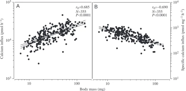
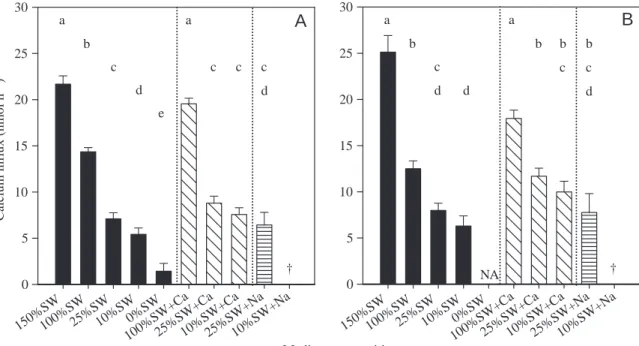
Effects of salinity and addition of Ca2+or Na+on Ca2+influx Whole-body calcium influx in 100%SW was strongly dependent on body mass (Cain=2588M0.496; Fig.·1A), even after normalisation to unit of body mass (Cain,M=2588M–504; Fig.·1B). The same relationship was observed 8·HAT to media of other salinities; the slope was similar but the y-intercept was proportional to the salinity (data not shown). Because the dependency of calcium influx on body mass was highly significant across the mass range of the larvae used in the experiments, subsequent data analysis considered mass as a covariate to remove its effect. When fish were transferred from 100%SW to 50%SW, adjustment to a lower calcium influx was noticeable between 4·h and 8·h after the onset of exposure (Fig.·2). By 8·HAT, the influx rate at 50%SW was 30% significantly lower than at 100%SW and was 65% lower by 16 HAT. Transfer of larvae from 100%SW to 150%SW led to a 50% increase in calcium influx by 8·HAT (Fig.·3A) and 100% by 24·HAT (Fig.·3B). Transfer to lower salinities lowered calcium influx 8·HAT by 50% at 25%SW and by 90% at 0%SW (Fig.·3A). A similar reduction was also found 24·HAT.
To establish whether salinity or ambient [Ca2+] determined the rate of calcium uptake, larvae were exposed to altered salinities and to different [Ca2+] and [Na+]. Larvae exposed to 100%SW to which [Ca2+] was adjusted to 17.6·mmol·l–1 (similar to that of 150%SW) had calcium influx rates at 8·HAT and 24·HAT similar to those in 150%SW (Fig.·3A,B). Similarly, transfer to 25% and 10% diluted seawater in which [Ca2+] had been adjusted to levels of 100%SW resulted in increased calcium influx rates. At 8·HAT, the increase was only significant at 10%SW+Ca (although influx rates did not reach those of 100%SW), but at 24·HAT both 25%SW+Ca and 10%SW+Ca had calcium influx rates similar to those found in 100%SW (Fig.·3B).
Placing larvae in diluted medium supplemented with NaCl Table 1. Ionic composition and chemical properties of seawater, seawater supplemented with marine salt, seawater diluted
with distilled water, seawater diluted with distilled water supplemented with calcium chloride or sodium chloride and freshwater alone
Osmolality
Media (%SW) Salinity (‰) (mOsmol·kg–1) Na+(mmol·l–1) Cl–(mmol·l–1) Ca2+(mmol·l–1)
150% 55 1674 696 678 17.1 100% 35.5 1070 454 520 11.4 50% 17 551 226 258 5.6 25% 9 268 123 175 3.2 10% 3.5 110 57 53 1.6 0% 0 3 1.4 1.3 0.5 100%+Ca 36 1097 442 534 17.6 25%+Ca 9.9 287 107 182 10.5 10%+Ca 3.7 112 51 79 9.2 25%+Na 36 1107 541 535 3.1 10%+Na 36 1101 502 527 1.5
to levels similar to 100%SW resulted in high mortality (Table·2), which was most severe in the 10%SW+Na group: all fish died within 2·HAT. Nevertheless, the surviving 25%SW+Na group had calcium influx rates similar to their respective controls (25%SW) at both 8·HAT and 24·HAT. Interestingly, fish transferred directly to 0%SW were able to adjust better than fish in 10%SW+Na, with 50% of the initial group surviving to the end of the 8-h period. To confirm that
calcium influx was related only to fish size and to water [Ca2+], a multiple regression analysis, which also included water osmolarity and [Na+] as independent variables, was carried out. We found strong colinearity between medium [Ca2+], [Na+] and osmolarity, implying some uncertainty as to the relative contributions between the three variables to calcium influx. The multiple linear regression equation, pooling data from 8·HAT and 24·HAT, was: log Cain=2.659+0.003[Na+]– 0.001Os+0.041[Ca2+]+0.679 log M, where Os is osmolarity. As can be observed, according to this model, the effect of [Ca2+] on Ca2+influx is at least one order of magnitude higher than that of [Na+] or osmolarity, suggesting that ambient [Ca2+] is a major modifier of Ca2+ influx. The relationships Body mass (mg) 10 100 C alciu m influx ( p mo l h – 1) 103 104 105 10 100 S p ecif ic c alciu m influx ( p mo l mg – 1 h – 1) 101 102 103 104 r0=0.685 N=355 P<0.0001 r0=–0.690 N=355 P<0.0001
A
B
Fig.·1. (A) Relationship between whole-body calcium influx and wet body mass. (B) The same relationship with calcium influx divided by body mass, referred to as specific calcium influx. Indicated are the regression lines (middle lines) and their 95% confidence limits, correlation coefficents (r0), statistical significance levels (P) and number of fish used (N).
Fig.·2. Time-course of whole-body calcium influx rate in fish maintained at 100%SW (filled circles) and in fish transferred to 50%SW (open circles) for a 16-h period. Each point is the mean least squares adjusted for body mass (10–50·mg), and the vertical lines are their standard errors. Asterisks represent statistically significant differences to 100%SW (*P<0.01; **P<0.001) at a given time.
Time (h) 0 2 4 6 8 10 12 14 16 Calc iu m i nflux ( n m ol h – 1) 2 4 6 8 10 12 14 16 100%SW 50%SW
*
**
Table 2. Cumulative mortality, M (%), and number of
surviving fish (N) recorded 8·h and 24·h after transfer to the media shown in Table·1
8·HAT 24·HAT Media (%SW) M (%) N M (%) N 150% 35 14 62 5 100% 3 55 7 24 25% 6 27 7 27 10% 9 24 15 14 0% 51 16 NA NA 100%+Ca 6 30 7 20 25%+Ca 10 20 11 22 10%+Ca 14 22 21 12 25%+Na 70 6 85 4 10%+Na 100 0 100 0
between calcium influx, body mass and medium [Ca2+] at 8·HAT and 24·HAT were as follows: Cain,8=283+M0.748+ [Ca2+]0.618(adjusted r2=0.511, N=213, P<0.001; range [Ca2+] 0.5–17.6·mmol·l–1; range M 8.6–82.8·mg) and Ca
in,24=813+ M0.575+[Ca2+]0.424(adjusted r2=0.511, N=128, P<0.001; range [Ca2+] 1.68–17.6·mmol·l–1; range M 10.0–57.6·mg).
Effect of salinity and addition of [Ca2+] or [Na+] on drinking As with calcium uptake, drinking rate was highly positively correlated to fish mass (at 100%SW DR=46.6M0.785; r
0=0.677, P<0.001, N=78). A marked salinity-dependent reduction in drinking was observed at 8·HAT (Fig.·4A). Fish that were exposed to media at lower salinities exhibited drinking rates Medium composition Cal cium in fl u x ( nmo l h –1) 0 5 10 15 20 25 30 0 5 10 15 20 25 30 a a b c c d c c d e † NA † b a d b a b c c d b c d
A
B
150%SW100%SW25%SW10%SW0%SW 100%SW +Ca 25%SW +Ca 10%SW +Ca 25%SW +Na 10%SW +Na 150%SW100%SW25%SW10%SW0%SW 100%SW +Ca 25%SW +Ca 10%SW +Ca 25%SW +Na 10%SW +NaFig.·3. Effect of salinity (filled bars) and of addition of calcium (bars with diagonal lines) or sodium (bars with horizontal lines) on sea bream larvae whole-body calcium influx after 8·h (A) or 24·h (B) exposure. At 10%SW and 25%SW, calcium and sodium were added to match levels at 100%SW. At 100%SW, calcium levels were added to match those at 150%SW (see Materials and methods and Table·1). Each bar represents the mean least squares adjusted for body mass and respective S.E.M. Identical letters indicate groups that do not differ statistically at P=0.05. The daggers indicate 100% mortality; NA, not tested.
0 200 400 600 800 Medium composition 150%SW100%SW25%SW10%SW0%SW 100%SW +Ca 25%SW +Ca 10%SW +Ca 25%SW +Na 10%SW +Na 150%SW100%SW25%SW10%SW0%SW 100%SW +Ca 25%SW +Ca 10%SW +Ca 25%SW +Na 10%SW +Na Dr in k in g r ate (n l h – 1) 0 200 400 600 800 a a b a b c c c c c d † NA † a b c a a b c a b a b b c a b
A
B
Fig. 4. Effect of salinity (filled bars) and of addition of calcium (bars with diagonal lines) or sodium (bars with horizontal lines) on sea bream larvae drinking rate after 8 h (A) and 24 h (B) exposure. See legend to Fig.·3 for further details.
that were significantly lower than those of control 100%SW fish. Fish exposed to 150%SW had drinking rates that were slightly higher but not statistically different from those kept at 100%SW. Drinking at 0%SW was significantly reduced in relation to all other tested media. After 24·HAT, similar relationships to that of 8·HAT were observed (Fig.·4B). Addition of calcium or sodium had no significant effects on drinking, although there was an indication that addition of calcium and especially sodium might stimulate drinking after 24·h. However, the large variability and low number of surviving fish meant this trend could not be confirmed.
Calcium imbibed was estimated from the drinking rate, and the extra-intestinal contribution to the whole-calcium uptake was determined (Fig.·5). Extra-intestinal contributions were higher in media with lower salinity and less calcium, accounting for ~93% of whole-body calcium entry at 10%SW and 0%SW and for ~85% at 25%SW. At 100%SW and above, the extra-intestinal contribution was only 45–50%. The effect of adding calcium to the water at lower salinities was to increase the relative contributions to levels close to those of 100%SW. However, addition of Na had little effect. The
change in the relative contribution of the two routes of calcium uptake is summarized in Fig.·6. The data further emphasise that increased calcium uptake at higher salinities is intimately associated with drinking but it is also dependent on environmental [Ca2+], since extra-intestinal uptake is also enhanced. It was not possible to separate the relative importance of drinking and [Ca2+] because of colinearity in the multiple regression.
Discussion
Gilthead sea bream grown in full-strength seawater show a linear increase in body calcium content, indicating a constant calcium accumulation related to skeletal growth. The relationship is allometric, possibly reflecting the higher density of the calcified skeleton. These requirements in calcium are met by an exponential increase in calcium uptake as the fish grows, and the positive relationship between fish size and calcium influx is maintained in larvae exposed to different salinities. The higher specific calcium uptake (i.e. expressed as influx per body mass) in larvae than in adults may be, at least
Calcium influx (nmol h
–1) 0 2 4 6 8 10 12 14 16 18 Medium composition 150%SW100%SW 25%SW 10%SW 0%SW 100%SW +Ca 25%SW +Ca 10%SW +Ca 25%SW +Na 10%SW +Na 0 2 4 6 8 10 12 14 16 † NA †
*
*
*
*
*
*
*
*
A
B
Fig.·5. Effect of salinity and of addition of calcium or sodium on estimated sea bream larvae intestinal (filled bars) and extra-intestinal (hatched bars) calcium uptake after 8·h (A) or 24·h (B) exposure. Modification in calcium and sodium in 10%SW, 25%SW and 100%SW is described in the legend to Fig.·3. Asterisks indicate statistically significant difference between intestinal and extra-intestinal fluxes at P=0.05. Daggers indicate 100% mortality; NA, not tested.
partially, due to the larger surface-to-volume ratio of larvae and/or the difference in epithelium permeability for calcium between larvae and adults (Hwang et al., 1994). The results also demonstrate that, as suggested previously (Flik et al., 1996), linear extrapolations from large to small fish are not acceptable.
Since extracellular calcium is maintained within tight limits, this indicates that gilthead sea bream larvae are able to regulate their calcium uptake (and loss) in order to comply with the calcium demands of their normal physiological processes as well as the extra demand imposed by the intense calcification period. This is also supported by the observation that fish adapted to low-calcium environments are capable of modulating transport mechanisms to sustain similar or greater accumulation rates of body calcium content during growth (Chou et al., 2002; Flik et al., 1986; Hwang et al., 1996; Mol et al., 1999; Vonck et al., 1998).
Gilthead sea bream larvae transferred from 100%SW to a hyposmotic environment (50%SW) require at least 16·h to adapt calcium transport mechanisms to the new environmental conditions. In juveniles of silver sea bream (Sparus sarba), abrupt hyposmotic exposure (from 33‰ to 6‰) resulted in a decline in serum total calcium at 24·HAT and a return to pre-exposure levels by 120·HAT (Kelly and Woo, 1999). A similar effect was reported by Mancera et al. (1993) for the gilthead sea bream, with complete recovery occurring after 30·days. In gilthead sea bream larvae, it was not possible to measure serum calcium, but a strong relationship between calcium influx and water salinity was found both at 8·HAT
and 24·HAT. Furthermore, [Ca2+] appears to be the main environmental factor determining calcium influx, as shown by the fact that fish exposed to CaCl2-enriched seawater had whole-body influx rates similar to those of control (non-enriched) seawater of similar calcium content. By contrast, salinity itself appeared to have little effect on calcium influx since NaCl-enriched diluted seawater had no effect on calcium influx. However, an effect of NaCl on calcium influx cannot be completely excluded since the low number of surviving larvae in the NaCl-treated group may not have allowed a more thorough analysis.
Few studies have described the effects of environmental calcium on calcium uptake in fish larvae. Tilapia larvae acclimated to freshwater with a high or low calcium content had a differential calcium influx when exposed to diverse calcium concentrations (Hwang et al., 1994). Tilapia larvae, in common with the sea bream larvae, had significantly lower calcium influxes immediately after transfer to water containing less calcium. Moreover, effluxes became progressively lower in the low-calcium group, indicating calcium retention to maintain a positive balance (Chou et al., 2002; Hwang et al., 1996). These observations further emphasise the ability of larval fish to accommodate their transporting mechanisms in response to changes in calcium.
An unexpected observation in the present study was that NaCl supplementation of diluted SW to obtain 36·g·l–1resulted in very high mortalities. This suggests that ion-exchange mechanisms may have been affected and that the ratio between Na+and/or Cl–and the other ionic components in the water are essential for maintenance of normal physiology in larvae. Potential explanations for the effects observed with excess Na+ could be (1) a Na+/K+ imbalance that would compromise the Na+/K+ pump or (2) slowing down or even reversal in direction of the Na+/Ca2+exchanger in the serosa side leading to an accumulation of intracellular Na+, which would be aggravated by the mentioned K+imbalance. However, further work will be required to clarify which mechanisms are affected by NaCl supplementation.
Drinking and calcium uptake
The results from the present study with gilthead sea bream larvae show a clear dependence of drinking rates on external osmolarity during the adaptation of gilthead sea bream to low-salinity environments. This is expected since the dehydrating strength of the medium determines the amount of water required to replace osmotic losses through body surfaces (Fuentes and Eddy, 1997). Other studies with marine and euryhaline fish larvae – herring (Clupea harengus), plaice (Pleuronectes platessa), cod (Gadus morhua; Tytler and Blaxter, 1988a), sea bass (Flik et al., 2002) and tilapia (Lin et al., 2001; Miyazaki et al., 1998) – Calcium (mmol l–1) 0 2 4 6 8 10 12 14 16 18 Calcium i nf lu x ( n mol h –1 ) 0 5 10 15 20 25 30 Intestinal Extra-intestinal Whole-body
Fig.·6. Relationship between adjusted means of whole-body (squares, broken line), intestinal (circles, continuous line) and extra-intestinal (inverted triangles, dotted line) calcium uptake and environmental calcium concentration for the data from Fig.·5. Regression lines are shown, and closed and open symbols indicate, respectively, 8·h and 24·h after transfer from 100%SW to other experimental salinities.
have similarly shown a decrease or increase in drinking when exposed to lower or higher salinities, respectively. In the sea bream larvae, exposure to 150%SW did not increase drinking rates significantly above those of fish in 100%SW, possibly because salt loads would be too high, as indicated by the higher mortality at 150%SW. Reduction in drinking at 55‰, compared with 34‰, has been observed in adult Aphanius dispar in the Dead Sea, probably as a means to reduce salt intake (Skadhaug and Lotan, 1974).
In seawater fish, the amount of calcium that enters the intestinal tract as a consequence of drinking is rather large, both due to high drinking rates and the elevated calcium concentration in water (Flik et al., 1995; Flik and Verbost, 1993; Sundell and Björnsson, 1988). There are no available data on calcium absorption from the intestine in larvae and only a few studies have considered this parameter in adult fish. Estimates of the contribution of intestinal calcium uptake are variable and range from almost zero to 20% in freshwater and seawater tilapia (Schoenmakers et al., 1993) to 40% in seawater cod (Sundell and Björnsson, 1988) and 70% in the euryhaline flounder Paralichthys lethostigma (Hickman, 1968). However, it is likely that intestinal Ca2+absorption rates vary with ambient salinity, Ca2+ or bicarbonate (Wilson et al., 2002) concentrations. Estimates in gilthead sea bream juveniles in 100%SW indicate that the intestinal calcium absorption rate can reach 90% (Guerreiro et al., 2002). Assuming an identical absorption rate in gilthead sea bream larvae, intestinal calcium transport could account for as much as 40% of the total calcium uptake. In the gilthead sea bream, the dependence of whole-body calcium uptake and environmental [Ca2+] is mainly due to a high dependence of the intestinal route on environmental conditions. The decrease in the intestinal contribution with the decrease in salinity most probably was a result of a limitation imposed by the amount of drinking, which can be enhanced or reduced in proportion to environmental [Ca2+]. At lower environmental [Ca2+], calcium influx is largely dependent on extra-intestinal transport. These results are in agreement with those obtained by Vonck et al. (1998) for branchial calcium influxes in juvenile tilapia raised at different salinities and water calcium content. The lack of parallelism in the relationship between extra-intestinal and intestinal calcium uptake as a function of [Ca2+] and the smaller slope for extra-intestinal uptake (Fig.·6) suggest that the dominant uptake transport mechanisms at the two sites are different, perhaps with active transport being an important component extra-intestinally and more passive or exchange mechanisms favoured intestinally when calcium gradients are favourable. In support for this hypothesis, a regulated low-affinity, high-capacity transport, possibly mediated by a carrier protein, have been described (Klaren et al., 1993). The question as to whether intestinal transport is just a consequence of drinking or provides a significant contribution to calcium balance remains to be clarified.
In fish larvae, the mitochondria-rich chloride cells, the major site for Ca2+and NaCl exchange in the external epithelia (Flik et al., 1995, 1996; Perry and Flik, 1988), are located throughout
the body integument, namely in areas where the interface between the bathing water and the extracellular fluids is thin. Seawater-adapted larvae have more chloride cells than those in freshwater, presumably to excrete excess NaCl (Flik et al., 2002; van der Heijden et al., 1999). As fish larvae develop, the distribution of the chloride cells changes and they become restricted to the gills in juveniles (see review by Kaneko et al., 2002; van der Heijden et al., 1999; Wales and Tytler, 1996). In 30-day-old gilthead sea bream larvae adapted to seawater, the highest density of chloride cells (MR-cells) is present in regions with high calcium requirements such as the jaw and fin epithelia, which are undergoing mineralisation (P.M.G., J.F., G.F., J.R., D.M.P. and A.V.M.C., unpublished results). The identification of extra-branchial MR-cells in these regions, as well as in the trunk and opercular surface, suggests that in gilthead sea bream larvae they may play an important role in extra-intestinal calcium uptake, providing for direct uptake in areas of high calcium demand, as previously suggested (Flik et al., 1995; Hwang et al., 1994; Varsamos et al., 2002).
In conclusion, the present study shows that gilthead sea bream larvae are able to accommodate their calcium-transporting mechanism according to the environmental conditions and that drinking and the intestine provide the main route for the increased calcium influx at higher salinities. The contribution of the intestine to the overall calcium uptake in diluted seawater and freshwater is negligible, and extra-intestinal mechanisms ensure adequate calcium uptake in calcium-depleted environments. By contrast, in calcium-rich waters and in seawater, the intestinal route becomes increasingly important. The mechanism by which calcium is taken up in the intestine remains to be elucidated but the strong uptake in response to added calcium and the relationship with salinity suggests that calcium transport is associated with or dependent on that of other ions.
This research has been carried out with the financial support of the Commission of the European Union, Quality of Life and Management of Living Resources specific RTD programme (Q5RS- Q5RS-2001-02904). P.M.G., J.F. and J.R. were in receipt of fellowships PRAXIS/BD/9207/96, PRAXIS/BPD/22033/99 and SFRH/BPD/1524/2000, respectively, from Fundação para a Ciência e Tecnologia (Portugal).
References
Alderdice, D. F. (1988). Osmotic and ionic regulation in teleost eggs and
larvae. In The Physiology of Developing Fish, vol. XI A (ed. W. S. Hoar and D. J. Randall), pp. 163-251. London: Academic Press.
Chou, M.-Y., Yang, C.-H., Lu, F.-I., Lin, H.-C. and Hwang, P.-P. (2002).
Modulation of calcium balance in tilapia larvae (Oreochromis
mossambicus) acclimated to low-calcium environments. J. Comp. Physiol. B 172, 109-114.
Faustino, M. and Power, D. M. (1998). Development of osteological
structures in the sea bream: vertebral column and caudal fin complex. J. Fish
Biol. 52, 11-24.
Faustino, M. and Power, D. M. (1999). Development of the pectoral, pelvic,
dorsal and anal fins in cultured sea bream. J. Fish Biol. 54, 1094-1110.
viscerocranial skeleton in sea bream: alternative ossification strategies in teleost fish. J. Fish Biol. 58, 537-572.
Flik, G., Fenwick, J., Kolar, Z., Mayer-Gostan, N. and Wendelaar Bonga, S. (1986). Effects of low ambient calcium levels on whole-body Ca2+flux
rates and internal calcium pools in the freshwater cichlid teleost
Oreochromis mossambicus. J. Exp. Biol. 120, 249-264.
Flik, G., Klaren, P. H. M., Schoenmakers, T. J. M., Bijvelds, M. J. C., Verbost, P. M. and Wendelaar Bonga, S. E. (1996). Cellular calcium
transport in fish: unique and universal mechanisms. Physiol. Zool. 69, 403-417.
Flik, G., Varsamos, S., Guerreiro, P. M., Fuentes, J., Huising, M. O. and Fenwick, J. C. (2002). Drinking in (very young) fish. In Osmoregulation and Drinking in Vertebrates (ed. N. Hazon and G. Flik), pp. 31-47. Oxford:
BIOS.
Flik, G., Verbost, P. and Wendelaar Bonga, S. (1995). Calcium transport
processes in fishes. In Cellular and Molecular Approaches to Fish Ionic
Regulation, vol. 14 (ed. C. Wood and T. Shuttleworth), pp. 317-342. San
Diego: Academic Press.
Flik, G. and Verbost, P. M. (1993). Calcium transport in fish gills and
intestine. J. Exp. Biol. 184, 17-29.
Fuentes, J. and Eddy, F. B. (1997). Drinking in Atlantic salmon presmolts
and smolts in response to growth hormone and salinity. Comp. Biochem.
Physiol. A 117, 487-491.
Guerreiro, P. M., Fuentes, J., Canario, A. V. M. and Power, D. M. (2002).
Calcium balance in sea bream (Sparus aurata): the effect of oestradiol-17β.
J. Endocrinol. 173, 377-385.
Guggino, W. B. (1980). Salt balance in embryos of Fundulus heteroclitus and Fundulus bermudae adapted to seawater. Am. J. Physiol. 238, R42-R49. Hickman, C. P., Jr (1968). Ingestion, intestinal absorption, and elimination
of seawater and salts in the southern flounder, Paralichthys lethostigma.
Can. J. Zool. 46, 457-466.
Hwang, P., Tung, Y. and Chang, M. (1996). Effect of environmental calcium
levels on calcium uptake in tilapia larvae (Oreochromis mossambicus). Fish
Physiol. Biochem. 15, 363-370.
Hwang, P. P., Tsai, Y. N. and Tung, Y. C. (1994). Calcium balance in
embryos and larvae of the fresh water-adapted teleost, Oreochromis
mossambicus. Fish Physiol. Biochem. 13, 325-333.
Kaneko, T., Shiraishi, K., Katoh, F., Hasegawa, S. and Hiroi, J. (2002).
Chloride cells during early life stages of fish and their functional differentiation. Fish. Sci. 68, 1-9.
Kelly, S. P. and Woo, N. Y. S. (1999). The response of sea bream following
abrupt hyposmotic exposure. J. Fish Biol. 55, 732-750.
Klaren, P. H. M., Flik, G., Lock, R. A. C. and Wendelaar Bonga, S. E.
(1993). Ca2+ transport across intestinal brush-border membranes of the
cichlid teleost Oreochromis mossambicus. J. Membr. Biol. 132, 157-166.
Lin, L. Y., Weng, C. F. and Hwang, P. P. (2001). Regulation of drinking
rate in euryhaline tilapia larvae (Oreochromis mossambicus) during salinity challenges. Physiol. Biochem. Zool. 74, 171-177.
Mancera, J. M., Perez-Figares, J. M. and Fernandez-Llebrez, P. (1993).
Osmoregulatory responses to abrupt salinity changes in the euryhaline
gilthead sea bream (Sparus aurata L). Comp. Biochem. Physiol. A 106, 245-250.
Miyazaki, H., Kaneko, T., Hasegawa, S. and Hirano, T. (1998).
Developmental changes in drinking rate and ion and water permeability during early life stages of euryhaline tilapia, Oreochromis mossambicus, reared in fresh water and seawater. Fish Physiol. Biochem. 18, 277-284.
Mol, J. H., Atsma, W., Flik, G., Bouwmeester, H. and Osse, J. W. M.
(1999). Effect of low ambient mineral concentrations on the accumulation of calcium, magnesium and phosphorus by early life stages of the air-breathing armoured catfish Megalechis personata (Siluriformes: Callichthyidae). J. Exp. Biol. 202, 2121-2129.
Pang, P. K. T., Griffith, R. W., Maetz, J. and Pic, P. (1980). Calcium uptake
in fishes. In Epithelia Transport in Lower Vertebrates (ed. B. Lahlou), pp. 122-132. Cambridge: Cambridge University Press.
Perry, S. F. and Flik, G. (1988). Characterization of branchial trans-epithelial
calcium fluxes in freshwater trout, Salmo gairdneri. Am. J. Physiol. 254, R491-R498.
Schoenmakers, T. J. M., Verbost, P. M., Flik, G. and Wendelaar Bonga, S. E. (1993). Transcellular intestinal calcium transport in freshwater and
seawater fish and its dependence on sodium calcium exchange. J. Exp. Biol.
176, 195-206.
Skadhaug, E. and Lotan, R. (1974). Drinking rate and oxygen consumption
in euryhaline teleost Aphanius dispar in waters of high salinity. J. Exp. Biol.
60, 547-556.
Sundell, K. and Björnsson, B. T. (1988). Kinetics of calcium fluxes across
the intestinal mucosa of the marine teleost, Gadus morhua, measured using an in vitro perfusion method. J. Exp. Biol. 140, 170-186.
Tytler, P. and Blaxter, J. H. S. (1988a). Drinking in yolk-sac stage larvae of
the halibut, Hippoglossus hippoglossus (L.). J. Fish Biol. 32, 493-494.
Tytler, P. and Blaxter, J. H. S. (1988b). The effects of external salinity on
the drinking rates of the larvae of herring, plaice and cod. J. Exp. Biol. 138, 1-15.
van der Heijden, A. J., van der Meij, J. C., Flik, G. and Wendelaar Bonga, S. E. (1999). Ultrastructure and distribution dynamics of chloride cells in
tilapia larvae in fresh water and sea water. Cell Tissue Res. 297, 119-130.
Varsamos, S., Connes, R., Diaz, J. P., Barnabé, G. and Charmantier, G.
(2001). Ontogeny of osmoregulation in the European sea bass
Dicentrarchus labrax L. Mar. Biol. 138, 909-915.
Varsamos, S., Diaz, J. P., Charmantier, G., Blasco, C., Connes, R. and Flik, G. (2002). Location and morphology of chloride cells during the
post-embryonic development of the European sea bass, Dicentrarchus labrax.
Anat. Embryol. 205, 203-213.
Vonck, A., Wendelaar Bonga, S. E. and Flik, G. (1998). Sodium and
calcium balance in Mozambique tilapia, Oreochromis mossambicus, raised at different salinities. Comp. Biochem. Physiol. A 119, 441-449.
Wales, W. and Tytler, P. (1996). Changes in chloride cell distribution during
early larval stages of Clupea harengus. J. Fish Biol. 49, 801-814.
Wilson, R. W., Wilson, J. M. and Grosell, M. (2002). Intestinal bicarbonate
secretion by marine teleost fish – why and how? Biochim. Biophys. Acta