Analysis of Factors Controlling the Onset of
Bacterial Biofilms
A Dissertation presented to the UNIVERSITY OF PORTO
for the degree of Doctor in Chemical and Biological Engineering
by
Joana M.R. Moreira
Supervisor: Prof. Filipe J. Mergulhão
Co-supervisors: Prof. Luís F. Melo and Prof. Manuel Simões
LEPABE – Laboratory for Process Engineering, Environment, Biotechnology and Energy Department of Chemical Engineering
Faculty of Engineering, University of Porto June, 2014
i
“Façamos da interrupção um caminho novo. Da queda um passo de dança, do medo uma escada, do sonho uma ponte, da procura um encontro.”
iii
First and foremost my thanks go to my supervisor Prof. Filipe Mergulhão for his guidance, great support, patience and encouragement throughout my research. He was and remains my best role model for a scientist, mentor, and teacher.
I am also very grateful to my co-supervisors Prof. Luís de Melo and Prof. Manuel Simões who spared me a lot of their valuable time and gave me constructive suggestions.
I would like to thank all my colleagues from LabE107 and E108, with special thanks to Carla Ferreira and Paula Araújo for the help and support in the situations of greatest work and need both inside and outside the lab. Special thanks to Luciana Gomes for being always available to help me and for her friendship and moral support.
I also would like to thank Paula Pinheiro, and Sílvia Faia for technical support. To the CEFT group, my colleague Ponmozhi, thank you for all your work. I also would like to acknowledge Dr. Manuel Alves, Dr. João Miranda and Dr. José Araújo for the numerical simulations.
I would like to acknowledge the financial support provided by the Portuguese Foundation for Science and Technology and European Community fund FEDER, trough Program COMPETE (Project PTDC/EBB-BIO/104940/2008).
To all those in the Department of Chemical Engineering and LEPABE, I would like to express my sincere thanks for providing excellent working facilities and possibilities to develop this work.
To my family, António, Helena, Rolando, Né, Xi, Adelaide and Sabino I would like to thank for the encouragement and motivation that you constantly gave me throughout my studies and for being good listeners.
To all my friends for providing the support and friendship that I needed. Special thanks to Cris for sticking by my side, even when I was irritable and depressed, for his encouraging attitude and for helping me with autocad.
Thank you!
v
Bacterial biofilms are often regarded as a problem in industrial and biomedical settings since their formation entails high costs and health risks. However, they can also be used advantageously in engineered systems where they should form rapidly and be stable at the operating conditions. The first step in biofilm formation consists on cell attachment to a pre-conditioned surface. Besides intrinsic factors pertaining to the particular microorganism, the main external factors controlling adhesion are the surface properties and the hydrodynamics. The main goal of this thesis was to understand the effect of those external factors in biofilm formation in order to enable the development of biofilm control strategies to delay the onset of detrimental biofilms or to promote the formation of beneficial biofilms. The Gram-negative bacteria Escherichia coli was chosen as a model organism due to its medical and industrial relevance.
Several in vitro platforms are currently used for biofilms studies including 96-well microtiter plates and flow systems. The hydrodynamic conditions inside them are often poorly understood and therefore computational fluid dynamics (CFD) was used to determine shear stresses and flow velocities in a semi-circular flow cell, in a parallel plate flow chamber (PPFC) and in a 96-well microtiter plate. After this study, the effect of different surfaces (conditioned surfaces and polymeric surfaces) on bacterial adhesion and biofilm formation was evaluated under selected shear stress conditions. The results have shown that these systems are suitable in vitro platforms to simulate biofilm formation in relevant biomedical and industrial scenarios. It was also observed that the average wall shear stress may be a suitable scale-up parameter between different platforms. Additionally, it was demonstrated that high flow rates should be used during cleaning and disinfection cycles because the increase in shear stress will promote biofilm detachment and also because the effect of biocides and other cleaning agents may be enhanced due to the increased mass transfer from the bulk solution to the surface of the biofilm.
Regarding the effect of the surface properties, this work followed two approaches. First, polystyrene surfaces were conditioned with components of the culture medium and cellular components since cell lysis may occur. Secondly, different polymeric materials were tested in order to find if cell adhesion could be correlated with thermodynamic surface properties. Conditioning studies have shown that nutrients rich in nitrogen and components of the cell architecture may have an inhibitory effect on biofilm formation. A correlation between bacterial adhesion and the ratio between the apolar Lifshitz van der Waals components (ᵞLW) and electron donor components (ᵞ-) of the total surface energy was found. Bacterial adhesion was reduced in surfaces with lower ᵞLW /ᵞ- ratio and enhanced otherwise. However, it was observed that the effect of the surface properties is modulated by the shear stress. This finding may be helpful in the design of new coatings by controlling ᵞLW /ᵞ- or in the selection of existing materials according to the desired application taking into consideration the prevailing hydrodynamic conditions.
vii
Os biofilmes bacterianos são muitas vezes vistos como um problema nos sectores industrial e biomédico uma vez que a sua formação implica elevados custos e acarreta um aumento do risco de saúde. Contudo, o seu uso pode ser vantajoso em aplicações onde a sua formação deve ser rápida e estável dentro das condições operacionais. A primeira etapa no processo de formação do biofilme consiste na adesão das células a uma superficie pré- condicionada. Para além dos factores intrinsecos a cada microorganismo, as apropriedades da superficie e as condições hidrodinâmicas, são os principais factores que controlam a adesão. O principal objectivo desta tese é entender o efeito destes factores externos na formação do biofilme de forma a desenvolver estratégias de controlo para atrasar o aparecimento dos biofilmes prejudiciais ou promover a formação dos benéficos. A bacteria Gram negativa Escherichia coli foi escolhida como organismo modelo devido à sua relevância medica e industrial.
Diversas plataformas in vitro como as microplacas de 96 poços e as células de fluxo, são usadas normalmente para realizar estudos de biofilmes. Esses estudos normalmente ignoram as condições hidrodinamicas dentro destas plataformas. Neste trabalho, foi usada a dinâmica de fluidos computacional (CFD) para determinar as tensões de corte e velocidades do fluido numa celula de fluxo semi-circular, numa câmara de fluxo de pequenas dimensões e numa microplaca de 96 poços. Após este estudo, foi avaliado o efeito de diferentes superficies (superficies condicionadas e poliméricas) na adesão de bactérias e na formação de biofilme em condições definidas de tensão de corte. Através dos resultados obtidos foi possivel verificar que estes sistemas são plataformas in vitro adequadas para simular a formação de biofilme em cenários biomédicos e industriais relevantes. Foi também observado que a tensão de corte média é um parametro adequado para fazer um aumento de escala entre diferentes plataformas. Foi ainda demonstrado que durante os procedimentos de limpeza e ciclos de desinfecção, devem ser usados caudais elevados porque um aumento da tensão de corte irá promover o desprendimento de biofilme e também porque o efeito dos biocidas e outros agentes de limpeza poderá ser aumentado devido ao aumento da transferência de massa do líquido para a superficie do biofilme.
Relativamente ao efeito das propriedades de superficie, este trabalho teve duas vertentes. Primeiro, foram condicionadas superficies de poliestireno com componentes do meio de cultura e componentes celulares devido à possibilidade de ocorrência de lise celular. Depois, foram testadas differentes superficies poliméricas de forma a perceber se a adesão celular pode ser correlacionada com as propriedades termodinamicas da superficie. Os estudos de condicionamento mostraram que os nutrientes ricos em azoto e que os componentes da arquitectura celular podem ter um efeito inibitório na formação de biofilme. Foi ainda encontrada uma correlação entre a adesão bacteriana e o racio entre o componente apolar (ᵞLW) e a componente dadora de electrões (ᵞ-) da energia total da superficie. A adesão bacteriana foi reduzida em superficies com menor racio ᵞLW /ᵞ- e aumentada no caso contrário. Contudo, foi observado que o efeito das propriedades de superficie é modulado pela tensão de corte. Estes resultados podem ser uteis no design de novos revestimentos de superficie através do contro do racio ᵞLW /ᵞ- ou até na selecção de materiais existentes de acordo com a aplicação desejada tendo em conta as condicções hidrodinamicas prevalecentes.
ix
Table of Contents
Acknowledgements ... iii
Abstract ... v
Resumo ... vii
List of Figures ... xiii
List of Tables ... xvii
List of Symbols and Acronyms... xix
Chapter 1 Introduction... 1
1.1 Relevance and motivation ... 3
1.2 Objectives and outline ... 4
1.3 References ... 6
Chapter 2 Literature Review ... 9
2.1 Microbial biofilms ... 11
2.2 Biofilm formation process ... 12
2.3 Biofilm control strategies ... 13
2.3.1 Surface properties ... 14
2.3.2 Hydrodynamics ... 20
2.4 In vitro platforms for biofilm studies ... 24
2.5 References ... 26
Chapter 3 Biofilm formation in a semi-circular flow cell: effect of hydrodynamics and mass transfer ... 35
3.1 Introduction ... 37
3.2 Materials and methods ... 38
3.2.1 Mass transport estimation and flow conditions ... 38
3.2.2 Flow cell system and culture conditions ... 39
x
3.5 References ... 44
Chapter 4 Cell adhesion in a PPFC: the combined influence of hydrodynamics and surface properties... 47
4.1 Introduction ... 49
4.2 Materials and methods ... 50
4.2.1 Numerical simulations ... 50
4.2.2 Bacteria and culture conditions ... 52
4.2.3 Surface preparation and flow chamber experiments ... 52
4.2.4 Surface hydrophobicity and free energy of adhesion ... 52
4.2.5 Data analysis ... 54
4.2.6 Statistical analysis ... 54
4.3 Results ... 55
4.3.1 Numerical simulation of the flow ... 55
4.3.2 Bacterial adhesion ... 59
4.4 Discussion ... 61
4.5 References ... 64
Chapter 5 Biofilm formation in agitated 96-well microtiter plates: hydrodynamic and nutrient concentration effects ... 69
5.1 Introduction ... 71
5.2 Materials and methods ... 72
5.2.1 Numerical simulations ... 72
5.2.2 Bacteria and culture conditions ... 73
5.2.3 Biofilm and glucose quantification ... 74
5.2.4 Statistical analysis ... 74
5.3 Results ... 75
5.3.1 Numerical simulation of the flow ... 75
5.3.2 Biofilm formation ... 77
5.4 Discussion ... 79
xi
Chapter 6 The effect of surface conditioning on bacterial adhesion and biofilm formation ... 87
6.1 Introduction ... 89
6.2 Materials and methods ... 90
6.2.1 Numerical simulations ... 90
6.2.2 Bacteria and culture conditions ... 91
6.2.3 Conditioning agents ... 91
6.2.4 Microtiter plate assay ... 92
6.2.5 Parallel plate flow chamber assay ... 93
6.2.6 Statistical analysis ... 93
6.3 Results ... 94
6.3.1 Numerical simulation of the flow ... 94
6.3.2 Bacterial adhesion and biofilm formation ... 95
6.4 Discussion ... 99
6.5 References ... 101
Chapter 7 Escherichia coli adhesion to surfaces – a thermodynamic assessment ... 105
7.1 Introduction ... 107
7.2 Materials and methods ... 108
7.2.1 Bacteria and culture conditions ... 108
7.2.2 Surface preparation ... 108
7.2.3 Surface characterization ... 109
7.2.4 Flow chamber experiments ... 110
7.2.5 Statistical analysis ... 110
7.2.6 Re-ploted data ... 110
7.3 Results and discussion ... 111
7.4 References ... 115
Chapter 8 Micro and macro flow systems to study E. coli adhesion on polymeric materials .. 119
8.1 Introduction ... 121
8.2 Materials and methods ... 122
8.2.1 Numerical simulations ... 122
xii
8.2.5 PPFC experiments ... 124
8.2.6 Microchannel experiments ... 125
8.2.7 Data analysis ... 125
8.3 Results and Discussion ... 125
8.4 References ... 129
Chapter 9 Conclusions and suggestions for future work ... 133
9.1 Conclusions ... 135
9.2 Suggestions for future work ... 136
xiii
List of Figures
Chapter 2
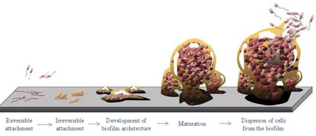
Figure 2.1 Life and times of a biofilm (adapted from Monroe (2007)). ... 12
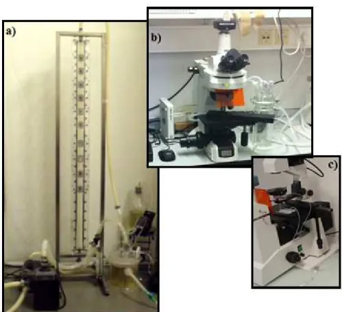
Figure 2.2 Flow systems: a) Semi-circular flow cell system, b) PPFC system, c) Microchannel system ... 25
Figure 2.3 Illustrative photograph of polystyrene microtiter plates used for biofilm formation: a) 6-well microtiter plate, b) 12-well microtiter plate c) 24-well microtiter plate and d) 96-well microtiter plate. ... 26
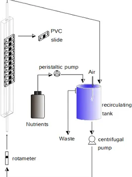
Chapter 3 Figure 3.1 Schematic representation of the biofilm producing system... 39
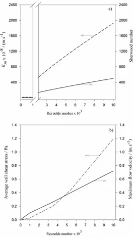
Figure 3.2 a) Calculated values using correlations for the Sherwood number (solid line) and for the external mass transfer coefficient Km (dashed line). Values for the transition zone (1000 ≤ Re ≤ 2100) were not represented due to the poor reliability of the results generated by empiric correlations in this zone. b) Average wall shear stress (dashed line) and maximum flow velocity (solid line) for Re ranging from 100 to 10000 predicted by CFD. ... 40
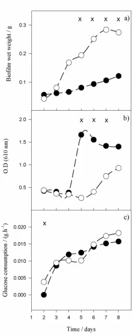
Figure 3.3 Time-course evolution of: a) biofilm wet weight, b) optical density in the recirculating tank, c) glucose consumption in the system. Closed symbols – higher flow rate (Re = 6720), open symbols – lower flow rate (Re = 4350). Time points marked with x are those for which a statistical difference was found between both conditions (confidence level greater than 95%, P ˂ 0.05) ... 42
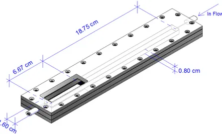
Chapter 4 Figure 4.1 Schematic representation of the PPFC. ... 50
Figure 4.2 Absolute velocity in the midplane of the cell. ... 55
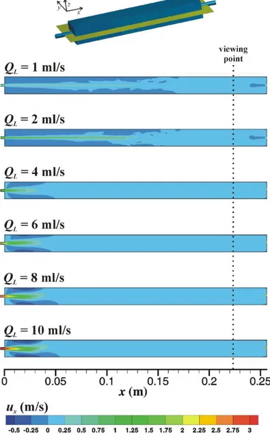
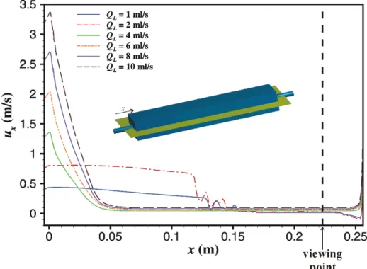
Figure 4.3 Axial velocity along the main axis of the cell. ... 56
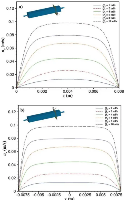
Figure 4.4 Velocity profiles in viewing region in a) z-direction and b) y-direction. ... 57
Figure 4.5 Wall shear stress in the bottom wall of the cell. ... 58
Figure 4.6 Wall shear stress along the axis of the bottom wall of the cell. ... 58 Figure 4.7 Adhesion of E. coli on PDMS (open symbols), on glass surfaces (closed
symbols) and the theoretical values predicted by the von Smoluchowski-Levich (SL) approximate solution (line), during 30 min for each flow rate: a) 1 ml.s-1, b) 2 ml.s-1,
xiv
from three independent experiments for each condition. Statistical analysis corresponding to each time point is represented with an * for a confidence level greater than 95% (P < 0.05). ... 60
Figure 4.8 Ratio between E. coli adhesion on PDMS and glass surfaces (circles) for
different flow rates (1, 2, 4, 6, 8, 10 ml.s-1). Average wall shear stress for each flow rate determined by CFD (triangles). A solid line was drawn to highlight the points where E. coli adhesion results are similar on both surfaces. These results are an average of those obtained from three independent experiments for each surface and flow rate. ... 61
Chapter 5
Figure 5.1 Grid that was used for the numerical simulations. ... 73 Figure 5.2 Free surface during a complete rotation (Dorb = 50 mm). ... 75 Figure 5.3 Average wall shear stress for both orbital diameters. ... 75 Figure 5.4 Wall shear stress for Dorb of 25 mm (upper row) and 50 mm (lower row). Wall shear stresses bellow 0.05 Pa are not represented... 76
Figure 5.5 Velocity field in a cross section of the well for Dorb of 25 mm (upper row) and 50 mm (lower row). ... 77
Figure 5.6 Time-course evolution of biofilm development and glucose concentration:
a) and c) 50 mm orbital shaking amplitude, b) and d) 25 mm orbital shaking amplitude. a) and b) Biofilm development, c) and d) glucose concentration. Closed symbols – high glucose concentration, (1 g.L-1), open symbols – low glucose concentration (0.25 g.L-1). These results are an average of those obtained from three independent experiments for each condition. Statistical analysis corresponding to each time point is represented with an * for a confidence level greater than 95% (P < 0.05). Error bars represent the standard deviation between the triplicates... 78
Chapter 6
Figure 6.1 Wall shear stress in a PPFC (A and B2) and in a well of a 96-well microtiter
plate (B1). A flow rate of 11 ml.s-1 was used for the simulation in the PPFC. A: wall shear stress in the bottom surface of the PPFC, the visualization plane is highlighted in the figure for clarity. B2: detail of the wall shear stress in the visualization zone. A shaking frequency of 150 rpm with an orbital shaking amplitude of 50 mm was used for the simulations in the
xv
well of a 96-well microtiter plate (B1). The well dimensions are indicated (D and H) as well as the liquid level at stationary condicions (S). ... 95
Figure 6.2 Biofilm formation after 24 h in microtiter plates pre-conditioned with
a) glucose, b) yeast extract, c) peptone, d) mannose, e) palmitic acid and f) BSA at different concentrations. Biofilm formed on unconditioned surface was used as control. The extent of biofilm formation was estimated by the crystal violet assay. Presented values are mean A570 nm ± standard deviation of three independent experiments with six replica wells per plate. Statistically significant differences are indicated with an asterisk. (*, P < 0.05) .... 96
Figure 6.3 Biofilm formation after 24 h in microtiter plates pre-conditioned with a) cellular
fragments, b) cytoplasm with cellular debris and c) periplasm at different concentrations. The extent of biofilm formation was estimated by the crystal violet assay. Presented values are mean A570 nm ± standard deviation of three independent experiments with six replica wells per plate. Biofilm formed on unconditioned surface was used as control. Statistically significant differences are indicated with an asterisk. (*, P < 0.05). ... 97
Figure 6.4 Number of adhered cells per cm2 in the PPFC after a) 24 h and b) 30 min on polystyrene pre-conditioned surface with peptone (PEP) at 2 g.L-1, yeast extract (YE) at 2 g.L-1, BSA at 0.3 g.L-1, palmitic acid (PA) at 0.025 g.L-1, cellular fragments (TCE) corresponding to a cellular concentration of 24.3×10⁸ cell.ml-1, cytoplasm with cellular debris (CCDE) corresponding to a cellular concentration of 24.3×10⁸ cell.ml-1 and periplasm (PE) corresponding to a cellular concentration of 0.38×10⁸ cell.ml-1. Cells adhered on unconditioned surface were used as control. Presented values are mean ± standard deviation of three independent experiments. Statistically significant differences are indicated with an asterisk. (*, P < 0.05)... 98
Chapter 7
Figure 7.1 Surfaces used and ᵞLW /ᵞ- tested in different works attempting to find a correlation between adhesion and thermodynamic properties. ... 113
Figure 7.2 Relationship between bacterial adhesion or protein adsorption and the ratio
between apolar Lifshitz van der Waals components (ᵞLW) and electron donor component (ᵞ-). a) E. coli adhesion on polymeric and glass surfaces b) Vibrio (circle), Cobetia (triangle) and P. fluorescens (square) adhesion on Ni – P coatings with TiO2 and PTFE and stainless steel, re-plotted from Liu et al. (2011a), c) Vibrio adhesion at 0.21 (circle), 0.46 (triangle), and 0.98 (square) mPa on Ni – P coatings with TiO2 and PTFE and stainless steel, re-plotted from Liu et al. (2011a), d) S. epidermis adhesion at 5 (circle), 50 (triangle) and 200 s-1
xvi
f) L. monocytogenes adhesion on synthetic surfaces, re-plotted from Cunliffe et al. (1999), g) Bovine serum albumin adsorption on synthetic surfaces, re-plotted from Cunliffe et al. (1999), h) Cytochrome c adsorption on synthetic surfaces, re-plotted from Cunliffe et al. (1999). Whenever a correlation was reported by the original authors it was also represented in this figure and the correlation factor (R2) is indicated (panels a, b and c). ... 114
Chapter 8
Figure 8.1 Wall shear stress: a) in the bottom wall of the PPFC (xy plan); b) in the viewing regions of the PPFC and microchannel; c) in the bottom wall of the microchannel (xy plan). ... 126
Figure 8.2 Bacterial adhesion rates on PA, glass, PDMS, CA and PLLA obtained in the
microchannel (black bars) and in the PPFC (white bars). Error bars shown for each surface represent the standard deviation from three independent experiments. ... 128
xvii
List of Tables
Chapter 2
Table 2.1 Summary of the work developed by several authors in which different platforms
are used under different operational conditions in order to evaluate the role of surface properties on bacterial adhesion or biofilm formation. ... 17
Table 2.2 Shear rate and shear stress in the human body, biomedical apparatus, industry
and others and the in vitro platforms which can be used to simulate the shear forces in each of these places. ... 22
Chapter 4
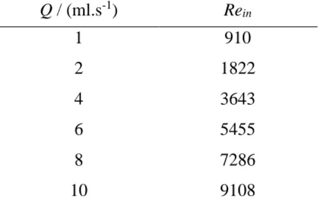
Table 4.1 Reynolds number at the inlet for each flow rate studied. ... 51 Table 4.2 The apolar (γLW ) and polar (γ AB ) components, the surface tension parameters (γ + and γ -) and the hydrophobicity (∆G) of two surfaces (glass and PDMS) and E. coli cells. ... 59
Table 4.3 Free energy of adhesion between E. coli and each surface, glass and PDMS.. 59
Chapter 7
Table 7.1 Surface thermodynamic properties and cell adhesion results. ... 111 Table 7.2 Summary of the work developed by other authors and in the present study. . 112
Chapter 8
Table 8.1 Microchannel and PPFC dimensions, operational data and numerical results. .... 127
Table 8.2 Contact angle measurements of each surface (bacteria, PLLA, PDMS, PA, CA,
glass) with the three liquids, water (θw), formamide (θform) and α-bromonaphtalene (θbr) and hydrophobicity (∆G). ... 127
xix
List of Symbols and Acronyms
Re – Reynolds number (ρ v d µ-1 – dimensionless)Sh – Sherwood number (Km d D -1 – dimensionless) Sc – Schmidt number (µ ρ -1 D-1 – dimensionless) Km – external mass transfer coefficient (L T-1) D – diffusivity (L2 T-1) d – diameter (L) V – velocity (L T-1) ρ – density (M L-3) µ – viscosity (M L-1T-1) cb – bacterial concentration (M L-3) rb – microbial radius (L)
h0 – height of the rectangular PPFC (L)
x – distance for which an average velocity variation below 15 % was determined (L) Dorb – orbital diameter (L)
Q – flow rate (L3 T-1)
ᵞ
LW–Lifshitz-van der Waals component of the surface energy (M T-2)ᵞ
AB –Lewis acid-base component of the surface energy (M T-2)ᵞ
-– electron donor parameter (M T-2)
ᵞ
+– electron acceptor parameter (M T-2)
ᵞ
Tot –total surface energy (M T-2)∆G – free energy of interaction (M T-2)
∆GAdh– free energy of adhesion (M T-2)
EPS – extracellular polymeric substance CFD – computational fluid dynamics PPFC – parallel plate flow chamber CIP – cleaning-in-place
SL – Smoluchowski-Levich CV – crystal violet
DNS – dinitrosalicylic colorimetric method OD – optical density
BSA – bovine serum albumin
CCDE – cytoplasm with cellular debris PE – periplasmic extract
TCE – total cell extract
PDMS – polydimethylsiloxane CA – cellulose acetate
PA – polyamide PLLA – poly-L-lactide PS – polystyrene
1
Chapter 1 Introduction
In this chapter, the relevance and motivation of this work are summarized and the main objectives presented. The thesis outline is explained.
3
1.1 Relevance and motivation
Biofilms can be described as a structured community of cells enclosed in a self-produced polymeric matrix and adherent to a surface (Van Houdt et al. 2005). This community is often regarded as a problem that can cause infections or deterioration of medical devices functionality, representing a cost of $5 billion annually in the US (Pace et al. 2006), or they can also have deleterious effects when formed in industrial systems such as pipes, heat exchangers and membranes, representing up to 30% of the total plant operating costs (Melo et al. 2010). Biofilms can also be used for human benefit in wastewater treatment or in the production of commodities (Vinage et al. 2003; Qureshi et al. 2005).
The accepted model for biofilm formation includes a reversible cell attachment to a pre-conditioned surface with macromolecules from the surrounding medium, irreversible attachment and development of the biofilm architecture, maturation and dispersion of cells from the biofilm (Habimana et al. 2014). This process is controlled by intrinsic factors (i.e. those concerning the microbial species involved, their genetics, metabolism and physiology) and also external factors that pertain to the particular environment where the biofilm is formed (Nikolaev et al. 2007). The existing flow conditions in each situation (environmental, physiological or engineered) and the properties of the surface which will be the docking place for bacteria have a profound influence on biofilm formation (Harding et al. 2014).
The effects of the surface material on the onset of a biofilm are still not clear. Researchers have been trying to understand the relation between the physicochemical surface properties and the bacterial adhesion process and further biofilm development (Chen et al. 2005). Electrostatic forces, van der Waals forces and hydrophobic interactions are involved either in the adsorption of the molecules that will constitute the conditioning film as well as in the reversible bacterial adhesion (Renner et al. 2011). The first candidates for surface conditioning agents are the components of the culture medium, cellular components and other cell-produced metabolites. Complex media often contains sources of polysaccharides and protein extracts and since the molecular size of these compounds is much smaller than that of bacterial cells, their diffusion to the surface is faster (Bruinsma et al. 2001). Furthermore, cell lysis occurs in bacterial cultivation, thus it is likely that cell-synthesized compounds or cellular structures, which are smaller than a whole cell, reach the surface first and start the conditioning process. The rate at which these macromolecules and bacteria are delivered to the surface, the time they reside in close proximity to the surface, oxygen and nutrient transport and the mechanical shear forces at the surface-fluid interface are all affected by the fluid hydrodynamics (Robert et al. 2010).
In environmental and biomedical systems, mass transport and shear stress generated by the fluid flow are dependent on the existing hydrodynamic conditions and thus, these conditions cannot be changed but should be taken in count since they can affect biofilm development (Gomes et al. 2013). Regarding the industrial field, mass transport and shear forces have been used as an effective tool in cleaning in place procedures and in the control of biofilm growth and stability (Liu et al. 2002; Jensen et al. 2005).
4
These external factors (surface properties and hydrodynamic conditions) often dictate the initial cell adhesion process and will also influence biofilm maturation and removal. Understanding the process of bacterial adhesion is key to control biofilm development either to inhibit or to delay the onset of detrimental biofilms or to promote beneficial biofilm development in engineered systems. Therefore, in order to develop more efficient control strategies and better understand the process of biofilm formation, in vitro biofilm simulation platforms are used. Among these, flow cells and 96-well microtiter plates are the most frequently used (Coenye et al. 2010). Microtiter plates have the advantage of enabling the simultaneous testing of many conditions in a high throughput fashion (Coenye et al. 2010). Flow cells have a lower throughput than 96-well microtiter plates, but they enable the use of coupons of different materials where biofilm formation is going to be simulated (Teodósio et al. 2011). Some flow cells also enable observation of this process in real time. However, the hydrodynamics and mass transfer features in such systems are still poorly understood, which is a drawback on the use of those systems in predicting biofilm behaviour at larger scales.
Although a solid body of evidence has accumulated regarding later stages of biofilm formation, information about early events is still scarce (Sauer et al. 2001; Simões et al. 2008). Since biofilm formation starts by reversible attachment to a pre-conditioned surface, one of the issues to be addressed is: what are the effects of the pre-conditioning phase on bacterial adhesion and biofilm development?
The effects of surface material on biofilm onset are also still not clear. The question to be answered is: what are the physicochemical surface properties that control the biofilm onset?
Since these surfaces are usually integrated into natural and engineered environments subjected to physiological, operational or environmental flow conditions, a question is raised: what is the importance of the hydrodynamic conditions on the biofilm onset and maturation?
1.2 Objectives and outline
The main objective of this work was to understand the processes entailed with biofilm formation in order to devise strategies to inhibit or delay detrimental biofilm formation or to obtain biofilms that are more resistant to operating conditions in biofilm reactors. In this thesis, the influence of initial external factors on the onset of bacterial biofilms and further development was assessed using the Gram-negative bacteria Escherichia coli as a model system due to the medical and industrial relevance of this organism.
The final purpose of this work was:
i) To assess the effect of surface conditioning agents on cell adhesion and biofilm development.
ii) To assess the influence of several starting variables such as different adhesion surfaces and different hydrodynamic conditions on bacterial adhesion and biofilm development.
5
iii) To devise guidelines for conditioning treatments and operational procedures to better control the properties of mature biofilms.
This thesis is outlined as follows:
Chapter 2 is a brief literature review describing the state of the art pertaining to this thesis.
In the following three chapters, a detailed characterization of the hydrodynamic conditions in three commonly used biofilm formation platforms was made.
In Chapter 3, the flow hydrodynamics in a semi-circular flow cell were characterized.
Average shear stresses and maximum flow velocities were determined by computational fluid dynamics (CFD). Additionally, correlations were used to characterize the mass transference in this system. The effects of two flow rates in the turbulent regime, which is often used in industrial settings, were assayed in a real semi-circular flow cell system in order to evaluate their effect on biofilm development.
In Chapter 4, the fluid hydrodynamics inside a parallel plate flow chamber (PPFC) were
characterized. Shear stresses and flow velocities were determined by CFD. The effect of six flow rates in bacterial adhesion on two surfaces with different physicochemical properties was evaluated.
In Chapter 5, the fluid hydrodynamics inside the wells of a 96-well microtiter plate were
characterized. Shear stresses and flow velocities were determined by CFD. The effect of two different hydrodynamic conditions on biofilm development in culture media with different glucose concentrations was assessed.
After this initial hydrodynamic characterization, the most suitable platforms were selected to conduct adhesion/biofilm assays in order to understand the factors which control biofilm development.
In Chapter 6, the effect of surface conditioning with medium components and cellular
extracts on bacterial adhesion and further biofilm maturation was assessed. A 96-well microtiter plate was used for screening purposes due to its high throughput and selected conditions were tested in a PPFC at the same shear stress. A flow cell system was chosen since it can mimic biofilms formed in real systems with similar fluid topology. The PPFC was chosen due to its reduced dimensions which makes it easier to handle, and it allows operation with a low hold-up which is ideal to test cellular extracts that are difficult to obtain. In this chapter, the scalability of the results obtained in these systems and the possibility of application to industrial settings were also discussed.
In Chapter 7, the PPFC was selected again, since it enables real time observation, to study
the effect of the physicochemical properties of polymers used in the biomedical field in bacterial adhesion. Additionally, this analysis was extended to published data from other authors which have studied bacterial adhesion or protein adsorption to different materials (soil minerals, synthetic materials, plasma treated surfaces and metallic materials) in different systems and operational conditions. The aim of this chapter was to find out a
6
selection criteria to predict bacterial adhesion to materials used in the industrial and biomedical fields.
In Chapter 8, bacterial adhesion to the polymeric surfaces used in the previous chapter
was evaluated in the PPFC and in a microfluidic system under physiological shear stress conditions. The microchannel was chosen as small scale platform and the PPFC as larger platform, since they enable testing of different surfaces and observation of bacterial adhesion in real time.
Finally, Chapter 9 contains the main conclusions of the work presented in this thesis and
some suggestions for future research.
1.3 References
Alan B, Buehler K, Schmid A. 2012. Biofilms as living catalysts in continuous chemical syntheses. Trends in Biotechnology. 30:453-465.
Bruinsma GM, van der Mei HC, Busscher HJ. 2001. Bacterial adhesion to surface hydrophilic and hydrophobic contact lenses. Biomaterials. 22:3217-3224.
Chen G, Zhu H. 2005. Bacterial adhesion to silica sand as related to Gibbs energy variations. Colloids and Surfaces B: Biointerfaces. 44:41-48.
Coenye T, Nelis HJ. 2010. In vitro and in vivo model systems to study microbial biofilm formation. Journal of Microbiological Methods. 83:89-105.
Gomes LC, Moreira JMR, Miranda JM, Simões M, Melo LF, Mergulhão FJ. 2013. Macroscale versus microscale methods for physiological analysis of biofilms formed in 96-well microtiter plates. Journal of Microbiological Methods. 95:342-349.
Habimana O, Semião AJC, Casey E. 2014. The role of cell-surface interactions in bacterial initial adhesion and consequent biofilm formation on nanofiltration/reverse osmosis membranes. Journal of Membrane Science. 454:82-96.
Harding JL, Reynolds MM. 2014. Combating medical device fouling. Trends in Biotechnology. 32:140-146. Jensen BBB, Friis A, Benezech T, Legentilhomme P, Lelievre C. 2005. Local wall shear stress variations predicted by computational fluid dynamics for hygienic design. Food and bioproducts processing 83:53-60. Liu Y, Tay J. 2002. The essential role of hydrodynamic shear force in the formation of biofilm and granular sludge. Water Research. 36:1653-1665.
Melo L, Flemming H. 2010. The science and technology of industrial water treatment. Taylor and Francis Group.
Nikolaev Y, Plakunov V. 2007. Biofilm - “City of microbes” or an analogue of multicellular organisms? Microbiology. 76:125-138.
Pace JL, Rupp ME, Finch RG. 2006. Biofilms, Infection and Antimicrobial Therapy. Boca Raton: CRC pres Taylor and Francis group.
Qureshi N, Annous B, Ezeji T, Karcher P, Maddox I. 2005. Biofilm reactors for industrial bioconversion processes: employing potential of enhanced reaction rates. Microbial Cell Factories. 4:24.
Renner LD, Weibel DB. 2011. Physicochemical regulation of biofilm formation. MRS Bulletin. 36:347-355. Robert JM, Salek MM. 2010. Numerical Simulations - examples and applications in computational fluid dynamics. Canada: InTech. Numerical simulation of fluid flow and hydrodynamic analysis in commonly used biomedical devices in biofilm studies.
7
Sauer K, Camper AK. 2001. Characterization of phenotypic changes in Pseudomonas putida in response to surface-associated growth. Journal of Bacteriology. 183:6579-6589.
Simões M, Simões LC, Vieira MJ. 2008. Physiology and behavior of Pseudomonas fluorescens single and dual strain biofilms under diverse hydrodynamics stresses. International Journal of Food Microbiology. 128:309-316.
Teodósio JS, Simões M, Melo LF, Mergulhão FJ. 2011. Flow cell hydrodynamics and their effects on E. coli biofilm formation under different nutrient conditions and turbulent flow. Biofouling. 27:1-11.
Van Houdt R, Michiels CW. 2005. Role of bacterial cell surface structures in Escherichia coli biofilm formation. Research in Microbiology. 156:626-633.
Vinage I, Rohr P. 2003. Biological waste gas treatment with a modified rotating biological contactor. I.Control of biofilm growth and long-term performance. Bioprocess and Biosystems Engineering. 26:69-74.
9
Chapter 2 Literature Review
In this chapter, a literature review describing the state of the art of the subjects presented in this thesis is made. The biofilm concept is presented and the main advantages and disadvantages of its formation are discussed. A brief description of the biofilm development process and the main factors which control its formation are also presented. Special emphasis is given to surface properties and hydrodynamic conditions which are of particular relevance to this thesis. A description of the in vitro platforms that are currently used for biofilm studies is also presented including a detailed description of the ones used in this study.
11
2.1 Microbial biofilms
Biofilms are structured communities of microorganisms attached to surfaces surrounded by a matrix of extracellular polymeric substances (EPS) which confers many advantages to biofilm cells that can develop synergistic interactions (Dufour et al. 2010). This matrix is mainly constituted by water (97%), polysaccharides (1-2%), proteins (< 1-2%) and nucleic acids (<1-2%) and is responsible for biofilm morphology, functional integrity, cohesion and structure (Sutherland 2001; Branda et al. 2005). The biofilm mode of living confers protection against harmful environments (nutrient deprivation, pH changes, oxygen radicals, hydrodynamic conditions, biocides, and antimicrobial agents), enables genetic material transference and facilitates the colonization of favorable and hostile niches (Nikolaev et al. 2007). It is estimated that more than 90% of bacteria in natural environments exist within a biofilm (Petrova et al. 2012). In industry, biofilms have been used in the production of chemicals, (e.g. ethanol, lactic acid, vinegar), bioremediation processes, waste-water treatment or even removal of volatile compounds from waste streams (Vinage et al. 2003; Qureshi et al. 2005; Singh et al. 2006; Alan et al. 2012). The use of biofilms in these processes enables higher cell concentrations and thus higher reaction rates and an easier separation between the final product and microorganisms which can be used for longer operational times (Qureshi et al. 2005). On the other hand, biofilm development is a common problem faced by the industrial (Rochex et al. 2007; Florjanic et al. 2011), environmental (Azevedo et al. 2006; Mahfoud et al. 2009; Cooper et al. 2010) and biomedical areas (Koseoglu et al. 2006; Silverstein et al. 2006). In the food industry, biofilms can lead to food spoilage by bioconversion (Shi et al. 2009; Van Houdt et al. 2010; Dourou et al. 2011), in industries with water process lines, besides causing problems in cleaning and disinfection, biofilms can reduce heat transfer in heat exchangers, reduce flow through blocked tubes and may contribute to the corrosion of various materials (Shi et al. 2009; Melo et al. 2010). It has been estimated that biofilm development in industrial process lines may represent up to 30% of the plant operating costs (Melo et al. 2010). In aquatic environments, biofilms can grow in ship hulls leading to an increase in fuel consumption that can reach up to US$ 400 h-1 for a ship travelling at 48 km h-1 (Cooksey et al. 1995). In the biomedical field, cells in biofilms are responsible for infections since they are typically more resistant to antimicrobial agents than planktonic cells and have a decreased susceptibility to host defense systems (Shunmugaperumal 2010). It has been reported that 65% of the hospital acquired infections are caused by biofilms which can grow in indwelling and other percutaneous medical devices and can cost $5 billion annually in the US (Pace et al. 2006; Bryers 2008). The development of biofilms in catheters, wound dressings, medical implants and medical devices is problematic since these biofilms can be reservoirs of pathogenic organisms, a source of disease spread and can cause material biodegradation, changes in surface properties and deterioration of the medical device functionality (Missirlis et al. 2004; Kaali et al. 2011). When such biofilms form in medical devices, sometimes the only solution is their surgical removal. However, the costs associated with the replacement of infected implants during revision surgery may triple the cost of the primary implant procedure (Busscher et al. 2012). Moreover, secondary implants
12
are further exposed to colonization by antibiotic resistant bacteria residing in the surrounding tissue which can proliferate and lead to new infections. (Busscher et al. 2012).
There is a need to better understand and control biofilms in order to promote the formation of beneficial biofilms or to facilitate the elimination or delay the onset of harmful biofilms.
2.2 Biofilm formation process
The currently accepted mechanism for biofilm development involves five stages (Figure 2.1) starting from reversible attachment of cells to a pre-conditioned surface, EPS production leading to irreversible attachment, early development of biofilm architecture, biofilm maturation and cell dispersion from the biofilm (Dunne 2002; Nikolaev et al. 2007; Goulter et al. 2009; Habimana et al. 2014)
A solid surface immersed in water is immediately covered by molecules (e.g. organic matter, proteins) from the liquid phase forming a conditioning film which may change the properties of this surface making it more or less suitable for bacterial anchorage. The formation of this layer of adsorbed molecules is the first stage, preceding the formation of a bacterial film. After planktonic cell transport from the bulk liquid to the substratum, cell adsorption at the surface followed by release or reversible adhesion takes place. Electrostatic forces, van der Waals forces and hydrophobic interactions are involved in the adsorption of the molecules that will constitute the conditioning film as well as in the reversible bacterial adhesion (Renner et al. 2011). The following stage begins when the cells become irreversibly attached to the surface. This step is mediated by stronger attractive forces such as covalent and hydrogen bonds and may be helped by cellular surface structures such as flagella and fimbriae (Renner et al. 2011). Then, the processes of cellular growth and EPS production begin. After biofilm maturation, biofilm growth and detachment/sloughing balance each other so that the total amount of biomass remains approximately constant in time (the steady-state is achieved).
13
Environmental factors and the properties of the cells affect the process of biofilm formation. The most important environmental factors are pH, salinity, temperature, osmolarity, oxygen partial pressure, accessibility to nutrient sources, surface properties (of both bacteria and substrate) and the force and type of liquid motion relative to this surface (Nikolaev et al. 2007). Biofilm cells differ from planktonic cells in gene expression, protein production and resistance to the immune system and antimicrobial agents (Petrova et al. 2012). This adaptive response depends on the surrounding fluid hydrodynamic conditions which will dictate shear forces and mass transference (oxygen, nutrients, cellular products, etc) (Purevdorj et al. 2002; Moreira et al. 2013). Thus, the biofilm architecture (thickness, porosity, etc) must adapt in order to resist to shear forces and to allow a better access to nutrients and oxygen.
2.3 Biofilm control strategies
Remedial approaches to eliminate biofilms usually consist in mechanical/chemical cleaning or material/equipment replacement in industry or medical device replacement and antibiotic treatment in the biomedical field (Simões et al. 2010; Van Houdt et al. 2010; Busscher et al. 2012). These processes have high costs and they are not always effective (Melo et al. 2010; Busscher et al. 2012). Moreover, it has been observed that bacteria have been developing resistance to antibiotics (Shunmugaperumal 2010). In many fields microorganisms are not a problem as long as they remain planktonic, and therefore the disinfection process would be facilitated if microorganism attachment could be prevented. This is because microorganisms embedded in a biofilm are 100–1000 times less sensitive to most antibiotics and biocides compared to planktonic cultures (Meyer 2003; Nikolaev et al. 2007). Therefore, a preventive strategy has been adopted to delay biofilm development by affecting bacterial adhesion (Van Houdt et al. 2010; Petrova et al. 2012; Campoccia et al. 2013a). Understanding the process of bacterial adhesion is key to control biofilm development either to inhibit the onset of detrimental biofilms or to promote beneficial biofilms in engineered systems. A number of studies has been performed to try to gain control over bacterial adhesion. Recently, with the increasing use of biodiesel as alternative to fossil fuels, some issues like the integrity of storage tanks which become compromised by the formation of biofilms is a concern. Restrepo-Flórez et al. (2014) studied the effect of biodiesel concentration on biofilm development on surfaces such as low-density polyethylene, cross-linked polyethylene, and a bilayer construction of linear-low density polyethylene and polyamide-11 under conditions similar to those found in an industrial fuel storage system. The authors verified that the composition of the biofilms developed is affected by the nature of the polymer and by the concentration of biodiesel used as a carbon source. These findings may be important in the design and management of efficient strategies to substitute diesel for biodiesel without comprising the integrity of the infrastructure. In the biomedical field, some diseases such as cancer have been the focus of research in this century. New strategies focused in the bacterial potential have been explored as alternatives to the conventional chemical treatments which have many detrimental side effects. Park et al. (2014) proposed a bacteria-based microrobot (bacteriobot) for theranostic activities against solid tumors. This bacteriobot acts as a
14
combination of microsensor, microactuator, and therapeutic agent and it can be considered as a new type of active drug delivery system. This system is based in the ability of mobile bacteria such as E. coli or Salmonella typhimurium to adhere to designed microsurfaces and originate a bacteriorobot that can move on human cells and have a higher affinity to cancer cells. The key for the success of this new theranostic approach is the strong attachment of the bacteria to the microstructure which is very important for its motility and stability in living tissues. Previous studies have shown that hydrophobic interactions can be important in the immobilization of bacteria on the microstructures and thus the use of new materials or surface conditioning with proteins can be important in the bacteriorobot success (Behkam et al. 2008; Park et al. 2010).
It is known that beyond the influence of the surface properties on the bacterial adhesion process, the hydrodynamic forces can also be crucial (Missirlis et al. 2004). Fang et al. (2012) made a study where they used the hydrodynamic forces (22, 110, 795 s-1) to tune the Staphylococcus aureus capture ability and direct bacteria to target regions of a poly(ethylene glycol) polymer brush. They verified that at a lower shear, the extension of bacterial adhesion was higher. At a high shear bacteria could adhere only on relatively rare “hot spots” and so the rate of bacterial adhesion on these spots was small but adhesively selective. Therefore bacterial adhesion to the “stickiest” surface regions is most selective at high shear. These findings may be important in the development of sensors in the biomedical field where bacteria can be selectively directed to targeted surface regions.
The shear forces can also have an important role in further contaminations by cells detached from mature biofilms which may adhere in new locations and originate new biofilms for instance in water distribution systems. The processes used to control biofilms in these systems have demonstrated limited efficacy. Thus, studies have been made in order to understand the factors that control the biofilm onset. Florjanic et al. (2011) investigated the effect of water hydrodynamics on surface colonization, biofilm growth and bacterial detachment. The authors concluded that hydrodynamic conditions have a significant influence on biofilm development. At a constant flow velocity, biofilm colonization and development was delayed, and a low number of bacteria detached from biofilm into the water. Additionally, they also observed that the primary biofilm acts as a constant reservoir of cells that after detaching (due to the flow shear) are able to occupy new surfaces very quickly.
Surface properties and hydrodynamic conditions are the two main factors which can be used in order to control biofilm formation in engineered systems. The other factors (temperature, pH, salinity, etc) may be dependent on the physiological conditions in the case of the human body or may be set by specific operational conditions in industrial systems.
2.3.1 Surface properties
Bacterial adhesion to a surface (substrate), the first step in the biofilm formation process, consists on the attraction of bacteria to the surface (natural or artificial) followed by adsorption and attachment. When immersed in aquatic systems, molecules at the surfaces tend to interact with molecules in the solution through physicochemical
15
interactions. The forces involved in this process are the Lifshitz van der Waals, electrostatic and Lewis acid-base interactions (Bos et al. 1999). The van der Waals forces have an electromagnetic nature and are usually attractive, the electrostatic elements originate from Coulomb interactions between the charged bacteria and the surface and the Lewis acid-base component is governed by the potential formation of covalent bonds between electron pairs (Perni et al. 2013). Thus, the surface energy is a measure of the interfacial attractive forces. A surface (from bacteria or substrate) can be classified into hydrophilic or hydrophobic (van Oss 1995). This classification is based in the interaction energy (∆G mJ.m-2) between molecules (from the surface) immersed in water. If the interaction between the two entities is stronger than the interaction of each entity with water,
∆G < 0 mJ.m-2, the material is considered hydrophobic, if ∆G > 0 mJ.m-2, the material is hydrophilic (van Oss 1995). Depending on the hydrophobicity of both bacteria and material surfaces, bacteria may adhere differently to materials with different hydrophobicities. Hydrophilic surfaces are usually more resistant to bacterial adhesion than hydrophobic surfaces due to a physical barrier known as hydration layer (An et al. 1998; Harding et al. 2014). This layer results from hydrogen bonding between functional groups at the surface and water molecules from the surrounding fluid which forms a type of scaffold that functions as a barrier (Harding et al. 2014).
Over the years, researchers have been trying to predict whether a bacteria will adhere to a surface through the variation of system (bacteria-substrate) Gibbs energy (Chen et al. 2005). Therefore, some theories concerning the forces involved in bacteria- substrate interactions were developed. In the thermodynamic approach the variation of the Gibbs energy of the system is based in the Lifshitz van der Waals forces and the Lewis acid-base interactions. In the DLVO theory, it is assumed that the energy of the system is the sum of the Lifshitz van der Waals forces and the electrostatic interactions, both depending on the separation distance between particles (Perni et al. 2013). However, since the Lewis acid-base interactions involved in bacterial adhesion process have been neglected by the DLVO approach (Azeredo et al. 1999; Bos et al. 1999; Perni et al. 2013), an extended DLVO (xDLVO) theory was developed taking into account the three interaction energies. Nowadays, both theories, the thermodynamic and xDLVO, have been applied to predict bacterial adhesion to different materials (Bos et al. 1999; Chen et al. 2005; Perni et al. 2013).
Researchers have been studying this interaction energies and they have been trying to find a relation between surface properties and bacterial adhesion (Liu et al. 2005). This knowledge would enable the manipulation of the surface energy and charge of the materials of process equipment and biomedical devices in order to promote or inhibit biofilm formation (Missirlis et al. 2004; Fernández et al. 2007). Intensive efforts have been focused in the fabrication of new surfaces, whether by new combinations of exiting materials (metal, glass, plastic) or by modification of their properties (Asan et al. 2013). Several surface modification techniques have been used in the construction of artificial surfaces and they can be categorized according to the surface coating or surface chemistry modifications (Asan et al. 2013). The most common techniques (Asan et al. 2013; Campoccia et al. 2013b; Alwiczek et al. 2014; Harding et al. 2014) are surface treatment with active gases and vapors (e.g. gas discharge, corona/ plasma discharge), solution
16
deposition (e.g polymer coatings, surfactant deposition), chemical treatment (e.g oxidation, chlorination) and physical adsorption of molecules (e.g proteins, peptides).
Recently, smart materials inspired in natural systems which have anti-fouling properties (e.g. lotus leaves and shark skin) are being created (Gu et al. 2014). The development of this new materials is based in the concept of self-cleaning coating. Moreover, some of them can quickly change their physicochemical properties in response to environmental stimulus such as pH, temperature, surrounding media, etc. These smart materials been developed based on silica nanoparticles, polymers (e.g. water-soluble synthetic polymers) and carbon nanotubes (Gu et al. 2014; Halake et al. 2014).
Another important factor in the interaction between surfaces and bacteria is the adsorption of molecules from the liquid medium where the surface is inserted. Proteins present in tears, blood, saliva, or in the milk in the industrial sector, and organic matter in natural systems are examples of molecules that are present in the medium where surfaces are inserted and can be adsorbed thus affecting surface interaction with bacteria (Bakker et al. 2003a; Dat et al. 2010; Lorite et al. 2011). Therefore, when a new material is created one should take in consideration the medium where this surface will be inserted, since its surface properties can be changed due to the adsorption of liquid native molecules. Based on this idea, researchers are exploring, the surface conditioning strategy to control biofilm formation. Loskill et al. (2013) have characterized the adhesion of Streptococcus mutans, Streptococcus oralis, and Staphylococcus carnosus on smooth, high-density hydroxyapatite surfaces, pristine (this material mimics the teeth surface) and pre- conditioned with a fluoride solution. These authors have observed that bacterial species exhibited lower adhesion forces after fluoride treatment of the surfaces highlighting the importance of fluoride as an effective caries-preventive agent.
Table 2.1 lists several studies assessing the effect of surface properties on cell adhesion performed in the last 30 years. In these studies, different materials (polymeric materials, coatings, plasma treated surfaces, metallic surfaces, etc) with applications in several fields (biomedical, industrial, etc) were tested under different conditions (hydrodynamics, temperature, etc) and operated in various platforms. The most used platforms in these studies were the flow cells systems and agitated microtiter plates. This table lists 25 studies and it is possible to observe that in some of them it was not possible to find a correlation between surface physicochemical properties and bacterial adhesion. In others it was possible to establish a correlation only for some particular cases. For the remaining studies, where a correlation between biofilm formation and surface properties was found, a unique parameter was not identified to correlate all the results. However, the parameters most often used were surface hydrophobicity (∆G) and free energy of adhesion (∆GAdh). This compilation highlights the difficulty in controlling cell adhesion by manipulation of the surface properties. Additionally it is also possible to verify that around 90% of these studies are focused in the reduction of the biofilm formation, showing that the majority of these authors are looking for antibacterial surfaces.
20
2.3.2 Hydrodynamics
Since the designed materials are usually integrated in natural or engineered environments with particular hydrodynamic conditions, the interaction between bacteria and the surface materials is subjected to the physiological, operational or environmental conditions (Harding et al. 2014). The flow conditions of each system (natural, biomedical or industrial) where the surface material is integrated, have a profound influence on the biofilm onset. The rate at which macromolecules (specific for each type of fluid) and bacteria are delivered to the surface, the time they reside in close proximity to the surface and the mechanical shear forces at surface-fluid interface is dictated by the flow hydrodynamics (Robert et al. 2010). There is an optimum flow rate for bacterial adhesion reflecting the balance between rate of delivery and the force acting on adhered bacteria (Missirlis et al. 2004). The interaction between bacteria and the surface will determine the shear forces that adhered bacteria will be able to withstand (Missirlis et al. 2004).
Several works have reported the importance of shear forces on bacterial adhesion. Katsikogianni et al. (2010) used a PPFC to study the effect of glass modified surfaces (with methyl (CH3), amino (NH2) and hydroxyl (OH) terminal groups) on the adhesion of two Staphylococcus epidermidis strains under four different shear rates (50, 500, 1000 and 2000 s-1). The authors observed that adhesion was higher on CH3 followed by NH2 and minimal on OH-terminated glass for both strains. However, the number of adhered cells on each surface decreased with increasing shear rates. Azevedo et al. (2006) used a 6-well microtiter plate to evaluate the adhesion of Helicobacter pylori to stainless steel 304 and polypropylene when exposed to increasing shear stresses (0, 0.138 and 0.317 Pa corresponding to 0, 146 and 334 s-1). These authors observed that the number of adhered cells on each surface decreased with increasing shear stresses. Patel et al. (2003) used a rotating disc model to observe the adhesion of S. epidermis on implant materials with surface modifying endgroups (SMEs) under physiological relevant shear stresses (0-1.8 Pa). They observed that bacterial adhesion was enhanced on materials modified with fluorocarbon SMEs and reduced on materials with polydimethyllsiloxane (PDMS) and polyethylene SMEs. However, although this adhesion trend has been observed for all tested shear stresses, the number of adhered microorganisms on each surface decreased with increasing shear stress. The authors concluded that bacterial adhesion on those surfaces was mediated by surface properties and shear stress. Skovager et al. (2012) studied the effect of flow shear stress (0.1 and 1.12 Pa) on the adhesion of different strains of Listeria monocytogenes to fine-polished stainless steel in a parallel-plate perfusion chamber. The authors observed that the initial adhesion rate for all strains (except for one), was significantly greater when high shear stresses were applied. Their results demonstrated that initial adhesion rates were dependent on the shear stress and strain type. Li et al. (2000) studied the effect of physiologic levels of shear stress (between 0.1 and 1.5 Pa) on the adhesion kinetics of S. aureus to three types of collagen surfaces in a parallel-plate perfusion chamber. The authors observed that S. aureus adhesion rate increased for all surfaces until a shear stress maximum between 0.3-0.5 Pa was reached and after this a decrease in the adhesion rate was observed. They have verified that although different
21
numbers of adhered cells were obtained on the different surfaces, the adhesion process was shear stress dependent.
It has been observed that, additionally to the relevant role that flow hydrodynamics has on the bacterial adhesion step, it is also one of the most decisive factors in the maturation of a biofilm (Gjersing et al. 2005). The fluid surrounding a biofilm, provides the primary source of nutrients and is the vehicle for cell by-products removal (Gjersing et al. 2005). An increase in flow velocity promotes the flux (in and out) of molecules (nutrients, cells, biocides, antibiotics, cellular products, etc) by changing the molecule concentration in the biofilm interface with the liquid phase. It also regulates the physiological properties of the biofilm by changing the mechanical shear stresses at the fluid-biofilm interface (Robert et al. 2010). It has been observed that higher shear forces led to the formation of a thinner and denser biofilm (Liu et al. 2002). Although higher flow velocities enhance molecular transport (convection), shear forces lead to denser biofilms which in turn reduces the diffusivity of molecules inside biofilms. Additionally, a higher flow velocity promotes stronger shear forces that can promote biofilm sloughing or detachment (Liu et al. 2002).
In the biomedical field, shear stress generated by the fluid flow is dependent on physiological conditions and thus these conditions cannot be changed but should be taken into account since they can affect biofilm development, morphology and susceptibility (Paramonova et al. 2009; Gomes et al. 2013). Kostenko et al. (2010) used a 6-well microtiter plate to investigate the impact of oscillatory surface physiological shear stresses (between 0.15 and 1.02 Pa) on S. aureus biofilm morphology and tolerance to antibiotics. These authors verified that local biofilm deposition and morphology correlate strongly with shear stress. Moreover, they observed a correlation between bacterial tolerance to antibiotics and the shear stress (in general, a higher tolerance was observed in high shear regions). Weaver et al. (2012) investigated a variety of clinical isolates of S. epidermis to determine the expression potential polysaccharide intracellular adhesin (PIA) in response to relevant fluid forces (mimicking wall shear stresses present in capillaries, venules and catheter lumens) experienced by S. epidermis during pathogenesis. Using a 96-well microtiter plate (in static conditions) and a microfluidic device (shear stresses between 0.065 and 1.14 Pa) they verified that bacteria that produced biofilms under static conditions increased their pathogenicity by secreting PIA due to the fluid shear. Additionally, in strains constitutively producing PIA the presence of fluid shear altered their metabolic profiles in the biofilm which led to a reduction of susceptibility to antimicrobial treatments enhancing the risk of infection. They concluded that catheter luminal design should consider physiological hydrodynamic conditions.
In the industrial field, shear forces have been used as an effective tool in cleaning in place procedures and in the control of biofilm growth and stability (Liu et al. 2002; Jensen et al. 2005b). Lelièvre et al. (2002) studied the effect of the wall shear stress (between 0.04 and 5.32 Pa) on Bacillus cereus removal from industrial equipment. They used stainless steel pipes with sudden and gradual expansion and contraction sites that represented complex equipment such as pumps or valves or even general pipes used in food processing lines. The authors observed that an increase in the flow rate induced a better cleaning of different zones as in sudden expansion pipes. However, in gradual contractions