1
UNIVERSIDADE DA BEIRA INTERIOR
Ciências
Identification of genetic mutations in patients
with familial central diabetes insipidus
Ângela Sofia Fernandes Alves Francisco
Dissertação para obtenção do Grau de Mestre em
Bioquímica
(2º ciclo de estudos)
Orientador: Prof. Doutor Manuel Carlos Loureiro de Lemos
Orientador: Prof.ª Doutora Isabel Maria Theriaga Mendes Varanda Gonçalves
ii
Para os meus pais
com imenso amor!
iii
Acknowledgement
Em primeiro lugar, quero agradecer ao Prof. Manuel Lemos e à Prof.ª Isabel Gonçalves por toda a dedicação a este projeto, pela ajuda e principalmente pelo incentivo a nunca desistir. É importante ter sempre alguém que nos oriente, mas também é importante ter quem confie no nosso trabalho e nos queira levar sempre para a frente, apesar de todos os desafios presentes durante o desenvolvimento desta tese. Muito obrigado por esta jornada, e por todos os conhecimentos que me transmitiram.
De seguida quero agradecer aos meus pais. Vocês sempre foram a minha âncora, o meu porto de abrigo. Vocês suportaram todas as lágrimas, todos os gritos, e todos os sorrisos que fui soltando durante este ano. Aconteça o que acontecer, sei que vão estar sempre lá para mim, e que sempre terei o vosso ombro para me apoiar, independentemente de todos os obstáculos que o futuro possa colocar no meu caminho. Tudo o que hoje sou e tenho, a vocês o devo, e durante toda a minha vida estarei imensamente grata por todo o amor que me deram durante a minha vida. Obrigado!
Ao meu querido irmão um muito obrigado por, mesmo longe, estar sempre presente, quer com telefonemas ou com mensagens. Obrigado por todos os empurrões, todos os incentives e todas as palavras que, tão queridas, me faziam sentir que estavas aqui comigo.
Tenho também de te agradecer Bruno. Este ano não foi fácil. Foram tantos os problemas, dificuldade e complicações, mas também foram bastantes os sorrisos e a felicidade. Simplesmente és a minha outra metade. Ajudaste-me a ultrapassar este longo ano, apoiaste-me em tudo e nunca apoiaste-me deixaste cair. Os teus conselhos sempre foram tão importantes para mim, e sempre me ajudaram a escolher o caminho certo. Obrigado por estares sempre aqui comigo.
Não posso deixar de agradecer aos amigos que descobri no laboratório. Mais do que colegas de trabalho, vocês foram uma força inesgotável de alegria e amizade. Eduarda, Catarina, Inês, Susana, Marina e Fernando, vocês deram um ar completamente diferente ao laboratório que, mais do que um local de trabalho, revelou-se como um local de amizade e entreajuda. O que quer que acontecesse, vocês estavam sempre lá para tornar as coisas mais simples, para não falar da quantidade de gargalhadas que os nossos momentos de convívio proporcionam. Obrigado por tudo!
Tenho também de agradecer aos meus amigos, quer os que me acompanham já desde os meus anos de infância, aos que conheci durante a adolescência e aos que criei já na universidade. Podemos conhecer muitas pessoas, mas só algumas é que se destacam no meio de tantas, e é a essas pessoas que quero agradecer por, mesmo longe, não permitirem que a distância separe corações que em certos tempos foram tão felizes juntos.
iv
Obrigado a todas as pessoas que, mesmo sem qualquer obrigação, dispensaram parte do seu tempo para me ajudaram no desenvolvimento deste projeto, transmitindo-me conhecimentos importantes, quer para este momento, quer para experiências futuras. Sempre que precisarem podem também contar com a minha ajuda.Por fim, quero agradecer o financiamento deste projeto por parte da FCT, através do programa COMPETE (PTDC/SAU-GMG/098419/2008 e PEst-C/SAU/UI0709/2011).
v
Resumo alargado
A diabetes insípida (DI) é uma doença rara, caracterizada principalmente pela excreção de elevados volumes de urina na forma diluída podendo, entre várias causas possíveis, ter origem num defeito genético.
O desenvolvimento da doença pode dever-se a quatro causas possíveis. A mais comum deve-se a uma deficiência na secreção da hormona antidiurética arginina vasopressina (AVP), sendo referida como DI central ou neurohipofisária. Outra possível causa da doença deve-se a uma insensibilidade, por parte das células renais, aos efeitos da AVP, sendo neste caso designada como DI nefrogénica. A DI pode também dever-se a uma excessiva ingestão de líquidos, que conduz à supressão da libertação da hormona AVP, sendo referida como polidipsia primária. Por fim, um aumento do metabolismo da hormona AVP durante a gravidez pode também ser uma causa da doença, designada por DI gestacional.
A hormona AVP é sintetizada nos neurónios magnocelulares. Estes têm origem no núcleo supra-óptico e para-ventricular do hipotálamo e os seus prolongamentos terminam na neurohipófise. A destruição destes neurónios resulta numa deficiência na produção da hormona, conduzindo à DI central. Esta destruição pode ter inúmeras causas, incluindo acidentes, cirurgias, doenças autoimunes, entre outras. Contudo, a doença também apresenta uma base familiar, correspondendo a 1% de todas as causas de DI central. A DI central apresenta sintomas persistentes de poliúria, polidipsia e sede, que geralmente se começam a manifestar vários meses ou anos após o nascimento.
A DI central familiar apresenta duas características principais: está associada a mutações num único alelo do gene que codifica a hormona (gene AVP), apresentando assim uma transmissão autossómica dominante; e é causada por uma deficiência progressiva pós-natal na secreção da hormona AVP, que se pensa resultar da degeneração seletiva dos neurónios magnocelulares.
O gene AVP é composto por 3 mil pares de bases e encontra-se localizado no braço curto do cromossoma 20. Este gene contém três exões que codificam para o péptido sinalizador, para a hormona AVP, para a neurofisina II (transportador da hormona) e ainda para um glicopéptido, conhecido como copeptina. Após sintetizados, a hormona, a neurofisina II e o glicopéptido são armazenados em vesiculas secretoras, nos terminais axonais dos neurónios, e são libertados após a ocorrência de estímulos. Após a entrada na corrente sanguínea, a hormona vai atuar a nível das células renais de modo a aumentar a sua permeabilidade para as moléculas de água, favorecendo assim a absorção de água no rim.
Até à data do início deste trabalho, a doença estava associada a 70 mutações diferentes no gene AVP localizadas ao longo de todo o precursor proteico. Pensa-se que estas mutações são a causa da doença uma vez que interferem na estabilidade da cadeia de aminoácidos, alterando a sua estrutura primária. Teoricamente, mutações que afetem a conformação de
vi
proteínas secretoras resultam no desenvolvimento de patologias devido ao seu impacto na função da proteína não conseguindo alcançar o seu destino, ficando retidas no reticulo endoplasmático. Contudo, a razão dos precursores AVP mutados serem tóxicos para os neurónios produtores de AVP está ainda por esclarecer.Existem, até ao momento, três teorias que tentam explicar o mecanismo da doença. O mecanismo não tóxico defende que há uma expressão simultânea dos precursores “wild-type” e dos precursores mutados resultando numa associação de ambos. Assim, o precursor “wild-type” é alterado, uma vez que ambos ficam retidos no reticulo endoplasmático. Contudo, este mecanismo não explica a morte dos neurónios magnocelulares. O mecanismo tóxico defende que a constante acumulação de precursores com conformações alteradas pode interferir com a produção de proteínas essenciais à sobrevivência celular, resultando assim na morte neuronal. Recentemente, um novo mecanismo foi proposto para explicar a patogénese da doença. Observou-se a formação de vesiculas autofágicas, após acumulação de precursores mutados, que resultam na destruição dos retículos endoplasmáticos danificados, juntamente com os agregados proteicos. Durante este processo, se as células forem expostas a insultos metabólicos e ambientais, pode ocorrer apoptose dependente de autofagia, resultando na destruição dos neurónios magnocelulares.
A DI central familiar apresenta uma natureza benigna, contudo é uma doença que apresenta uma intensa pesquiza em torno dos seus mecanismos moleculares uma vez que se trata de um modelo de interesse para o estudo de doenças neuro-endócrinas e de transmissões autossómica dominante.
O presente estudo tem por objetivos fazer uma revisão das mutações descritas na literatura científica para o gene AVP, aumentar o número de mutações descritas com a análise de novos pacientes diagnosticados com DI central familiar e caracterizar as consequências funcionais das novas mutações identificadas.
Para alcançar os objetivos descritos, utilizou-se a seguinte metodologia: a revisão de todas as mutações descritas até à data, através de pesquisa bibliográfica de artigos científicos; realização de estudos genéticos, baseados na amplificação por PCR e na posterior sequenciação dos três exões do gene AVP de 9 pacientes diagnosticados com DI central familiar; inserção das novas mutações num vector de expressão contendo o cDNA do gene AVP, através de técnicas de clonagem, digestão enzimática e mutagénese dirigida; e finalmente a realização de estudos funcionais, por otimização das técnicas de transfecção e imunocitoquímica com o vector de expressão AVP “wild-type”.
Os resultados obtidos mostraram que as 3 famílias apresentam mutações no gene AVP. O paciente III-1, da família A, apresenta a alteração de uma timina para uma citosina na posição 154 do cDNA (c.154T>C) que origina a substituição de uma cisteína por arginina na posição 52 da proteína (p.C52R). O paciente II-1, da família B, apresenta uma alteração de
vii
citosina para guanina na posição 289 do cDNA (c.289C>G) que resulta na substituição de uma arginina por glicina, na posição 97 da proteína. O paciente II-4 da família C apresenta a alteração de uma guanina para uma timina na posição 343 do cDNA (c.343G>T) que resulta na substituição de um ácido glutâmico por um codão de terminação na posição 115 da proteína. As três mutações estão em heterozigotia e as duas mutações encontradas no exão 2 correspondem a mutações novas, enquanto a mutação presente no exão 3 já se encontra descrita na literatura.Um vector de expressão contendo o cDNA do gene AVP (pRc/RSV-AVP), foi-nos gentilmente oferecido por investigadores da área. O cDNA do gene AVP contido no vector de expressão (pRc/RSV-AVP) foi sub-clonado no vector pVAX/lacZ e, através de mutagénese dirigida, as mutações desejadas (c.154T>C e c.289C>G) foram introduzidas no cDNA. Assim, o cDNA com as mutações está pronto a ser inserido no plasmídeo de expressão. Os ensaios de transfecção e imunocitoquímica foram otimizados para o vector de expressão “wild-type”, uma vez que foi observada marcação para a neurofisina II nos prolongamentos dos neurónios após transfecção de uma linha celular neuronal (N2A) e marcação com anticorpos específicos.
Com este estudo, o número de mutações descritas para o gene AVP aumentou de 70 para 72 e mais três famílias fazem parte do número total de famílias estudadas com DI central familiar. É importante continuar o desenvolvimento de estudos funcionais, de modo a obter respostas sobre os mecanismos moleculares responsáveis pelo desenvolvimento da doença uma vez que estas serão importantes não só para a DI central familiar, mas também para o esclarecimento de outras doenças que apresentem mecanismos moleculares semelhantes.
Palavras chave
Diabetes insípida central, mutações AVP.
viii
Abstract
Diabetes insipidus (DI) is associated with defects that involve the secretion and the action of hormone arginine vasopressin (AVP) resulting in the excretion of abnormally large volumes of diluted urine. The most common defect that results in disease development is the deficient secretion of the hormone AVP and the disease is referred to as central or neurohypophyseal DI. The AVP hormone is synthesized in magnocellular neurons, that originate in the supraoptic and paraventricular nuclei of the hypothalamus and are projected to neurohypophysis, and the destruction of these neurons leads to a deficiency of AVP hormone, resulting in neurohypophyseal DI. The familial form of disease represents 1% of all causes of neurohypophyseal DI and the main points of the disease are: it is associated with mutations in one allele of the AVP gene, and it is caused by postnatal development of deficient AVP secretion, proposed to result from selective degeneration of the magnocellular neurons.
The aims of this thesis are: to review AVP mutations described in the scientific literature, to expand the spectrum of mutations through the analysis of additional patients with DI and to characterize the functional consequences of identified novel AVP mutations. To achieve these aims a bibliographic research was developed; genetic studies were performed to amplify and to sequence the three exons of the AVP gene in 9 patients; an expression vector containing the desired mutations was constructed by subcloning, site-directed mutagenesis and enzymatic digestion; and the functional studies were initialized by optimization of transfection and immunocytochemistry assays for WT AVP cDNA expression vector.
Three mutations were identified: c.154T>C, c.289C>G and c.343G>T. The first two mutations are novel and the last mutation is already described in the scientific literature. The AVP cDNA from the expression vector was subcloned in the pVAX/lacZ vector and the mutations were inserted in the AVP cDNA by site-directed mutagenesis and enzymatic digestion. The mutated AVP cDNAs were sequenced and have been prepared to be inserted in the expression vector. The transfection and immunocytochemistry protocols have been optimized for WT AVP cDNA expression vector.
This study allowed the increase in the number of mutations from 70 to 72 different mutations, although further work is necessary in order to understand the molecular mechanisms responsible for the development of the disease and to give help and information to patients affected with this disease.
Keywords
ix
Table of Contents
1. Introduction ... 1
1.1. Definition and classification of Diabetes Insipidus ... 1
1.2. Clinical aspects of Familial Neurohypophyseal Diabetes Insipidus (FNDI) ... 3
1.3. AVP gene and AVP processing ... 5
1.4. Genetic basis of adFNDI ... 9
1.5. Pathogenesis of adFNDI ... 10
1.6. Diagnosis and treatment ... 14
1.7. Future perspectives ... 15
1.8. Aims of the thesis ... 16
2. Methods ... 17
2.1. Literature search of AVP gene mutations ... 17
2.2. Subjects and clinical procedures ... 17
2.3. DNA extraction ... 17
2.4. Amplification of the AVP gene by polymerase chain reaction ... 19
2.5. Gel electrophoresis ... 20
2.6. AVP gene sequencing ... 21
2.7. pRc/RSV Sequencing ... 22
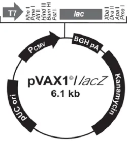
2.8. Cloning of AVP cDNA into pVAX1/lacZ ... 23
2.9. Competent cells ... 24
2.10. Transformation of competent cells with recombinant pVAX1/lacZ ... 25
2.11. Site – directed mutagenesis ... 26
2.12. Cloning of Frag3 and Frag CB ... 30
2.13. Expression of the normal AVP gene in Neuro 2A cells ... 31
2.14. Immunocytochemistry ... 32
3. Results ... 35
3.1. Reported mutations in the AVP gene ... 35
3.2. Identification of kindred with mutations ... 41
3.3. Identification of mutations in the AVP gene ... 41
x
3.5. Site-directed mutagenesis as a method to introduce desired mutations in AVP cDNA 463.6. Cloning of mutations in pVAX/AVP ... 48
3.7. Analysis of AVP WT gene expression in N2A cells ... 49
4. Discussion ... 51
5. Conclusion ... 53
xi
List of Figures
Fig. 1. Structural organization of the AVP gene and the protein vasopressin precursor ... 5
Fig. 2. Structural organization of the AVP pro-hormone ... 7
Fig. 3. Model of the regulation of water permeability in renal collecting duct cells ... 8
Fig. 4. Synthesis and processing of secretory proteins like AVP ... 11
Fig. 5. A proposed model for the molecular basis of adFNDI... 13
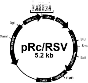
Fig. 6. Expression vector pRc/RSV ... 23
Fig. 7. Cloning vector ... 24
Fig. 8. AVP cDNA with primers used in site-directed mutagenesis for the first mutation (c.154T>C) ... 28
Fig. 9. AVP cDNA with primers used in site-directed mutagenesis for the second mutation (c.289C>G) ... 29
Fig. 10. Percentage of each type of protein change caused by the mutations in 104 reported families. ... 35
Fig. 11. Unique mutations described in the human AVP gene ... 41
Fig. 12. Pedigrees of three families with adFNDI. Index cases are marked by an arrow... 42
Fig. 13. Electrophoresis of PCR products for each AVP gene exon ... 43
Fig. 14. Electropherograms from fragments of the AVP gene of one healthy individual and three clinically affected subjects with novel identified mutations ... 44
Fig. 15. Analysis of purified pDNA after enzymatic digestion with XbaI ... 45
Fig. 16. Confirmation of AVP cDNA in the recombinant pVAX/AVP ... 45
Fig. 17. First PCR reactions for site directed mutagenesis for both mutations ... 46
Fig. 18. Subsequent PCR reactions for both mutations ... 47
Fig. 19. Electropherograms from site-directed mutagenesis fragments ... 47
Fig. 20. SmaI/FseI digestion of FragCB, Frag3 and pVAX/AVP ... 48
Fig. 21. Electropherograms from the sequences of the mutated pVAX/AVP vector ... 49
Fig. 22. Cellular localization of NPII protein in transiently transfected N2A cells as visualized by confocal laser microscopy ... 50
xii
List of Tables
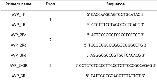
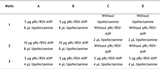
Table 1. Sequence of the primers used for amplification of AVP exons. ... 20 Table 2. Sequence of primers used for site-directed mutagenesis. ... 27 Table 3. Summary of trasfection assay conditions. ... 32 Table 4. Antibodies used for immunocytochemistry assays with dilutions and incubation times ... 33 Table 5. Immunocytochemistry assay. ... 34 Table 6. Reported mutations in the AVP gene causing FNDI ... 36
xiii
List of Acronyms
AC Adenylyl cyclase
adFNDI Autosomal dominant familial neurohypophyseal diabetes insipidus
AMP Adenosine monophosphate
AQP2 Aquaporin 2
AQP3 Aquaporin 3
AQP4 Aquaporin 4
AQP2-P Phosphorilated aquaporin 2
AVP Arginine vasopressin
bp Base pair
BSA Bovine serum albumin
cAMP cyclic
cDNA Complementary DNA
DDAVP Des-amino-D-arginin vasopressin
DI Diabetes insipidus
DMEM Dulbecco’s modified eagle’s medium
DNA Deoxyribonucleic acid
dNTP Deoxynucleotide
ddNTPs 2’,3’-dideoxyribonucleoside triphosphates
E. coli Escherichia coli
FBS Fetal bovine serum
Fig. Figure Frag1 Fragment 1 Frag2 Fragment 2 Frag3 Fragment 3 FragAB Fragment AB FragBB Fragment BB FragCB Fragment CB GP glycopeptide
HSP70 70 kilodalton heat shock proteins
kb Kilo bases
MRI Magnetic resonance image
N2A cells Neuro 2A cells
NPII Neurophysin II
PCR Polymerase chain reaction
pDNA Plasmid DNA
PKA Protein kinase A
xiv
SP Signal peptide
UV Ultra-violet
V2 Arginine vasopressin type 2 receptors
1
1. Introduction
1.1. Definition and classification of Diabetes Insipidus
Blood osmolality in healthy individuals is maintained within restricted limits by a series of complex mechanisms. Adjustments in water balance determine the level of that osmolality. These adjustments are mediated by delicate alterations in the thirst mechanism plus the capacity of the kidney to alter urine flow rate and its osmolality in response to small changes in the plasma concentration of the hormone arginine vasopressin (AVP) [1]. Thus, through all
these mechanisms, healthy humans can conserve their osmotic level despite extreme climatic conditions and, to a certain degree, when water supply is inadequate [1].
However, alterations in these mechanisms can occur and lead to one of two main states: inappropriate accumulation of water in organism, which is recognized as hypoosmolar states, and loss of renal water, which is recognized as hyperosmolar syndrome [1].
Diabetes insipidus (DI) is a rare disease and is characterized by excretion of abnormal large volumes, known as polyuria (>50mL/Kg/day) of dilute urine (<300mmol/Kg) [2, 3]. This
definition allows the exclusion of osmotic diuresis, which occurs when an excess of solute is being excreted, like in the case of glucose in patients with diabetes mellitus and this is the main difference between the two disorders [3]. This disorder can be acquired as a result of
various injuries or diseases, but can also be idiopathic or have a genetic origin [2].
In DI, the magnitude of the abnormality in concentration and excretion of the urine varies according to some factors like the severity of the defect which results in the disorder, the age of the patient, and the rate of solute and water intake [2].
Four basic defects are responsible for the development of DI. The first and the most common defect that occurs in this disorder is the deficient secretion of the hormone AVP, and in this case the DI is referred as neurohypophyseal, neurogenic, central or hypothalamic [3]. This
form of DI can be completely controlled by administration of AVP or its analogue, des-amino-D-arginin vasopressin (DDAVP) [4]. The second type of DI is caused by renal defects, where the
cells of the kidneys are insensitive to the antidiuretic effects of AVP and is referred as nephrogenic DI. In this case, the patients are unresponsive or poorly responsive to the administration of AVP or DDAVP [3, 4]. In both forms of disease, the thirst mechanism remains
normal to regulate water balance [1]. Another defect that causes DI is excessive water intake
(polydipsia) that leads to suppression of AVP release and consequent polyuria. This form of DI is called primary polydipsia and may be due to defects in the thirst mechanism or to cognitive impairment. Hormone supplements like AVP and DDAVP can reduce the symptoms of polyuria,
2
although these treatments should not be used because they can originate water intoxication, since they reduce polyuria more than polydipsia, which results in rapid retention of excess water and development of hyponatremia [1, 3, 4]. The fourth type of DI is due to increasedmetabolism of AVP during pregnancy resulting in gestational DI. This form of DI can be treated with DDAVP but is unresponsive to AVP. This occurs because the analogue of AVP is much less susceptible to degradation by placental vasopressinase [3, 4].
Depending on the cause that originates DI, the deficiency in vasopressin action or secretion can be partial or nearly total. Thus, the deficiency may or may not be associated with concentration of the urine after a fluid-deprivation test or in response to other strong stimuli like in the case of nausea, severe hypovolemia or severe hypotension [2].
Differentiating between the forms of DI is relatively easy if patients have severe deficiency in either the secretion or action of AVP. In both cases, the patients undergo dehydration induced by fluid deprivation, but the urine remains dilute [4]. This first result excludes the
possibility of primary polydipsia since in this form of disease, a fluid deprivation results in concentration of urine because the hormonal mechanism remains normal and the problem resides in excess of water intake. The next step to differentiate nephrogenic DI from neurohypophyseal and gestational DI is the injection of AVP and DDAVP and measurement of the urinary response [4]. Patients with nephrogenic DI do not respond to treatment since their
problem resides in renal insensitivity to AVP and not in hormonal deficiency and their urine remains dilute. However, patients with neurohypophyseal or gestational DI are able to concentrate their urine when AVP or DDAVP are administered because of the increased plasma levels of AVP. If fluid deprivation results in concentration of urine, other tests are necessary to differentiate between primary polydipsia and a less severe deficiency in the secretion or action of AVP [4]. The most reliable way to make this distinction is to measure
plasma AVP and to relate the results to the plasma and urine osmolality during a fluid deprivation and/or hypertonic saline infusion test [4].
However, with time the diagnosis becomes more complicated and the forms of the disease can be confused. After prolonged periods of polydipsia, a decrease in maximal urine-concentration ability occurs in the kidneys, regardless of the primary cause [5]. The passage of
large amounts of dilute urine through the distal nephron removes existent solutes from the renal medullary interstitium, a process known as washout phenomenon, and results in the decrease of osmotic gradient across the collecting tubular cells [1]. Since this gradient is
essential for the antidiuretic action of AVP, any mechanism responsible for DI may lead to an additional defect at the renal level that complicates the interpretation of diagnostic tests based on indirect analyses of the antidiuretic action of AVP [1].
3
1.2. Clinical aspects of Familial Neurohypophyseal Diabetes
Insipidus
AVP is synthesized in magnocellular neurons that originate in the supraoptic and paraventricular nuclei of the hypothalamus, which project down through the diaphragma sellae to form the neurohypophysis [3, 4]. In contrast to the adenohypophysis, the
neurohypophysis does not synthesize hormones but functions as a reservoir for the storage and release of hormones synthesized in the hypothalamus [6].
Destruction of magnocellular neurons results in a deficiency of AVP, leading to neurohypophyseal DI. This neuronal destruction can have a variety of causes, including trauma from surgery or accident, infections, autoimmune disease, congenital brain malformations, aneurysms, and others [6]. However, neurohypophyseal DI can also occur on an
inherited, or familial, basis representing 1% of all causes of neurohypophyseal DI [7]. Usually,
the disease has an autosomal dominant transmission, however in 1996 an X-linked recessive form was discovered [4].
There is another type of neurons that can produce and segregate AVP, known as parvocellular neurons [4]. The projections of these neurons are located in the median eminence of the
hypothalamus [4]. In some studies, it was observed that these projections are apparently
unaffected in patients with neurohypophyseal DI and this fact may explain the preservation of normal circadian rhythm and pituitary-adrenal function in these patients [4] because these
neurons also produce corticotrophin releasing factor, which is thought to interact with AVP in the regulation of adrenocorticotropic hormone secretion [8]. Since these neurons are not
affected by neurohypophyseal DI, it is believed that the two types of AVP-producing neurons have very different susceptibilities to the cytotoxic effects of the genetic mutations that lead to development of the disease [4].
Autosomal dominant familial neurohypophyseal DI (adFNDI) is a rare disease with persistent symptoms of polyuria, polydipsia and thirst which usually manifest several months or years after birth [9]. Studies performed with mice with AVP gene mutations revealed some
differences, when compared with mice with the normal AVP gene: mice with a mutated AVP gene consumed larger volumes of water, they excreted much more urine and the volume of urine excreted is worse over time, and their urine osmolality is lower [9]. Thus, like in humans
with adFNDI, mice with certain mutations produce excessive amounts of dilute urine but compensate by increasing water intake and so they can avoid severe dehydration and this fact demonstrates that thirst mechanisms remain intact in the presence of the disease [9]. Despite
these symptoms, adFNDI causes relatively few and well-tolerated symptoms. Nocturia (elimination of urine at night, disturbing sleep) is common and in children may present as
4
enuresis (urine during sleep) [4]. Physical exams and routine laboratorial analyses are usuallywithin normal limits and hypernatremia or signs of hypertonic dehydration are minimal or absent, except in the case of patients that are comatose, have an impaired thirst mechanism or if the patients are unable to increase fluid intake [4].
The symptoms of severe polyuria and polydipsia, which segregate in an autosomal-dominant pattern and respond to exogenous DDAVP, show several intriguing features. First, the affected family members show a completely normal water balance at birth and during early infancy but develop progressive symptoms of compulsive drinking at some point in childhood
[10]. Second, in some studies it was demonstrated that during repetitive fluid-deprivation
tests, the secretion of AVP is normal before the onset of the disease but then starts diminishing during early childhood [11]. Finally, once fully developed, the symptoms of
polyuria and polydipsia continue throughout life [10]. Occasionally, spontaneous remissions of
polyuria and polydipsia during middle-age are observed, even though the patients continue to have a deficient AVP secretion as severe as in their symptomatic kin. However, this remission mechanism remains unexplained [4].
Magnetic resonance image (MRI) exams have been used to investigate neurohypophysis anatomy in patients with adFNDI [4]. Some authors found that anterior and posterior lobes of a
normal pituitary gland have different signal intensities in images of magnetic resonance. The posterior lobe presents a well-defined oval or round area of hyperintensity in both normal and abnormal pituitary glands, although there are some variations in size and shape of the signal from one person to another [6]. Some patients with adFNDI lack the characteristic bright spot,
or high-intensity signal [4, 12], which is common in the posterior lobe of the pituitary in
52%-100% of healthy adults [6]. Signal intensity seems to be correlated closely with posterior lobe
function as it is suspected to result from neurovesicles in axon endings of AVP-producing neurons [7, 13]. If neurovesicles really are responsible for the bright spot, it is not clear why
oxytocin-containing vesicles or vesicles located in the hypothalamus do not cause a high-intense signal, maybe their concentration is insufficient [7]. Thus, the absence of the bright
spot in the posterior pituitary lobe could result from a neurotoxic accumulation of precursor proteins that consequently lead to cell death [7, 12, 13].
However, the significance of MRI results is uncertain because the exact cellular source of the signal is not yet known [4]. It is believed that the bright spot is absent in all patients with
neurohypophyseal DI due to destructive or unidentified pathological processes [6], however to
date relatively few patients with adFNDI have been studied and the results have been conflicting since in some affected individuals the bright spot has been observed [4]. A positive
bright spot in a patient with adFNDI can be caused by a defect in hormone release from the posterior pituitary leading to accumulation of neurovesicles and, thus, to a normal MRI instead of defects in intra-axonal transport or processing of proteins [7]. However, at present,
5
absence of AVP-producing neurons in the posterior pituitary [4]. Thus, it is important thatadditional MRI studies including adFNDI patients are performed to clarify the importance and significance of the high-intensity signal found in some people [4].
1.3. AVP gene and AVP processing
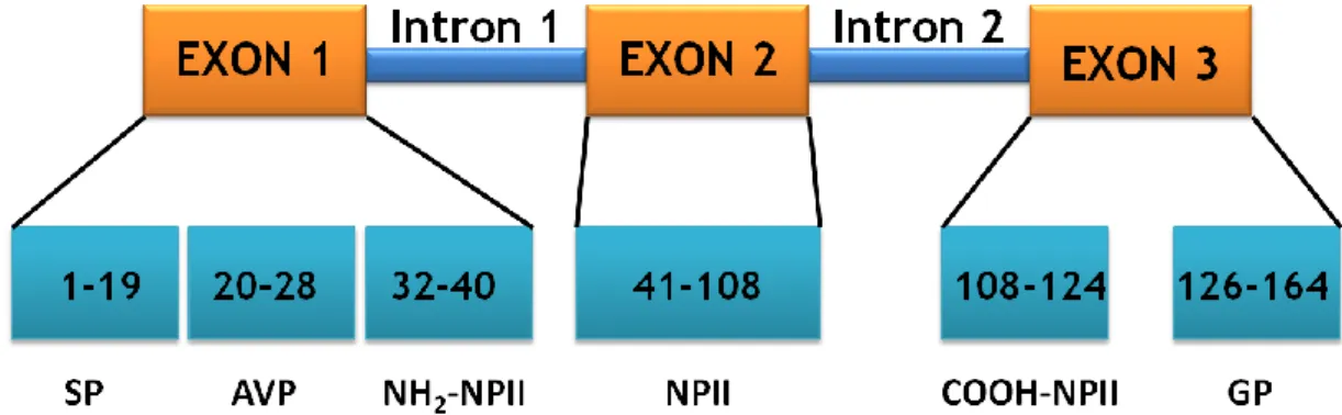
adFNDI is caused by mutations in one allele of the gene that encodes for AVP. The gene has approximately 3 kb (Gene ID: ENSG00000101200, Esemble) and is located on the short arm of chromosome 20 (20p13) [3]. It consists of 3 exons and 2 introns and encodes AVP and
neurophysin II (NPII), the carrier of AVP. The first exon encodes the signal peptide, the hormone AVP, and the NH2-terminal region of NPII [3]. The second exon encodes the highly
conserved central region of NPII and the third exon encodes the COOH-terminal region of NPII and the glycopeptide, which is known as copeptin [3, 14]. The small size of the AVP gene
facilitates the mutational analysis [3] and the study of the mutations at the protein level (Fig.
1).
Fig. 1. Structural organization of the AVP gene and the protein vasopressin precursor. The gene is
composed of 3 exons and 2 introns. The signal peptide contains 19 amino acids, AVP hormone contains 9 amino acids, NPII contains 93 amino acids and copeptin contains 39 amino acids. SP, signal peptide; AVP, arginine vasopressin; NPII, neurophysin II; GP, glycopeptide.
Several studies to analyze the expression of the AVP gene were performed using AVP transgenes derived from some animals. These results support the hypothesis that cell-specific enhancers and/or silencers that restrict expression of the AVP gene to specific neuronal cell-types in the hypothalamus are present in the regions either downstream or upstream of the AVP gene [15]. Recently, some authors demonstrated that DNA sequences in a 178 bp region
immediately downstream of exon 3 of the AVP are necessary for cell-specific expression of AVP in rat hypothalamus [16].
6
AVP and NPII are synthesized as a single precursor, prepro-vasopressin. The prepro-hormone presumably is translated on ribosomes in the cytosol and translocated across the membrane of the rough endoplasmic reticulum [4]. Once inside the endoplasmic reticulum, it is supposedthat the signal peptide remains attached noncovalently to the inner surface of the membrane via the positive charges at its N-terminal. This ligation is thought to facilitate accurate cleavage of the signal peptide by ensuring proper alignment with the signal peptidase [4].
However, the presence of certain small and neutral amino acids at the -1 and -3 positions immediately adjacent to the cleavage site of the signal peptide are required for efficient and accurate cleavage [4]. In this case, the signal peptide of AVP gene has an alanine and serine at
-1 and -3 positions, respectively [4]. The pro-hormone is generated by removal of the signal
peptide from the prepro-hormone and from by addition of a carbohydrate chain to the copeptin [3, 9]. There are no certainties that the glycosylation process and copeptin are
important for protein proper folding, trafficking or further processing, however, it seems possible that copeptin glycosylation plays an important role by assisting refolding of misfolded AVP pro-hormone monomers through its interaction with the calnexin-calreticulin system in the endoplasmic reticulum [17]. This system monitors protein folding and interacts principally
with the sugars of glycosylated proteins and places these proteins into the proximity of a glycoprotein-specific member of the protein disulfide isomerase family [17].
After removal of the signal peptide, the precursor generally must fold and dimerize correctly in the lumen of the endoplasmic reticulum [4], where the unique oxidizing environment allows
the formation of disulphide bridges [10], before they can proceed through the Golgi apparatus [4]. If folding is not correct, usually precursors are retained in the endoplasmic reticulum,
where they may be taken up by chaperones or heat shock proteins and degraded [4]. In vitro
studies suggest that the stability of folding is dependent on binding of the N-terminal of the hormone to a specific site located in the N-terminal of NPII [4]. Also, in the case of AVP and
NPII, like in others proteins, the correct folding of AVP-NPII in the endoplasmic reticulum probably also depends of the position of critically situated amino acids, like in the case of glycine or proline residues that enable the molecule either to rotate freely or to form a rigid bend. Cysteine positions are also very important because, under the action of a disulphide isomerase found in endoplasmic reticulum, they form specific disulphide bridges, which also serve to stabilize the molecule in the correctly folded conformation [4]. Binding of AVP to NPII
also facilitates self-association of the folded pro-hormone into dimmers which are then transported to the Golgi apparatus [4]. Here, final glycosylation takes place and the correctly
folded pro-hormones are finally packaged into dense granules which are transported along the axon to the posterior pituitary [4, 9]. The pro-hormone has different cleavage signal sequences.
Whereas the hormone is followed by a sequence of three residues, glycine-lysine-arginine, NPII is followed by a monobasic cleavage site, an arginine residue (Fig. 2) [10]. During axonal
transport, additional posttranslation processing occurs inside granules yielding AVP, NPII and the glycopeptides in separated forms [3]. This posttranslation process consists of two
7
successive cleavages; the first occurs between the hormone and the NPII by the action of a dibasic endopeptidase, and the second between NPII and copeptin by a monobasic endopeptidase [18]. Then, these molecules are stored within neurosecretory vesicles in thenerve terminals and released into the blood in response to osmotic stimuli [9]. Inside the
vesicles, reversible noncovalent interactions between AVP and NPII persist until these complexes are secreted into the bloodstream and they dissociate into free hormone and NPII
[4]. NPII can be seen as a chaperone-like molecule facilitating intracellular transport in
magnocellular cells [3], protecting AVP from proteolytic degradation during axonal transport of
the secretory granule to the posterior pituitary [13].
Fig. 2. Structural organization of the AVP pro-hormone. Each rectangle represents the individual
domains of the pro-hormone. The amino acid sequence of the hormone and the cleavage sites are represented.
AVP controls serum osmolality by altering renal water absorption. Its release is a calcium-mediated process of exocytosis when the axon is depolarized by an appropriate stimulus [2, 19],
which is determined mainly by the osmotic pressure of the plasma and extracellular fluid of the body [2]. There are specialized hypothalamic cells, called osmoregulatory neurons, which
mediate the secretion of the hormone by responding to extremely small alterations in the plasma concentration of sodium and other exogenous solutes [2]. The secretion of AVP is
stimulated by increases in serum osmolality, like in the case of hypernatremia, and by more pronounced decreases in extracellular fluid [3].
The antidiuretic function of AVP can be summarized in few steps. After AVP release into the systemic circulation, it binds to arginine vasopressin type 2 receptors (V2) on the basolateral
membrane of the collecting ducts cells of the kidneys, initiating a signal-transduction cascade
[3, 10]. The V
2 receptor is coupled to a Gs protein and when AVP is present, the V2 receptor
activates the α subunit of the G protein which stimulates the adenylyl cyclase leading to an increase in cyclic AMP (cAMP) inside collecting ducts cells and to the consequent activation of protein kinase A (PKA) [2, 3]. The activation of the cAMP-PKA pathway originates two related
mechanisms: it increases the expression of a specific water pore, known as aquaporin 2 (AQP2) [2, 20], and it leads to the phosporylation of homotetrameric AQP2, which results in the
fusion of AQP2-containing vesicles with the luminal membrane of these cells [2, 21]. When
8
cells and exit through the basolateral sides via different water channels, called aquaporin 3 and 4 (Fig. 3) [21].Fig. 3. Model of the regulation of water permeability in renal collecting duct cells. AVP binds to its
receptor (V2) which activates adenylyl cyclase (AC), increasing the cyclic AMP (cAMP) concentration.
This intermediate activates protein kinase A (PKA) which stimulates aquaporin 2 (AQP2) synthesis and its phosporylation, leading these transporters to the apical membrane in renal cells. AQP3, aquaporin 3; AQP4, aquaporin 4; AQP2-P, phosphorilated aquaporin 2.
The described process is the molecular basis of the vasopressin-induced increase in the water permeability of the apical membrane of the collecting tubule leading to a decrease in renal water excretion [3]. In the absence of AVP stimulation, the cells of the collecting duct remain
impermeable to water and the large volumes of diluted urine that enter the collecting tubules pass unmodified [2]. Thus, the excretion of urine reaches high rates and low
osmolarity [2].
AVP also increases the water reabsorption capacity of the kidney by regulating the urea transporter in the collecting duct and the permeability of principal collecting duct cells to sodium [20]. Thus, in the absence of AVP stimulation, the collecting duct cells have very low
permeability to water, sodium and urea, allowing the excretion of large volumes of hypotonic urine [10].
Some patients with adFNDI retain some limited capacity to secrete AVP during severe dehydration, however in most cases the deficiency of AVP secretion progresses and eventually becomes so severe that the organism can no longer concentrate urine, even during severe hypertonic dehydration [3, 4]. Symptoms of the disease usually appear after the first year of
9
receptors or in AQP2 and the symptoms are present during the first week of life [3]. In the firstyears of life, AVP deficiency can be partial and patients can concentrate their urine during a fluid deprivation test [4]. Thus, this result can lead to a misdiagnosis of primary polydipsia and
to a delay in effective treatment [4].
1.4. Genetic basis of adFNDI
Until now, adFNDI has been associated with several different mutations in the AVP gene and all, except two, are located in the coding region [10] (Human Gene Mutation Database). Most
of the mutations are single base substitutions, few are dinucleotide substitutions and the remaining are deletions of 1 or 3 nucleotides [10].
Although varied in location and nature, mutations appear to have several characteristics in common. The first similarity is that mutations appear to result in a similar clinical phenotype. Second, most of the mutations affect residues that are in hydrophilic regions of the molecule. Finally, all except one of the mutations are predicted to alter or remove one or more residues that are important for folding and self-association of the pro-hormone [4, 10, 22], changing its
primary structure. Production of an abnormal precursor caused by changes in its primary structure may be due to three types of mutations: those predicted to interfere with binding of the AVP and NPII, those predicted to alter the flexibility, rigidity and disulphide bridge formation of the pro-hormone and mutations predicted to encode a truncated NPII by introducing premature stop codons [4, 10]. Mutations that interfere with binding of AVP and
NPII can result from changes in the N-terminal of AVP, like mutations that impair or misdirect cleavage of the signal peptide or mutations that alter any of the first three amino acids of AVP, or can result from alterations in the shape of the NPII binding pocket [4]. The second
type of mutations is the most common, and these mutations modify pro-hormone characteristics by replacing, deleting or creating de novo a glycine, proline or cysteine residue [4]. Mutations that delete glycine residues would be expected to interfere with folding
of the pro-hormone due to a loss of flexibility at those sites, mutations that replace or create proline residues increase the rigidity of the molecule and mutations that replace or delete cysteine residues are likely to impair folding by eliminating or modifying one or more of the eight disulphide bridges that normally stabilize the pro-hormone into its proper conformation
[4, 13].
The mutations responsible for disease development are distributed throughout the precursor protein [12]. Several mutations modify the signal peptide but the substitution of a alanine for
threonine at position 19 (A19T) is the most common mutation described in adFNDI and has been found in several unrelated families around the world [10, 14]. This mutation is caused by a
single base substitution (guanine to adenine) in exon 1 [14] and gives rise to an aberrant
10
cleavage by the signal peptidase [9, 23]. In mouse studies, mice that had this mutation did notdevelop an apparent DI phenotype and the authors did not detect loss of AVP-producing neurons, even in homozygous mice. Thus, like in humans, this mutation originates a relatively mild phenotype in mice [9]. As for mutations in the NPII domain, a number of different
mutations have been identified, including missense mutations, nonsense mutations and a single amino acid deletion [9]. There is evidence suggesting that the age of onset of the
symptoms is lower in several kindreds with mutations in the NPII domain than in those with the A19T mutation in the signal peptide [4]. This fact can be explained because mutations
affecting the signal peptide cleavage site would be expected to allow the formation of some normal pro-hormone from the mutant alleles, whereas the NPII mutations would not [14].
Mutations that alter the AVP hormone were also found [10]. No mutations predicting changes in
the linker regions connecting the pro-hormone domains or in the copeptin domains have been identified, apart from the premature stop codons, which also truncate copeptin together with distal portions of the NPII domain [4, 10]. Recently, Hedrich et al. identified one variant in the
copeptin domain which predicts a replacement of guanine by adenosine. However, individuals carrying this nucleotide substitution alone do not show disease symptoms and authors concluded that this alteration seems to be a rare polymorphism and not a disease-causing mutation [13]. To date, the intronic mutation, found by Tae and colleagues [24], is the only
described mutation that does not occur in the exon regions of the AVP gene and is predicted to cause retention of intron 2 during mRNA splicing. This mutation causes a frameshift from position +1 of intron 2 and the introduction of a premature stop codon in exon 3 [24]. The
aberrant protein formed consists of 167 amino acids that lack the C-terminal of NPII due to a premature codon insertion, whereas the protein translated by normal mRNA sequence of AVP gene consists of 164 amino acids that include signal peptide, the hormone AVP, NPII and copeptin [24].
All of the mutations described seem to be completely penetrant, although a few mutations might not result in appearance of adFNDI until late adolescence [2].
1.5. Pathogenesis of adFNDI
The pathogenesis of adFNDI has been studied in different model systems during the past few years [10]. The main points of the disease are: the disease is associated with mutations in one
allele of the AVP gene and is caused by postnatal development of deficient AVP secretion proposed to result from selective degeneration of the magnocellular neurons that produce the hormone in normal conditions [10].
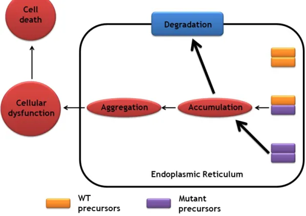
Theoretically, mutations that affect the folding of secretory proteins result in loss-of-function phenotypes due to their direct impact on protein function because these mutant proteins are prevented from reaching their final destination [3]. Thus, mutant proteins that fail to fold
11
correctly are retained initially in the endoplasmic reticulum [3], as this organelle has theability to recognize, retain and degrade misfolded, incompletely folded or partially assembled copies of the proteins in a mechanism known as endoplasmic reticulum quality control [25],
and subsequently the proteins are degraded either by proteasomes or by another degradation mechanism (Fig. 4) [3]. However, why AVP misfolded mutants are cytotoxic to AVP-producing
neurons is a question without answer, for now [3].
Fig. 4. Synthesis and processing of secretory proteins like AVP. mRNA and respective ribosomes migrate
to endoplasmic reticulum. Then, ribosomes attach to endoplasmic reticulum by a signal recognition peptide (SRP) and the SRP receptor (SR). The growing peptide passes through the membrane via a translocon (TR). Proteins with the correct fold are stored in vesicles which will proceed to the Golgi apparatus. Misfolded proteins are initially retained in the endoplasmic reticulum, but then they are translocated to the cytosol and degraded by proteasomes. C, vesicle coat protein. Adapted from [3].
Mutant hormone precursors that do not fold and self-associate correctly probably do not move from the endoplasmic reticulum to the Golgi apparatus and to neurosecretory granules, finally, where processing mechanisms that leads to NPII, copeptin and active AVP normally occur [4]. The block in trafficking and processing of the precursor could completely eliminate
AVP production from the mutant allele [4]. However, the other allele remains normal and a
simple block in processing of the mutant allele would be insufficient to cause the clinical symptoms that patients with adFNDI develop, especially because the deficiency of AVP secretion is much greater than 50% [4]. This means that the mutations also interfere with the
12
mechanism can occur at any level like in transcription, translation, precursor processing and in molecule secretion [4]. Based on some studies, the processing and secretion of mutantprecursors are delayed relative to processing of the wild type (WT) precursors [12] and this
fact can explain the accumulation of the mutant precursor in the endoplasmic reticulum. Evidence that proves the existence of misfolded proteins in the endoplasmic reticulum is the induction of a molecular chaperone called BiP, a member of the 70 kilodalton heat shock proteins (HSP70) family [9]. This chaperone binds to misfolded proteins whose transport from
the endoplasmic reticulum is blocked and BiP expression is increased as part of the unfolded protein response [9].
There are at least two mechanisms by which retention of misfolded mutant precursors in the endoplasmic reticulum could impair production of AVP from the normal allele [4]. In the first
place, there is a ‘nontoxic’ mechanism when the mutant precursor is expressed at the same time as the WT precursor leading to the association of both precursors to form abnormal heteroligomers [4]. Thus, the mutant precursor impairs the trafficking of the WT precursor and
both precursors are retained in the endoplasmic reticulum where they can be degraded or otherwise eliminated by the cell, leading to a decrease in protein activity of the WT precursors [4, 26]. With time and the high rate of mutant precursors/dimers accumulation in
endoplasmic reticulum, together with the rapid degradation by the cytosolic proteasome of these heterodimers, this mechanism could easily result in the development of a severe AVP deficiency, even though the normal allele remains to be expressed at its usual rate [4, 10]. The
formation of heterodimers and homodimers between mutant and WT AVP pro-hormones was already shown, such as the impairment of WT precursor trafficking by the mutant precursor during heterologous expression in cell cultures [26], resulting in formation of abnormally
configured heterodimers that are retained in the endoplasmic reticulum [10].
However, the nontoxic mechanism does not explain the autopsy evidence for selective degeneration of AVP-producing magnocellular neurons [4]. Thus, it is postulated that the
continuous accumulation of unfolded or misfolded mutant precursors in the endoplasmic reticulum prevents expression of the normal allele by interfering with the production of essential proteins that are important for survival of these neurons leading to a toxic mechanism [4]. However, there is little evidence of cell death caused by apoptosis, suggesting
that it may occur by other pathways [9]. Some studies using immunohistochemical analyses to
detect cell death of AVP-producing neurons were negative to apoptosis by using apoptosis markers [9]. But, this observation does not mean that apoptosis really does not occur in these
neurons because given the small number of AVP-producing neurons and the progressive loss of cells over weeks to months, these assays may not be sensitive enough to detect apoptosis of a small number of neurons [9].
The nontoxic and the toxic theories are not mutually exclusive and together they could explain some facts like the delayed onset of the disease and its occurrence despite the
13
presence of a normal allele [9]. On the other hand, these two mechanisms can representdifferent phases of the same pathologic process (Fig. 5) [4].
Fig. 5. A proposed model for the molecular basis of adFNDI. Functional and physical interactions occur
between WT and mutated precursor that are retained in endoplasmic reticulum, impairing the transport and processing of WT. These aggregates will lead to cellular toxicity and to posterior cell death. Adapted from [26].
The hypothesis of toxicity caused by misfolded proteins (misfolding-neurotoxicity hypothesis) is consistent with all clinical, hormonal and biochemical existent data [4]. However, it is based
on several factors that remain to be tested and validated and they include the following: the disease is always linked to a mutant AVP gene that originates a mutant precursor that does not fold and self-associate like the WT precursor and is not carried from the endoplasmic reticulum to the Golgi apparatus; the endoplasmic reticulum retention results in lethal accumulation and/or aggregation of mutant precursors; the decrease in AVP secretion is associated with selective degeneration of the producing magnocellular neurons; and AVP-producing parvocellular neurons are not affected by mutations that cause death in magnocellular neurons due to a much lower rate of AVP production by these cells, and/or more robust elimination mechanisms to eliminate misfolded proteins [4, 10].
Recently, new studies suggested a new mechanism that explains the pathogenesis of adFNDI. Castino et al. [27] have shown that some mutations result in accumulation of mutated
14
precursores in the endoplasmic reticulum, forming insoluble aggregates [27]. This accumulationresults in the development of a pathology characterized by a grossly deranged endoplasmic reticulum which contains both mutated and WT protein [27]. With the aid of morphological
observations, Davies et al. suggest that these structures represent autophagic vesicles [28].
Autophagy results in organelle destruction together with the WT AVP, resulting in a progressive AVP deficiency [27]. Under these circumstances, autophagy is a cell survival
mechanism that removes the deranged structures [27]. However, the cells are continuously
exposed to environmental and metabolic insults that can lead the cell to an autophagy-dependent apoptosis once the neurons are frail, already undergoing autophagy in order to clear mutant proteins [29]. This hypothesis does not exclude the misfolded-neurotoxicity
hypothesis as, although authophagy may be responsible for the initiation of adFNDI’s symptoms, it does not exclude the possibility that degeneration of the AVP-producing magnocellular neurons can be a long-term consequence [10].
The accumulation and cellular death caused by cytotoxicity of mutated precursors is a slow and prolonged process, which explains some facts like the progressive onset of the symptoms of the disease and the AVP deficiency [3, 14]. Autopsy studies performed in adFNDI patients
show a selective loss of AVP-producing magnocellular neurons in the supraoptic and, to a lesser extent, in the paraventricular nucleus along with loss of their axonal extensions into the neurohypophysis [4, 30]. These studies also show atrophy of the neurohypophysis and gliosis [4, 30].
Some authors suggest that cell survival depends on its efficiency to degrade unfolded or incompletely folded proteins. Thus, degradation-resistant proteins that accumulate in the endoplasmic reticulum cause a more profound cytotoxic effect than proteins that are not resistant to the degradation process [12].
It is difficult to determine significant differences in the severity of the disease produced by the various AVP gene mutations [14]. The number of patients available for careful evaluation is
very limited and there is a high degree of variability, even for patients of the same family, like the debut of symptoms, severity of polyuria and the degree of AVP deficiency [14]. Thus,
these factors result in a lack of genotype-phenotype correlation which could help to determine the best treatment for the patients.
1.6. Diagnosis and treatment
The clinical diagnosis of DI can be made easily by measuring urine osmolality during a fluid-deprivation test, at least when the disease is present in its complete form, as described above [10]. However, with the development of knowledge related with the disease, some
15
performed in all patients with familial occurrence of DI symptoms [10]. Once the moleculardiagnosis is established in adFNDI kindreds, it is easier to screen other family members for the same mutations [10]. This fact has particular importance in infants at risk of inheriting the
mutation as this method allows the presymptomatic diagnosis, relieving years of parental concern about the evolution of the disease in their offspring [10]. As adFNDI presents very few
symptoms and allows a normal quality of life, at least when offered an appropriable treatment, and because there is little evidence for an associated risk of severe central nervous system sequelae [31] compared with nephrogenic DI, a prenatal diagnosis seems not to
be indicated [10].
The treatment of adFNDI is relatively simple as the administration of the AVP analogue, DDAVP, 2 to 3 times daily eliminates symptoms [32]. Patients with adFNDI have preservation of
the osmoregulation of thirst, thus only minor fluctuations in plasma osmolality are seen even during irregular pharmacological treatment and the risk of inducing hyponatremia is very small in these patients [10]. To date, no other V
2 receptor agonist has been introduced in the
treatment of adFNDI but several delivery methods have been investigated and they are available in nasal sprays, in common tablets and more recently in sublingual instant melting tablet [10, 33]. However, it remains unknown if each delivery method results in better control
of polyuria and polydipsia or if it is only a matter of preference [10].
In an ideal perspective, the treatment of adFNDI should be able to provide a long-lasting antidiuretic effect with the possibility to provide escape in case of higher-than-required fluid intake, like in case of social reasons. This treatment can be obtained with gene therapy which provides constantly high levels of AVP through the expression of the AVP gene contained in a viral vector [10]. Many studies have shown the efficiency of gene therapy in AVP-deficient rats
using electroporation [34]. The next step in this treatment is the escape from the constant
antidiuresis induced by gene therapy and this can be achieved using the recently developed V2 receptor antagonists [34]. However, there are several diverging opinions relative to the
safety of such viral approaches [10] and a further work is needed to clarify all the questions
around gene therapy.
1.7. Future perspectives
adFNDI is a disease with low morbidity and an effective treatment but, despite its benign nature, the disease has been subject of intense research. This fact occurs due to its potential value as a model for studies of neuroendocrinological diseases and for studies of dominant negative mutations and due to its importance in the understanding of the effects of such mutations on the folding of hormone precursors and the role of the protein quality control machinery in the cellular handling of misfolded protein [35].
16
Russel et al. [9] proposed that adFNDI could be considered a neurodegenerative disorder likeAlzheimer disease, Parkinson disease and others [9]. This suggestion is due to accumulation of
cytotoxic precursors inside neuronal cells in adFNDI, as in the above diseases, leading to the posterior death of the cell.
A possible therapeutic approach to diseases caused by accumulation of misfolded proteins inside the endoplasmic reticulum can be the use of pharmacologic chaperones to promote the escape of proteins from this organelle [3]. Thus, the proteins can proceed their transport to its
target cells. In this case, without trafficking impairment, the mutant proteins could be sufficiently functional if the problem resided in the transport of the proteins [3].
It is very important to proceed with genetic and molecular studies of adFNDI as the results can give answers not only about adFNDI, but also help to explain other diseases that have the same molecular mechanism like the case of neurodegenerative diseases or other pathologies that involve protein misfolding or aggregates. On the other hand, it is necessary that patients are informed about their state more deeply, principally in case of genetic diseases that are transmitted through several generations.
1.8. Aims of the thesis
The present study is based on three main aims. First, to review AVP mutations described in the scientific literature. Second, to expand the spectrum of mutations through the analysis of additional patients with DI. Third, and last, to characterize the functional consequences of identified novel AVP mutations.
17
2. Methods
2.1. Literature search of AVP gene mutations
A database of the described and published AVP gene mutations was constructed by searching the National Center Of Biotechnology Information Pubmed literature ~database for articles in English, using the keywords AVP, mutation and Neurohypophyseal Diabetes Insipidus.
A total of 61 articles that described 70 different mutations were identified and evaluated. The most relevant information was analyzed and a new nomenclature was assigned to each mutation, based on recommendations from the authors Dunnen and Antonarakis [36]. Beyond
the alteration in AVP cDNA, others changes were also taken into account like the exon in which mutations occur, the alterations caused at the protein level (amino acid changes), protein domain, the population and the existence of functional studies.
2.2. Subjects and clinical procedures
A total of 9 patients diagnosed with neurohypophyseal DI, consisting of 3 familial cases and 6 sporadic cases, gave their informed consent for genetic studies of their AVP gene, in order to identify possible mutations which could be responsible for their disease. Diagnosis of patients was performed at the Endocrinology, Diabetes and Metabolism Service (University Hospital from Coimbra, Portugal) and was based on a fluid deprivation test followed by DDAVP administration.
The present study was approved by the Ethics Committee of the Faculty of Health Sciences at the University of Beira Interior.
2.3. DNA extraction
When a blood sample is collected to perform molecular analysis, like the identification of genetic mutations, the first step in laboratorial procedure is DNA extraction. The method chosen for DNA extraction from peripheral blood is very important as it is necessary to obtain a highly purified DNA without fragmentation. Some points are very important when a particular technique is chosen like technical requirements, the time required to develop the protocol, the efficiency of the method and its monetary cost [37]. Several methods are used
to extract DNA, including the use of organic solvents, but the contamination with proteins is a frequent problem [38]. Miller et al. published, in 1987, a new method to extract DNA that