Pedro Miguel Oliveira Soares
Determination of the importance of iron
acquisition for
Staphylococcus epidermidis
biofilm survival in human blood
Pedro Miguel Oliveira Soares
Determination of the importance of iron
acquisition for
Staphylococcus epidermidis
biofilm survival in human blood
Dissertação de Mestrado
Mestrado em Bioengenharia
Trabalho efetuado sob a orientação de
Doutor Nuno Cerca e coorientação de
Doutora Ângela França
iii
DECLARAÇÃO
Nome: Pedro Miguel Oliveira Soares
Endereço eletrónico: pedro.mo.soares@gmail.com
Título da dissertação: Determination of the importance of iron acquisition for Staphylococcus epidermidis biofilm survival in human blood
Orientador:
Doutor Nuno Miguel Dias Cerca Co-orientadora:
Doutora Ângela Maria Oliveira de Sousa França
Ano de conclusão: 2015 Mestrado em Bioengenharia
É AUTORIZADA A REPRODUÇÃO INTEGRAL DESTA DISSERTAÇÃO APENAS PARA EFEITOS DE INVESTIGAÇÃO, MEDIANTE DECLARAÇÃO ESCRITA DO INTERESSADO, QUE A TAL SE COMPROMETE.
Universidade do Minho, _____/_____/_________
v
A
CKNOWLEDGEMENTS
/
ACKNOWLEGDEMENTS/AGRADECIMENTOS
vii
Durante o desenvolvimento desta tese de mestrado, diversas pessoas contribuíram direta ou indiretamente para que esta etapa da minha formação fosse concluída. É a estas pessoas que quero agradecer pelo apoio demonstrado.
Em primeiro lugar, agradeço aos meus orientadores, ao Doutor Nuno Cerca por me ter aceitado neste projeto, pela partilha de conhecimento e orientação ao longo de todo o trabalho realizado, e à Doutora Ângela França pela disponibilidade para ajudar demonstrada desde o primeiro momento, seja para esclarecer dúvidas ou acompanhar em algumas das experiências realizadas. Aos dois, o meu sincero obrigado!
Aos meus colegas da PBMS pelo bom ambiente de trabalho e fácil integração neste laboratório de investigação. Aos meus colegas sob orientação do Doutor Nuno Cerca, agradeço todas as críticas construtivas durante a apresentação do trabalho realizado nas diversas reuniões de grupo.
Aos meus amigos e colegas de mestrado, Carlos, Sara, Paulina, Andreia, Rita e Joana pelo apoio em momentos de stress e momentos de boa disposição durante os longos dias passados no DEB. Aos meus amigos de sempre, em especial ao Rui, Veloso e Maria João pelos conselhos, partilha de experiências e momentos de boa disposição.
Agradeço à Marta, por todo o apoio, carinho e pela paciência para ouvir todos os meus problemas ao longo desta etapa.
Finalmente, à minha família, aos meus pais e irmão, pelo suporte que foram durante todos os anos da minha vida e por me tentarem sempre mostrar qual o melhor caminho a seguir.
ACKNOWLEGDEMENTS/AGRADECIMENTOS
viii
Financial support
This thesis was supported by European Union funds (FEDER/COMPETE) and by national funds (FCT) under the project with reference FCOMP-01-0124-FEDER-041246 (EXPL/BIA-MIC/0101/2013) and by the project RECI/BBB-EBI/0179/2012 (FCOMP-01-0124-FEDER-027462).
ix
ABSTRACT
xi
Nosocomial infections are a worldwide concern due to their impact on patients’ health and costs to the health care system. The majority of these infections are associated with the use of indwelling medical devices, which serve as scaffold for biofilm formation by bacteria or other microorganisms. Due to its remarkable capability to form biofilms on medical devices, Staphylococcus epidermidis, a commensal bacterium of healthy human skin and mucosae, has emerged as one of major causes of medical devices-associated infections, being particularly associated with vascular catheters. As a consequence, S. epidermidis biofilms are frequently associated with the emergence of bloodstream infections. Nevertheless, the interplay between S. epidermidis biofilms and human blood is poorly understood.
Recently it was reported that S. epidermidis biofilms increase the transcription of genes associated with iron utilization when in contact with human blood. Iron is an important element for bacterial growth and low free-iron environments, like human blood, may be detrimental. Hence, in order to determine the importance of iron utilization for S. epidermidis, we have assessed the influence of different iron concentrations on the bacterium growth rate and biofilm formation capacity. In addition, the susceptibility of S. epidermidis biofilms to humans’ blood bactericidal activity and the quantification of the transcription of genes involved in iron detoxification (hssR and hrtA genes) were also addressed.
The results obtained showed that the increasing iron concentrations tested had no significantly effect on S. epidermidis growth rate. In contrast, two of the four strains used showed increased biofilm formation capacity in the presence of high concentration of iron (500 µM FeCl3). Interestingly, 32 to 73%
of the biofilm cells were able to survive the exposure to human blood. Nevertheless, the transcription of hssR gene was only found significantly increased in one of the strains used (clinical isolate PT11003). Although it was not possible to detect the hrtA gene, it is probably being transcribed since the activation of the hrtAB operon is hssR-dependent. In the future, iron-deprivation experiments should be performed in order to better understand iron utilization in S. epidermidis biofilms.
RESUMO
xii
Nos últimos anos, as infeções nosocomiais têm sido uma fonte crescente de preocupação devido ao seu elevado impacto na qualidade de vida dos pacientes e no orçamento do sistema de saúde. A maioria destas infeções está associada ao uso de dispositivos médicos invasivos, nos quais bactérias e outros microrganismos são capazes de formar biofilmes. Um dos principais agentes etiológicos das infeções associadas ao uso de dispositivos médicos é a bactéria Staphylococcus epidermidis, habitual colonizadora da pele e mucosas humanas saudáveis, que se destaca pela sua notável capacidade para formar biofilme neste tipo de superfícies, particularmente em cateteres vasculares.
Uma descoberta recente reportou um aumento na transcrição de genes envolvidos na utilização de ferro por biofilmes de S. epidermidis em contacto com sangue humano. O ferro é um elemento essencial para a sobrevivência e crescimento de microrganismos, e ambientes com quantidades mínimas de ferro disponível na sua forma livre, como o sangue humano, levantam problemas à sobrevivência do microrganismo. Com base nestes factos, e para perceber quão determinante é a aquisição de ferro para a sobrevivência de S. epidermidis, foi analisado o efeito de diferentes concentrações de ferro na taxa de crescimento e formação de biofilme, assim como a quantificação da suscetibilidade dos biofilmes de S. epidermidis ao contacto com sangue humano e da transcrição de genes envolvidos na utilização de ferro (hssR e hrtA).
Os resultados obtidos revelaram que as crescentes concentrações de ferro testadas não causaram efeitos significativos na taxa de crescimento de S. epidermidis. Contudo, em duas das estirpes testadas verificou-se, na maior concentração de ferro (500 µM FeCl3), um aumento significativo da formação de
biofilme. Após a incubação com sangue humano, 32 a 73 % das células foram capazes de sobreviver, tendo também sido registado um aumento significativo da expressão do gene hssR numa das estirpes (PT11003). Apesar de não ter sido possível detetar o gene hrtA é provável que este esteja a ser expresso, uma vez que o aumento da expressão do hssR promove a transcrição do operão hrtAB. Futuramente, e para perceber melhor o metabolismo do ferro em biofilmes de S. epidermidis, experiências com quantidades residuais ou mesmo na ausência de ferro deverão ser efetuadas.
Palavras-chave: Staphylococcus epidermidis, Biofilmes, Ferro, Sangue humano, Expressão genética.
INDEX
xiii
I
NDEXAcknowledgements/ Agradecimentos ... v
Abstract/Resumo ... ix
List of figures ... xvii
List of tables ... xix
List of abbreviations ... xx 1. Chapter 1 – Introduction ... 1 1.1 Biofilm-associated infections ... 3 1.2 Staphylococcus epidermidis ... 4 1.3 S. epidermidis biofilms ... 5 1.3.1 Attachment ... 5 1.3.2 Biofilm maturation ... 6 1.3.3 Biofilm detachment... 8
1.4 S. epidermidis biofilms interaction with human blood ... 10
1.5 Main objectives of this work ... 12
2. Chapter 2 – Materials and Methods ... 13
2.1 Bacteria ... 15
2.2 The effect of iron on planktonic cells growth ... 15
2.3 The effect of iron on biofilm formation... 15
2.4 Blood collection ... 16
2.5 The effect of human blood on S. epidermidis biofilms ... 16
2.6 PCR ... 16
2.7 Gene expression quantification ... 18
2.7.1 RNA extraction ... 18
2.7.2 DNase treatment and total RNA quantity and purity ... 18
2.7.3 complementary DNA synthesis ... 19
2.7.4 Quantitative PCR... 19
INDEX
xiv
3. Chapter 3 – Results and Discussion ... 21
3.1 Effect of iron on S. epidermidis planktonic cells growth rate... 23
3.2 Effect of iron on S. epidermidis biofilm formation ... 24
3.3 S. epidermidis biofilm cells cultivability upon interaction with human blood... 26
3.4 Gene expression quantification ... 27
4. Chapter 4 – Conclusions and future approaches ... 31
5. Chapter 5 – Bibliography ... 35
xv
L
IST OF FIGURES
,
TABLES AND
LIST OF FIGURES, TABLES AND ABBREVIATIONS
xvii
L
IST OF FIGURES
Chapter 1 - Introduction
Figure 1 – Biofilm development in S. epidermidis. Biofilm development can be divided into initial
attachment, maturation and detachment. Attachment can occur in abiotic or biotic surfaces, each one with different factors involved. Cells start to divide producing exopolysaccharides (such as PNAG) and adhesion proteins (such as Bhp and Aap), which constitute, among other molecules, the biofilm matrix and promote intracellular aggregation. Disruptive forces are involved in biofilm structuring, channel formation and detachment. Figure withdrawal of Otto 2012 (Otto, 2012). ... 5
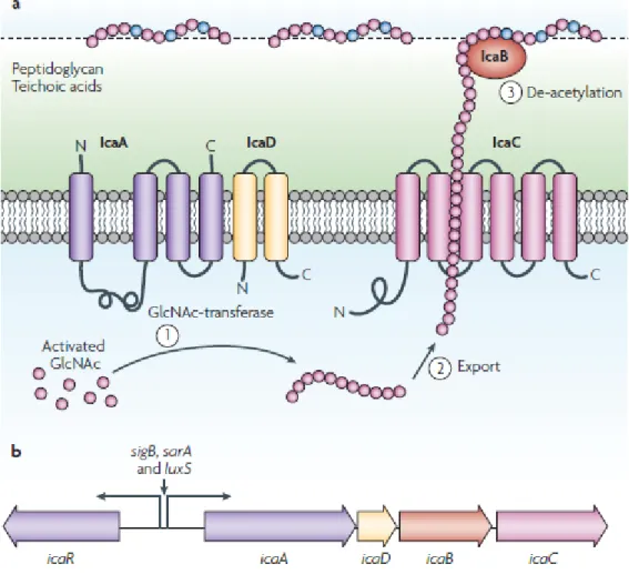
Figure 2 – The exopolysaccharide poly-N-acetylglucosamine (PNAG). a| Process of synthesis,
exportation and modification of PNAG and the products of ica operon involved. b| The ica operon, that encodes the Ica proteins, is constituted by icaA, icaD, icaB and icaC genes. Figure withdrawal of Otto 2009 (Otto, 2009). ... 7
Figure 3 – Hemin sensing by the S. aureus HssRS two-component system. 1| HssS senses
hemin through a direct binding or an indirect mechanism. 2| Autophosphorylation of HssS at histidine 249 triggered by HssS activation. 3| HssS phosphorylates HssR by transferring the phosphate group to the aspartate 52 of HssR. 4| After phosphorylation, HssR binds to a repeat DNA sequence within the hrtAB promotor. 5| Increased transcription of hrtAB, resulting in increased production of HrtAB efflux pump. 6| HrtAB reduces stress associated with the accumulation of hemin inside bacterium by exporting excess cytosolic hemin or a by-product of hemin-mediated toxicity. Figure withdrawal from Stauff et al. 2007 (Stauff et al., 2007). ... 11
Chapter 3 - Results and Discussion
Figure 4 –S. epidermidis biofilm formation after 24 hours of incubation with TSB (control) or TSB supplemented with different concentrations of FeCl3 (5, 50 or 500 µM). The values
represent the average and the bars the standard deviation of, at least, 3 independent experiments. Statistical differences between the different concentrations of iron and the control were determined using
LIST OF FIGURES, TABLES AND ABBREVIATIONS
xviii
Multiple T-test. * indicates statistical significance (p = 8.84E-06 for 10B and p = 7.61E-08 for PT11003). ... 25
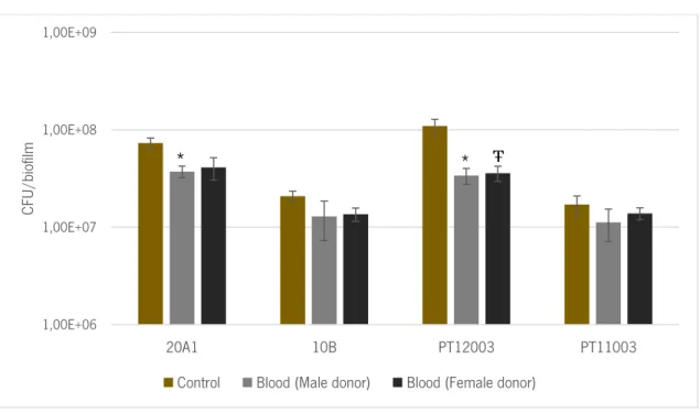
Figure 5 – The number of colony forming units (CFU per biofilm after 2 hours of incubation with TSB (control) or human blood. The values represent the average and the bars the standard deviation of 6 independent experiments. Statistical differences between both conditions were determined using Multiple T-test. * indicates statistical significance between control and the values obtained when using male donors (p = 0.039 and p = 0.016 for 20A1 and PT12003, respectively). Ŧ indicates statistical difference between control and the values obtained when using female donors (p = 0.018 for PT12003). ... 26
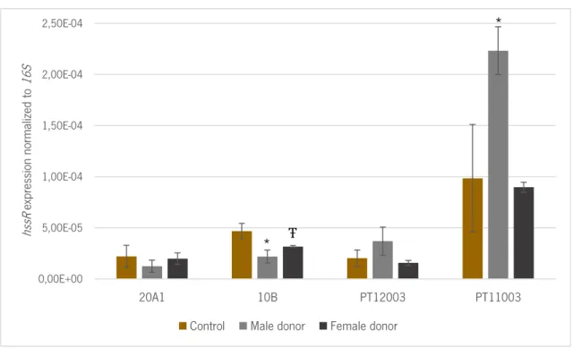
Figure 6 – The change in the expression of hssR gene after 2 hours of incubation with blood from male and female donors, as compared with biofilms incubated with TSB. The values
represent the average and the bars de standard deviation of 2 independent experiments. Statistical differences between both conditions were assessed using Multiple T-test. * indicates statistical significance between control and values obtained when using male donors (p = 0.003 and p = 0.020 for 10B and PT11003, respectively). Ŧ indicates statistical significance between control and values obtained when used female donors (p = 0.044 for 10B). ... 29
Appendix
Figure A1 – Amplification of hssR (A) and hrtA (B) by qPCR using cDNA from a blood sample of S. epidermidis 20A1, showing that hrtA has not been amplified, and consequently, not detected. ... 47
LIST OF FIGURES, TABLES AND ABBREVIATIONS
xix
L
IST OF TABLES
Chapter 2 – Materials and methods
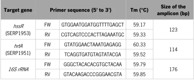
Table 1 – Primers used for hssR, hrtA and 16S rRNA detection by PCR and quantification by qPCR. The set of primers presented were designed using the genome of S. epidermidis strain RP62A (GenBank accession number: NC_002976.3).Tm - melting temperature, bp - base pairs. ... 17
Chapter 3 - Results and discussion
Table 2 – Doubling time (expressed in hours) of S. epidermidis isolates after planktonic growth in TSB (control) and TSB supplemented with different concentrations of FeCl3 (5, 50 or 500 µM). Statistical differences between commensal and
clinical isolates were accessed by Multiple t-test. * indicates statistical difference between doubling time of commensal isolates (p = 3.9E-05). ... 23
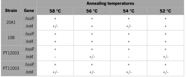
Table 3 – PCR results for hssR and hrtA amplification at different annealing temperatures. Symbols indicate the presence (+), absence (-) or poorly defined (+/-) bands in
agarose gel. ... 28
Table 4 – qPCR reaction efficiency with both hssR and hrtA primers at different annealing temperatures. (ND – non-detected). ... 28
LIST OF FIGURES, TABLES AND ABBREVIATIONS
xx
L
IST OF ABBREVIATIONS
Aap – accumulation-associated protein Agr – accessory gene regulator
AMP’s – antimicrobial peptides Bhp – biofilm homologue protein
CoNS – coagulase-negative staphylococci eDNA – extracellular DNA
Embp – extracellular matrix binding protein FeCl3 – Iron (III) chloride
Fnbp – fibrinogen-binding protein gDNA – genomic DNA
HrtAB – heme-regulated transporter efflux pump HssRS – heme-sensor system
ICU – intensive care units
MSCRAMMs – microbial surface components recognizing adhesive matrix molecules NaCl – Sodium chloride
OD – optical density
PCR – polymerase chain reaction
PIA – polysaccharide intercellular adhesion PNAG – poly-N-acetylglucosamine
PSM’s – phenol-soluble modulins
qPCR – quantitative polymerase chain reaction TSA – tryptic soy agar
TSB – tryptic soy broth
1
1.
C
HAPTER
1
CHAPTER 1–INTRODUCTION
3
1.1 Biofilm-associated infections
Nosocomial infections are infections acquired in healthcare facilities during the time that a patient is admitted in the hospital and until discharge, more accurately, an infection that was not present or incubating at the moment of patient admission at a hospital. These hospital-acquired infections represent a worldwide concern since they affect 5 to 15 % of patients during their stay in the hospital (Eggimann and Pittet, 2001), and it is recognized that intensive care units (ICU) patients are 5 to 10 times more susceptible than other hospitalized patients, due to their severe health condition and the use of several invasive medical devices (Weber et al., 1999).
Indwelling medical devices are frequently colonized by microorganisms that form biofilms on their surface being this association responsible for 60 % of nosocomial infections diagnosed (Darouiche, 2004). Biofilms are bacterial surface-attached communities involved by a self-produced and hydrated extracellular matrix (Costerton et al., 1999). Its function is to protect bacteria from external stresses (Costerton et al., 1999; Otto, 2012) being able to protect bacteria from chemicals such as antibiotics (Hoiby et al., 2010), which are very important in the treatment of bacterial infections. Indwelling medical devices like catheters, artificial heart valves or bone and joint prostheses are frequently affected by biofilm-forming microorganisms (Otto, 2012). The microorganisms recurrently associated with biofilm related infections are bacteria such as Enterococcus faecalis, Staphylococcus aureus, Staphylococcus epidermidis, Streptococcus viridans, Escherichia coli, Klebsiella pneumoniae, Proteus mirabilis, Pseudomonas aeruginosa, as well as some yeasts, like Candida spp (Davey and O’Toole, 2000). In addition, the infections caused by these microorganisms have become more severe due to biofilm increased tolerance to antibiotics (Guggenbichler et al., 2011). The mechanisms that provides tolerance to antibiotics in biofilms are not the traditional mechanisms of antibiotic resistance (Anderl et al., 2000; Stewart and Costerton, 2001). Indeed, a bacterium sensitive to a particular antibiotic can increase their tolerance to the same antibiotic up to 100-1000 fold when in biofilm (Hoiby et al., 2010). There are multiple hypotheses for antibiotic tolerance in biofilms including: 1) diffusional limitations of antibiotics in biofilms (De Beer et al., 1994; Hoyle et al., 1992; Suci et al., 1994), 2) slow growth of bacterial cells within the biofilm (Tuomanen et al., 1986), 3) appearance of biofilm-specific phenotype (Mah and O’Toole, 2001) and persister cells (Spoering and Lewis, 2001). Nevertheless, it is still unclear if there are any other mechanisms responsible for the biofilm-inherent tolerance to antibiotics.
CHAPTER 1–INTRODUCTION
4
Among all the microorganisms capable of forming biofilm two organisms have arisen as the major causative agents of medical devices-associated infection: S. aureus and, mainly, the coagulase-negative staphylococci (CoNS) S. epidermidis (Mack et al., 2013; Uçkay et al., 2009).
1.2
Staphylococcus epidermidis
S. epidermidis is a gram-positive bacteria and a colonizer of the healthy human skin and mucous membranes, being predominantly isolated from axillae, head and nares (Kloos and Musselwhite, 1975). This CoNS differs from the coagulase-positive staphylococci, to which belongs S. aureus, due to the lack of production of the enzyme coagulase (Otto, 2009).
Although S. epidermidis has a low pathogenic potential and rarely causes disease, in the past decades, it has emerged as one of the most common causes of nosocomial infections (Lim and Webb, 2005) due to its ability to form biofilms, which is considered its main virulence factor (Vuong and Otto, 2002). For bacteria that colonize human skin, such as S. epidermidis, biofilm provides protection against washing or scraping, as well as dramatic environmental changes, like osmotic pressure or humidity (Otto, 2012). When bacteria transpose the epithelial barrier and start a colonization and infection stage, biofilm can protect them from antibiotics and from the host immune system effectors, such as phagocytes and antimicrobial peptides (AMP’s) (Otto, 2010).
In the last surveillance report of healthcare-associated infections in Europe, it was reported that 28.5 % of ICU-acquired bloodstream infections are caused by coagulase-negative staphylococci, being 96 % of these infections due to S. epidermidis (European Centre for Disease Prevention and Control, 2012). In the USA, very similar values were observed, being S. epidermidis biofilms formed on the top of central venous catheters responsible for 22 % of the ICU-acquired bloodstream infections diagnosed (Otto, 2009). Despite the clear human consequences, S. epidermidis biofilm formation on vascular catheters also impact the health system outgoings as it results, only in the USA, in extra 2 billion US dollars, per year, for the diagnosis and treatment of vascular catheters-related infections such as bacteraemia (Dimick et al., 2001; Rogers et al., 2009). Hence, the mechanisms involved in S. epidermidis biofilm formation have been extensively study in the last decades.
CHAPTER 1–INTRODUCTION
5
1.3
S. epidermidis
biofilms
The formation and development of S. epidermidis biofilms can be divided into 3 main phases: attachment, maturation and detachment (figure 1). Each of these phases have specific molecular factors involved (O’Toole et al., 2000).
Figure 1 – Biofilm development in S. epidermidis. Biofilm development can be divided into initial attachment, maturation and detachment. Attachment can occur in abiotic or biotic surfaces, each one with different factors involved. Cells start to divide producing exopolysaccharides (such as PNAG) and adhesion proteins (such as Bhp and Aap), which constitute, among other molecules, the biofilm matrix and promote intracellular aggregation. Disruptive forces are involved in biofilm structuring, channel formation and detachment. Figure withdrawal of Otto 2012 (Otto, 2012).
1.3.1 Attachment
Attachment of free-floating cells can occur in abiotic or biotic surfaces. Attachment to an abiotic surface depends, mainly, on physicochemical characteristics, essentially hydrophobic and electrostatic interactions, of both surfaces and bacteria (Otto, 2012). In addition to these interactions, there are specific adhesion proteins that contribute to the hydrophobic character of S. epidermidis cell surface (Otto, 2009). This group of proteins include the bifunctional adhesion and autolysin AtlE, an abundant surface protein that mediates the initial attachment to uncoated polystyrene surfaces (Heilmann et al., 1997), and the biofilm homologue protein (Bhp). It is believed that teichoic acids can also help in the attachment process since in S. aureus it was shown that teichoic acids affect the attachment to abiotic surfaces through the binding of autolysins (Gross et al., 2001).
In the case of abiotic surfaces covered with host matrix proteins like indwelling medical devices that are rapidly covered by human proteins after implantation, attachment is achieved by entirely different
CHAPTER 1–INTRODUCTION
6
interactions (Otto, 2012). S. epidermidis express a vast array of surface-anchored proteins, which bind specifically to host matrix proteins, called microbial surface components recognizing adhesive matrix molecules (MSCRAMMs). MSCRAMMs can bind proteins like fibrinogen or fibronectin as well as many others host matrix proteins (Patti et al., 1994). SdrG (also known as Fbe) and SdrF surface proteins bind to fibrinogen and collagen, respectively (Arrecubieta et al., 2007; Hartford et al., 2001), while the autolysins AtlE and Aae show a less-specific interaction binding to fibrinogen, fibronectin and vitronectin (Heilmann et al., 2003, 1997). Teichoic acids can also mediate the attachment to these surfaces, namely by interacting with fibronectin-coated surfaces (Hussain et al., 2001).
1.3.2 Biofilm maturation
After initial attachment cells start to divide giving rise to the complex and characteristic tridimensional structure of biofilm, which is achieved through a particular equilibrium between adhesive and disruptive forces. Adhesive forces keep bacteria together during their proliferation, and disruptive forces, which tear the cells apart, are essential to form channels within the biofilm. These channels ensure that nutrients and water reach the deeper layers of the biofilm, as well as the transportation of wastes outside the biofilm (O’Toole et al., 2000). The hallmark of biofilm maturation phase is the production of several extracellular polymeric substances that constitute the biofilm matrix (Otto, 2009). S. epidermidis biofilm matrix is mainly composed by polysaccharides, proteins and extracellular DNA (eDNA) (Boles and Horswill, 2011). Nevertheless, the composition of biofilm matrix is not precise and can even vary among strains of the same species (Boles and Horswill, 2011).
The major S. epidermidis biofilm matrix component is the exopolysaccharide poly-N-acetylglucosamine (PNAG), also termed polysaccharide intercellular adhesion (PIA) (Mack et al., 1996). Almost all S. epidermidis strains produce and use PNAG to surround and connect cells within a biofilm (Mack et al., 1996). PNAG production and its role in S. epidermidis biofilms is probably associated with environmental conditions, like anaerobic growth, presence of sub-inhibitory concentrations of antibiotics, high temperatures and osmolarity, as well as other environmental stresses (O’gara, 2007). It is recognized that S. epidermidis have a dominant mechanism for biofilm formation mediated by PNAG, which has prompt the study of the particular genetic features and function of PNAG in the past decades (Ziebuhr et al., 2006). PNAG is synthesized, exported to the cell wall surface and modified by the products
CHAPTER 1–INTRODUCTION
7
of the ica operon, which includes icaA, icaD, icaB and icaC genes (Cramton et al., 1999; Heilmann et al., 1996) (figure 2).
Figure 2 – The exopolysaccharide poly-N-acetylglucosamine (PNAG). a| Process of synthesis, exportation and modification of PNAG and the products of ica operon involved. b| The ica operon, that encodes the Ica proteins, is constituted by icaA, icaD, icaB and icaC genes. Figure withdrawal of Otto 2009 (Otto, 2009).
IcaA and IcaD form a membrane-located N-acetyltransferase responsible for the extension of PNAG, by the addition of active N-acetylglucosamine residues to the growing peptide chain, whereas icaC product acts as an exporter of PNAG (Gerke et al., 1998). More recently, it was proposed that IcaC is an O-succinyltransferase that adds succinyl groups to the PNAG molecule (Atkin et al., 2014). Partial de-acetylation of the molecule is then accomplished by the cell surface-located enzyme, product of icaB gene (Vuong et al., 2004a). The de-acetylation of PNAG is very important since it introduces positive charges, to otherwise neutral molecule, that are crucial for the polysaccharide to adhere to the surface of the bacterium and for intercellular adhesion (Otto, 2012). PNAG has also been associated with immune evasion since it is able to reduce the activity of AMP’s and the killing by human neutrophils (Vuong et al., 2004a). Its ability to protect S. epidermidis from AMP’s found on human skin (Vuong et al., 2004c) such
CHAPTER 1–INTRODUCTION
8
as cathelicidin/hCAP18 (LL-37) (Turner et al., 1998), human β-defensin 3 (hBD3) (Harder et al., 2001) and dermicin (Schittek et al., 2001) is based on electrostatic repulsion mechanism, resulting from the interaction between the positive charges introduced by de-acetylation process and the cationic nature of the AMPs (Vuong et al., 2004c). Additionally, Cerca et al. demonstrated by using opsonic antibodies anti-PNAG, that cells within a biofilm are more resistant to opsonic killing by phagocyte cells than planktonic cells due to the higher production of PNAG (Cerca et al., 2006). This suggests that the accumulation of PNAG prevents antibody binding and bacterial signalling, which is needed for efficient opsonic killing (Cerca et al., 2006).
Despite the importance of PNAG in the biofilm formation and accumulation there are some strains of S. epidermidis without ica genes that are still capable of forming biofilms, indicating that PNAG is not essential for biofilm formation (Kogan et al., 2006; Rohde et al., 2007). In this case, it was suggested that proteins, the second major component of S. epidermidis biofilm matrix, and eDNA would substitute PNAG as a structural matrix component and in intercellular adhesion (Boles et al., 2010; Izano et al., 2008; Lauderdale et al., 2010). Hence, there is a growing list of proteins with adhesion functions, that among others, include the accumulation-associated protein (Aap) (Rohde et al., 2005), extracellular matrix binding protein (Embp) (Christner et al., 2010) and fibrinogen-binding proteins (FnbpA and FnbpB) (O’Neill et al., 2008). In S. epidermidis, PNAG-independent biofilms (absence of ica locus), the biofilm formation is mainly mediated by specific surface proteins namely Aap (Hussain et al., 1997) and Bhp (Tormo et al., 2005). The 140 kDa surface protein Aap was shown to form polymeric fibrils on S. epidermidis cell surface, mediating intracellular adhesion in biofilms (Banner et al., 2007). Aap requires proteolytic activation (Rohde et al., 2005) and zinc ions (Conrady et al., 2008) to play its role in biofilm formation. Bhp is a 239 kDa protein frequently found in S. epidermidis strains which also play a role in biofilm adhesion and accumulation through a still unknown mechanism (Christner et al., 2010). eDNA is the most recently described component of S. epidermidis biofilm matrix. The genomic DNA released from lysed bacteria seems to contribute to cell adhesion during biofilm maturation (Boles and Horswill, 2011). However, eDNA is only a minor component of S. epidermidis biofilm matrix (Izano et al., 2008).
1.3.3 Biofilm detachment
Biofilm detachment is still not well understood in S. epidermidis as the attachment or biofilm maturation phases (Otto, 2009). It was shown that biofilm detachment is controlled by the accessory
CHAPTER 1–INTRODUCTION
9
gene regulator (Agr) quorum sensing system since biofilms with a deficient Agr system forms thicker biofilms with clear defects in cells detachment mechanism (Vuong et al., 2004b, 2003). The Agr quorum sensing system is a regulatory mechanism that controls gene expression in a cell-density-dependent manner (Otto, 2012). When biofilm reach the stationary growth phase, the Agr system upregulates the expression of toxins and degradative exoenzymes, such as proteases, and downregulates the expression of many surface adhesion proteins (Otto, 2012). The activity of this quorum sensing system is mainly in the superficial layers of a biofilm, where the higher levels of agr expression leads to cells detachment (Yarwood and Schievert, 2003). Nevertheless, recently it was shown that agr expression in S. aureus is not only confined to the superficial layers of biofilms, but also in the deeper layers, where it is crucial for the efficient formation of biofilm channels (Periasamy et al., 2012). Accordingly, assuming that Agr system is a common staphylococcal mechanism (Otto, 2009), we can undertake that agr is also expressed in the deeper layers of S. epidermidis biofilms.
There are two major mechanisms promoting biofilm detachment in S. epidermidis: the production of extracellular enzymes and surfactants molecules, both controlled by the Agr system (Otto, 2009). S. epidermidis produce several exoproteases with low substrate specificity that may be involved in degradation of surface proteins and consequent release of cells (Dubin et al., 2001; Ohara-Nemoto et al., 2002; Teufel and Gotz, 1993), but so far no PNAG degrading enzyme was found (Otto, 2009). Surfactants are detergent-like molecules that degrade and solubilize adhesive components of the biofilm matrix (Boles and Horswill, 2011). These molecules are capable of disrupting electrostatic and hydrophobic interactions between the cationic PNAG and anionic surface polymers or between hydrophobic regions of the bacterial surface (Otto, 2009) aiding, consequently, in the release of biofilm cells. It has been proposed that phenol-soluble modulins (PSMs), a family of amphipathic and α-helical peptides produced by S. epidermidis have surfactant functions (Wang et al., 2011, 2007) when in the monomer form (Solano et al., 2014). Under certain conditions, this PSMs can also form aggregates in an amyloid-like fibres and contribute to biofilm development, a fact demonstrated in S. aureus (Schwartz et al., 2012; Solano et al., 2014).
Hence, as can be imagined, the detachment phase has important clinical repercussions since the release of S. epidermidis cells into the bloodstream, for instance, can originate systemic dissemination of infection (Otto, 2012). As referred before, S. epidermidis biofilm formation on vascular catheters are responsible for 22% of the bloodstream infections detected in the USA and, therefore, is necessary to
CHAPTER 1–INTRODUCTION
10
better understand the interactions between biofilms and human blood to define an effective strategy to avoid the pathologic events of biofilm detachment.
1.4
S. epidermidis
biofilms interaction with human blood
Human blood is a complex milieu with several types of immune cells and soluble factors that act against invading microorganisms. Despite the high antimicrobial properties of human blood, pathogens involved in systemic infections develop mechanisms to survive in this hostile environment (Fradin et al., 2003).
The characterization of the transcriptome of S. epidermidis biofilms upon contact with human blood can provide important information about the alterations in gene expression during infection. This information would be essential to understand how this bacterium survive and cause systemic infection (França et al., 2014). Nevertheless, there is very little information on this matter. Recently, França et al. showed that, in contact with human blood, S. epidermidis biofilms increase the transcription of genes involved in the biosynthesis and metabolism of amino acids, small molecules, carboxylic and organic acids, as well as cellular ketones (França et al., 2014). However, the most remarkable finding was the increase of genes involved in iron uptake, recognition and sequestration (França et al., 2014).
Iron is an essential element for microorganisms growth, since it is cofactor in basic metabolic pathways (Jakubovics and Jenkinson, 2001; Jordan and Reichard, 1998), being a nutrient that pathogenic bacteria must acquire during a process of infection in human body (Bullen and Griffiths, 1999). Therefore, the immune system has in iron sequestration a first defence against bacterial infections, a mechanism known as nutritional immunity (Skaar, 2010), resulting in very low levels of free iron available for bacterial growth (Chaffin et al., 2012; Caroline Massonet et al., 2006). This is a topic poorly explored in S. epidermidis, while the majority of studies are concentrated in S. aureus. In addition to the production of siderophores, a molecule capable of chelating free iron and removing iron bound to transferrin or other iron-transporting molecules(Modun et al., 1994; Wandersman and Delepelaire, 2004), it has been described that S. aureus uses the two component Heme-sensor system (HssRS) for iron utilization and detoxification (Torres et al., 2007).
S. aureus is able to sequestrate heme from hemoglobin (Mazmanian et al., 2003; Torres et al., 2006), and although heme and its oxidized form hemin constitute a great iron source, high levels of these
CHAPTER 1–INTRODUCTION
11
molecules can be toxic (Torres et al., 2007). However, S. aureus evolved in order to be able to grow in the presence of higher concentration of hemin, through a pre-exposure to sub-toxic concentrations of hemin, which is facilitated by HssRS (Torres et al., 2007). This sensor, in the presence of heme, hemin, hemoglobin or blood, activates the expression of heme-regulated transporter efflux pump (HrtAB), which controls hemin-associated toxicity and is an important factor in heme homeostasis (Friedman et al., 2006; Torres et al., 2007). HssRS is composed by a histidine kinase HssS that senses hemin and undergoes autophosphorylation, which then phosphorylates the response regulator HssR. Once phosphorylated, HssR binds to a repeat DNA sequence within the hrtAB promotor, leading to an increase in the expression of hrtAB, and consequently, an increase in HrtAB production. HrtAB, composed by an ATP-binding protein (HrtA) and permease (HrtB), is an efflux pump that reduces heme-associated toxicity (Friedman et al., 2006; Stauff et al., 2007; Torres et al., 2007). An overview of this process is schematized on figure 3.
Figure 3 – Hemin sensing by the S. aureus HssRS two-component system. 1| HssS senses hemin through a direct binding or an indirect mechanism. 2| Autophosphorylation of HssS at histidine 249 triggered by HssS activation. 3| HssS phosphorylates HssR by transferring the phosphate group to the aspartate 52 of HssR. 4| After phosphorylation, HssR binds to a repeat DNA sequence within the hrtAB promotor. 5| Increased transcription of hrtAB, resulting in increased production of HrtAB efflux pump. 6| HrtAB reduces stress associated with the accumulation of hemin inside bacterium by exporting excess cytosolic hemin or a by-product of hemin-mediated toxicity. Figure withdrawal from Stauff et al. 2007 (Stauff et al., 2007).
CHAPTER 1–INTRODUCTION
12
S. aureus hssRS and hrtAB orthologues were found in many Gram-positive bacteria, including S. epidermidis, S. saprophyticus, Listeria monocytogenes, Bacillus anthracis and B. cereus (Stauff et al., 2007). The conservation of these genes among Gram-positive bacteria indicates that HssRS could be a conserved host-sensing strategy among microorganisms that contact with vertebrate blood (Stauff et al., 2007). Similar molecules to HssRS and HrtAB were also found in S. epidermidis (Juarez-Verdayes et al., 2012), and it was seen that upon contact with blood, hssR gene is upregulated (França et al., 2014). An increase of transcripts for receptors that specifically recognize transferrin in human blood was also seen, a fact that suggests the importance of iron acquisition in the survival of S. epidermidis biofilms in human blood (França et al., 2014). The requirement for iron ensures that the systems involved in iron uptake are located at the bacterium surface. Thus, the inactivation of these receptors has been proposed as interesting vaccine candidates for some pathogens including Neisseria meningitides (Thompson et al., 2003), Hemophilus ducreyi (Afonina et al., 2006), S. aureus (Stranger-Jones et al., 2006) and E. coli (Alteri et al., 2009).
Hence, in order to better understand the importance of iron uptake in S. epidermidis biofilms, since there is little information about this theme on this bacterium, the expression of heme sensor system and heme-regulated transporter need to be characterized in more detail and in different S. epidermidis clinical and commensal isolates.
1.5 Main objectives of this work
The main goal of this thesis was to evaluate iron utilization/detoxification in S. epidermidis biofilms survival after contact with human blood. This was motivated by the recent findings relating an increase in the transcription of genes involved in iron utilization/detoxification when S. epidermidis biofilms are in contact with human blood. This was not surprising since iron is essential for microorganisms, mainly in stringent situations as an infection process.
Four different S. epidermidis strains, two commensal strains and two clinical isolates, were used in this work. In a first stage, the effect of iron in the growth rate and biofilm formation of S. epidermidis was evaluated. Then, the susceptibility of S. epidermidis biofilms upon contact with human blood was determined. Finally, the expression levels of important genes, involved in iron utilization and detoxification during an infection process, was determined in biofilms exposed to human blood.
13
2.
C
HAPTER
2
CHAPTER 2–MATERIAL AND METHODS
15
2.1 Bacteria
In order to determine the importance of iron utilization in S. epidermidis, two commensal (20A1 and 10B) and two clinical isolates (PT11003 and PT12003) were used. These strains belong to a culture collection of bacteria isolated and identified during the scope of the research project PTDC/BIA-MIC/113450/2009. Each isolate was individually propagated by inoculating a single colony, from Tryptic Soy Agar (TSA) plates (Liofilchem, Roseto degli Abruzzi, Italy), into 2 mL Tryptic Soy Broth (TSB) (Liofilchem) and incubated overnight at 37 °C in an orbital shaker at 200 rpm.
2.2 The effect of iron on planktonic cells growth
Planktonic cultures were started by inoculating 20 µL of an overnight culture (grown as described above) into 10 mL of TSB, TSB supplemented with 5, 50 or 500 µM FeCl3 (Sigma-Aldrich, St. Louis, MO,
USA) and incubating, in a 25 mL erlenmeyer, at 37 °C in an orbital shaker at 120 rpm for 24 hours. Every 2 hours, 100 µL aliquots were collected and diluted in 900 µL of TSB for optical density (OD) measurements, at 640 nm, in a JASCO V56 spectrophotometer (JASCO, Easton, MD, US). The final OD values were obtained multiplying the OD measured in the spectrophotometer by the dilution factor (10 ×).
The influence of iron on planktonic cells growth was determined through the calculation of population doubling time. Growth curves were constructed with the OD values obtained for each time point and for each strain used. The doubling time was then calculated during the exponential phase of growth.
2.3 The effect of iron on biofilm formation
Biofilm formation was prepared as previously described (Cerca et al., 2007), with minor modifications. Briefly, biofilms were formed in 96-well plates (Orange Scientific, Braine-l’Alleud, Belgium) by inoculating 2 µL of an overnight culture (grown as described above), previous diluted (1:5), into 198 µL of TSB supplemented with 0.4 % glucose (TSBG), TSBG with 5, 50 or 500 µM FeCl3. The plates were
then incubated at 37 °C in an orbital shaker at 120 rpm for 24 hours. After 24 hours of growth, biofilms were washed once and then suspended in 200 µL of 0.9 % NaCl by scrapping the biofilms from the
CHAPTER 2–MATERIAL AND METHODS
16
bottom of the plate. For each condition and strain under study, 3 biofilms were pooled together. Quantification of biofilm formation was assessed by measuring OD at 640 nm (JASCO V56 spectrophotometer) as previously described (Freitas et al., 2013).
2.4 Blood collection
Peripheral blood was collected from six healthy adult volunteers (three men and 3 women) by venepuncture into BD Vacutainer® lithium heparin tubes (Becton Dickinson, NJ, US). This procedure was performed according to the Oviedo Convection and Declaration of Helsinki, as well as under a human subjects protocol approved by the ethics subcommittee for life and health sciences of the University of Minho (SECVS – 002/2014).
2.5 The effect of human blood on
S. epidermidis
biofilms
Biofilms, formed as described in the subsection 2.3 but without iron supplementation, were washed once with 0.9 % NaCl and 100 µL of TSB with heparin (TSB was transferred into blood collection tubes in order to obtain the same concentration of heparin as blood samples since heparin can influence bacterium survival and gene expression (Makrides, 1998; Mollnes et al., 2002)) or 100 µL of whole human blood were added to the respective wells. Biofilms were then incubated at 37 °C in an orbital shaker at 120 rpm for 2 hours.
After incubation with TSB or blood, biofilms were washed twice with 0.9 % NaCl and then suspended in 200 µL of 0.9% NaCl by scrapping the biofilms from the bottom of the plate. A pool of 3 biofilms was performed for each condition. Biofilm cells cultivability was then determined by the standard colony forming units (CFU) counting. Briefly, 10-fold dilutions were performed from -1 to -7 in a final volume of 1 mL and 4 drops, of 5 µL, of the dilutions -4 to -7, and 2 drops of the -3 dilutions were plated on TSA plates and incubated at 37 °C overnight.
2.6 PCR
In order to test hssR, hrtA and 16S ribosomal RNA primers binding efficacy and specificity, PCR reactions were performed using both genomic DNA (gDNA) and complementary (c) DNA as template.
CHAPTER 2–MATERIAL AND METHODS
17
Primers were designed using Primer 3 software (Rozen and Skaletsky, 2000) (table 1) having S. epidermidis RP62A (PubMed accession number NC_002976.3) as template. gDNA from each S. epidermidis isolate was obtained by lysing the cells through a thermal shock. In brief, bacteria were transferred from TSA plates to a 1.5 mL tube with 100 µL of water. Thermal shock was applied by placing the cells 10 minutes at 100 °C and 10 minutes on ice. Thereafter, a 10 minutes centrifugation at 16000 g was performed in order to collect the supernatant containing gDNA. cDNA was synthesized from total RNA as described below, in the subsection 2.8.3.
Table 1 – Sequence of the primers used for hssR, hrtA and 16S rRNA detection by PCR and quantification by qPCR. The set of primers presented were designed using the genome of S. epidermidis strain RP62A (GenBank accession number: NC_002976.3).Tm - melting temperature, bp - base pairs.
Target gene Primer sequence (5' to 3') Tm (°C) amplicon (bp) Size of the
hssR (SERP1953) FW GTGGAATGGATGGTTTTGAGCT 59.17 123 RV CGTCAGTCCCACTTAGAAATGC 59.33 hrtA (SERP1951) FW GTATGGAACTAAATGAGAGG 60.33 114 RV TCAGGTGATGTAGTATACGA 59.52 16S rRNA FW GGGCTACACACGTGCTACAA 59.79 176 RV GTACAAGACCCGGGAACGTA 59.85
Each PCR reaction contained 5 µL of DreamTaq™ PCR Master Mix (Thermo Scientific, Waltham, MA, US), 0.5 µL of forward and reverse primers at 100 µM, 2 µL of water and 2 µL of gDNA or cDNA (diluted 1:100 in water). Negative (without template) and positive (λϕ phage) controls were prepared for each PCR.
PCR run was performed on a MyCyclerTM Thermal cycler (Bio-Rad, Hercules, CA, USA) with the
following cycle parameters: 10 minutes at 95 °C and 40 cycles of 15 seconds at 95 °C, 30 seconds at 52, 54, 56 or 58 °C and 30 seconds at 72 °C. Final PCR products were mixed with 6 × loading dye (40% sacarose (Fisher Scientific) and 0.25% Bromophenol blue (Fisher Scientific)) and loaded into 1 % agarose (Lonza, Rockland, ME, USA) gel stained with midori green (Nippon genetics, Europe GmbH, Dueren, Germany). Electrophoresis was the carried-out in 1 × of Tris Acetate EDTA buffer at 80V during 1 hour and, in the end, DNA in agarose gel were visualized in ChemiDoc XRS (Bio-Rad, Hercules, CA, US).
CHAPTER 2–MATERIAL AND METHODS
18
2.7 Gene expression quantification
2.7.1 RNA extraction
After exposure to either whole human blood or TSB, biofilms were washed twice and immediately suspended in 1 mL of RNA protect™ bacteria reagent (QIAGEN, Hilden, Germany) previously diluted 2:1 in RNase-free water (Gibco, Grand Island, NY, US). RNA extractions were performed using chemical (phenol) and mechanical lysis (glass beads) along with silica membrane columns from E.Z.N.A. bacterial RNA kit (Omega Bio-Tek, Norcross, GA, US), as optimized before (França et al., 2012). In brief, bacteria were harvested by 10 minutes centrifugation at 16000 g and 4 °C and the pellets suspended in 500 µL of TRK Lysis buffer and 500 µL of phenol solution (Applichem, Darmstadt, Germany). This suspension was then transferred into a 2 mL safe lock tube containing 0.4 g of acid-washed 150–212 µm silica beads (Sigma-Aldrich, St Louis, MO, US) and the cells lysed using the FastPrep® 24 cell disruptor (MP Biomedicals, Santa Ana, CA, US) with the following parameters: 35 seconds with 6.5 meter/seconds of speed. The tubes were then cooled on ice for 5 minutes and the lysis step repeated 2 times more. Afterwards, the tubes were centrifuged at 16000 g for 1 minute to pellet down the beads and cellular debris. Supernatants were transferred into a new RNase-free 2 mL tube and mixed with equal volume of 70% ethanol, which was prepared in RNase-free water (Gibco). The lysate, including any remaining precipitate, was transferred into the silica membrane columns and the manufacturer’s instructions were strictly followed. RNA was eluted in 50 µL of water and immediately placed on ice or store at – 20 ºC until further use.
2.7.2 DNase treatment and total RNA quantity and purity
Genomic DNA co-purified with total RNA was digested with DNase I enzyme (Thermo Scientific). To 50 µL of total RNA were added 5 µL of DNase I buffer (10 ×) and 2 µL of DNase I, followed by 30 minutes incubation at 37 °C. In order to inactivate DNase I, 5 µL of EDTA were added to each sample, which were then incubated for 10 minutes at 65 °C.
CHAPTER 2–MATERIAL AND METHODS
19
Concentration and purity of total RNA was determined using a NanoDrop 1000™ (Thermo Scientific). The absorbance ratios A260/280 and A260/230 were used, respectively, as indicators of proteins
and polysaccharides, phenol and/or chaotropic salts contamination.
2.7.3 complementary DNA synthesis
Complementary (c) DNA synthesis was completed in a MyCyclerTM Thermal cycler (Bio-Rad,) in the
presence of the reverse transcriptase (RT) H minus enzyme (Thermo Scientific) and using random primers (NZYTech, Lisbon, Portugal) as priming strategy.
Firstly, total RNA quantity from control and blood samples was normalized to the highest concentration possible and mixed with 1 µL of random primers and water up to 10 µL. The samples were incubated at 65 °C for 5 minutes and then on ice for the same period. The cDNA synthesis master mix containing 1 µL of water, 2 µL of buffer, 1 µL of dNTPs (Fermentas), 0.25 µL of RiboLock (RNase inhibitor, Thermo Scientific) and 0.75 µL of RT enzyme (Thermo Scientific) was prepared separately. Five µL of cDNA master mix were added to 5 µL of RNA samples prepared previously. In order to assess the level of gDNA carry-over and reagents contamination, control reactions lacking, respectively, reverse transcriptase enzyme or template were performed. To complete cDNA synthesis, all samples were incubated, in a thermal cycler, for 5 minutes at 25 °C, 60 minutes at 42 °C and 10 minutes at 70 °C.
2.7.4 Quantitative PCR
The expression, in biofilm cells, of both hssR and hrtA genes was determined by quantitative (q) PCR, using the primers presented on table 1. qPCR analysis was performed in a CFX96™ (Bio-Rad) using Luminaris Color HiGreen qPCR Master Mix (Thermo Scientific).
As indicated by the manufacturer, 25 µL reactions were prepared by mixing together 5 µL of nuclease-free water, 1.25 µL of forward and reverse primers at 10 µM, 12.5 µL of qPCR master mix and 5 µL of cDNA (diluted 1:200). Thereafter, samples were incubated for 2 minutes at 50 °C, 10 minutes at 95 °C, followed by 40 cycles of 15 seconds at 95 °C, 30 seconds at 58 °C and 30 seconds at 72 °C. At the end of each run, in order to confirm that only the desired sequences were amplified, a melting curve was performed with readings ranging from 65 °C to 95 °C with increments of 1 °C in each 5 seconds. Reaction efficiency was determined by the dilution method (Pfaffl, 2004), testing a range of annealing temperatures from 50 to 60 °C. At 58 ºC the amplification efficiency of the qPCR reactions
CHAPTER 2–MATERIAL AND METHODS
20
containing both hssR and 16S rRNA primers was around 100%, while hrtA primer was not detected with the different annealing temperatures tested. The quantification of the specific transcripts for each condition under study was determined using the delta cycle threshold method (EΔCt), a variation of the
Livak method (Livak and Schmittgen, 2001).
2.8 Statistical analysis
Statistical analysis was performed using GraphPad Prism® version 6.05 (GraphPad Software, Inc., San Diego, CA, USA). The analysis of the effect of iron on planktonic cells growth was attained using multiple t-test and 2 way ANOVA, while the influence of iron or whole human blood in biofilms was determined using Multiple t-test. All tests were performed with a confidence level of 95 %.
21
3.
C
HAPTER
3
CHAPTER 3–RESULTS AND DISCUSSION
23
The main objective of this work was to determine the importance of iron uptake in S. epidermidis biofilms survival in human blood. For that, the response of different commensal and clinical isolates upon interaction with human blood was evaluated.
First, in order to verify if iron had any effect on the growth rate as well as in the ability of commensal and clinical isolates to form biofilms, different and increasing concentrations of iron were added to the bacterial growth medium. Then, biofilms were incubated with human blood to evaluate the cultivability of biofilm cells after interaction with the soluble and cellular immune factors of human blood but also, and more importantly, to understand if the contact with human blood caused alterations in the expression of genes involved in the iron utilization/detoxification.
3.1 Effect of iron on
S. epidermidis
planktonic cells growth rate
The effect of iron on S. epidermidis planktonic cells growth rate was evaluated by determining the doubling time of the bacterium in the presence of increasing concentrations of FeCl3. The results obtained
(table 2) suggested, by the slight variations observed in the doubling time determined, that the different concentrations of iron tested did not significantly affect S. epidermidis growth rate.
Table 2 – Doubling time (expressed in minutes) of S. epidermidis isolates after planktonic growth in TSB (control) and TSB supplemented with different concentrations of FeCl3 (5, 50 or 500 µM).
Statistical differences between commensal and clinical isolates were accessed by Multiple t-test. * indicates statistical difference between doubling time of commensal isolates (p = 3.9E-05).
Condition Strains 20A1 10B PT12003 PT11003 Control 68.4 ± 24.6 * 91.2 ± 9.6 * 77.4 ± 18.6 76.8 ± 21.0 5 µM 72.0 ± 18.6 * 92.4 ± 7.2 * 72.6 ± 20.4 79.2 ± 19.2 50 µM 73.8 ± 20.4 * 90.6 ± 9.0 * 86.4 ± 9.6 79.2 ± 21.6 500 µM 72.6 ± 20.4 * 94.8 ± 6.6 * 81.0 ± 16.8 82.8 ±
Although no differences were found between the clinical isolates tested (PT12003 and PT11003) (1-7 minutes between doubling time), when comparing the commensal isolates with each other (20A1
CHAPTER 3–RESULTS AND DISCUSSION
24
and 10B) differences of about 20 minutes in the doubling time were observed (p = 3.9E-05, Multiple t-test).
A limitation of the previous observations was related with cell clusters formation by the isolates 20A1 and PT12003after 6 to 8 hours of growth. This may had effect on the optical density (OD) measured as the formation of clusters will decrease the OD in the suspension and, consequently, affect the determination of the doubling time. Thus, the doubling time determined for 20A1 and PT12003 would probably be lower if no aggregates were present. A possible solution for this problem could be the sonication of the suspensions; however, the sonication of cells every two hours would probably have negative effects on bacteria growth. Furthermore, it has been shown that even a few minutes after sonication, some S. epidermidis strains form clusters affecting, therefore, OD measurements (Freitas et al., 2014).
Nevertheless, the variations found did not follow a pattern of increased or decreased doubling time with increasing iron concentration, indicating that S. epidermidis planktonic cells growth rate is not influenced by high levels of iron. On the other hand, other studies have shown that the absence or presence of small quantities of iron in the growth medium significantly influence this bacterium growth (Massonet et al., 2006; Matinaho et al., 2001). In the study of Matinaho et al., for instance, where culture medium without or with iron was used, they observed that while small concentrations of iron (1-2 µM iron aminopolycarboxylate complex) promoted bacterial growth, increasing concentrations of iron did not produced such effect (Matinaho et al., 2001). Massonet et al. showed the same results by growing bacteria without and with 1 µM of FeCl3 (Massonet et al., 2006). Hence, it can be concluded that the
absence or presence of small concentrations of iron have more influence on S. epidermidis growth than high iron concentrations.
3.2 Effect of iron on
S. epidermidis
biofilm formation
S. epidermidis ability to form biofilm was tested in the presence of increasing iron concentrations. As can be seen in figure 4, the commensal isolate 10B and the clinical isolate PT11003, showed a significant increment in biofilm formation (p = 8.84E-06 and p = 7.61E-08, respectively) in the highest concentration of iron tested (500 µM). Nevertheless, no differences were found in the other S. epidermidis isolates tested.
CHAPTER 3–RESULTS AND DISCUSSION
25 Figure 4 –S. epidermidis biofilm formation after 24 hours of incubation with TSB (control) or TSB supplemented with different concentrations of FeCl3 (5, 50 or 500 µM). The values represent the
average and the bars the standard deviation of, at least, 3 independent experiments. Statistical differences between the different concentrations of iron and the control were determined using Multiple T-test. * indicates statistical significance (p = 8.84E-06 for 10B and p = 7.61E-08 for PT11003).
The effect of iron in biofilm formation is controversial. In one study, Deighton and Borland have shown that iron-limiting conditions increased biofilm formation by S. epidermidis (Deighton and Borland, 1993). However, other study referred that in media where iron is bound to iron-transporting molecules such as transferrin, the presence of catecholamines, which promote the release of iron from transferrin (Freestone et al., 2000), improved biofilm formation (Lyte et al., 2003). Herein, we have tested iron-replete medium and no differences were detected, with the exception of the strains 10B and PT11003. The fact that no differences were seen among the iron concentrations tested may be related with the culture medium used as it already have sufficient iron ions for bacterial growth, and therefore, an increment of iron did not affect bacterial growth.
In our study, in which only four strains were tested, we observed different responses to the iron concentrations tested, namely with the higher concentration used (500 µM). This demonstrates that different strains have a different reaction to the same stimulus. It was previously shown that different S. epidermidis strains showed distinct behaviour in terms of biofilm production when cultivated with monoclonal antibodies against PNAG (França et al., 2013). While one strain (RP62A) showed a decrease in biofilm formation, two other strains tested showed an increase in biofilm production in the presence
0,00 0,10 0,20 0,30 0,40 0,50 0,60 20A1 10B PT12003 PT11003 OD/b io film (6 40 nm ) CT 5 µM 50 µM 500 µM
*
*
CHAPTER 3–RESULTS AND DISCUSSION
26
and with the increment of antibodies concentration (França et al., 2013). Many other studies demonstrate that different S. epidermidis strains had different susceptibility to some antibiotics as well (Cherifi et al., 2014; Martins et al., 2013; Monzon et al., 2001; Najar-Peerayeh et al., 2014; Sahal and Bilkay, 2014).
3.3
S. epidermidis
biofilm cells cultivability upon interaction with human
blood
As previously shown, iron uptake and detoxification plays an important role on bacterial survival in human blood (Bullen and Griffiths, 1999). Therefore, the next experimental step was to assess how biofilms of different clinical and commensal isolates would react to the contact with human blood, an iron depleted environment (Skaar, 2010). On a first approach, the susceptibility of S. epidermidis biofilms after contact with whole human blood was determined by CFU counting. Due to the particularities associated with female and male donors, the results are depicted by gender.
Figure 5 –The number of colony forming units (CFU per biofilm after 2 hours of incubation with TSB (control) or human blood. The values represent the average and the bars the standard deviation of 6 independent experiments. Statistical differences between both conditions were determined using Multiple T-test. * indicates statistical significance between control and the values obtained when using male donors (p = 0.039 and
p = 0.016 for 20A1 and PT12003, respectively). Ŧ indicates statistical difference between control and the values obtained when using female donors (p = 0.018 for PT12003).
1,00E+06 1,00E+07 1,00E+08 1,00E+09 20A1 10B PT12003 PT11003 CFU /bio film
Control Blood (Male donor) Blood (Female donor)
CHAPTER 3–RESULTS AND DISCUSSION
27
Being human blood a complex milieu with immune cells and soluble factors that attack invading microorganisms, it was expected that upon incubation with blood a decrease in the number of cells within the biofilm (França et al., 2014). Not surprisingly, the cultivability of the cells within the biofilms formed by all S. epidermidis isolates tested was reduced upon 2 hours of incubation with human blood (figure 5). The reduction detected was statistically significant in the case of 20A1 (with male blood) and PT12003 (with both male and female blood), however it cannot be considered biologically significant, since it was not verified a reduction of 1 log. PT12003 was the isolate with the highest decrease upon contact with human blood, with a reduction in CFU counting of 68 ± 26 %, followed by 20A1 (46 ± 21 %), 10B (36 ± 25 %) and PT11003 (27 ± 15 %).
Despite the decrease in the number of CFU, the majority of cells were able to withstand the high microbicide activity of human blood as previously described (França et al., 2014). Interestingly, no particular pattern was observed when comparing commensal and clinical isolates profiles. Also, no significant differences were found between the results obtained with blood from male or female donors, suggesting that the intrinsic factors associated with gender may not have influence in the cultivability of S. epidermidis biofilm cells.
3.4 Gene expression quantification
In order to better understand the role of iron in S. epidermidis biofilms survival in human blood, the expression of hssR and hrtA genes, which are involved in iron utilization and heme detoxification (Torres et al., 2007), was determined by qPCR.
Before proceeding to the qPCR experiments, the presence of the genes hssR and hrtA as well as the specificity of the primers designed was assessed in all the isolates under study. For that, using gDNA as template, standard PCR reactions were performed at different annealing temperatures, ranging from 52 °C to 58 °C, taking into account the melting temperature defined in silico (annealing temperature is 2 to 5 oC below the melting temperature defined).
As can be seen in table 3, the primers designed for the amplification of hssR gene were able to bind to the target sequence in all the annealing temperatures and S. epidermidis isolates used. However, regarding hrtA, the results were considerably different. Poorly defined bands were observed in almost all the isolates and annealing temperatures tested. Indeed, in some cases, no product was detected.
CHAPTER 3–RESULTS AND DISCUSSION
28
Table 3 – PCR results for hssR and hrtA amplification at different annealing temperatures. Symbols indicate the presence (+), absence (-) or poorly defined (+/-) bands in agarose gel.
Annealing temperatures Strain Gene 58 °C 56 °C 54 °C 52 °C 20A1 hssR + + + + hrtA +/- + +/- + 10B hssR + + + + hrtA + + + + PT12003 hssR + + + + hrtA - +/- - +/- PT11003 hssR + + + + hrtA +/- +/- +/- +/-
These results suggest that this set of primers did not efficiently bind to hrtA gene. However, despite these results, and since qPCR has a higher sensitivity than standard PCR, qPCR reaction efficiency for the two set of primers was also determined at different annealing temperatures (54, 56, and 58°C).
Table 4 – qPCR reaction efficiency with both hssR and hrtA primers at different annealing temperatures. (ND – non-detected).
Annealing temperatures Gene 58 °C 56 °C 54 °C
hssR 100 ± 5 % 103 ± 7 % 96 ± 18 %
hrtA ND ND ND
The cDNA used for reaction efficiency determination was from the commensal strain 20A1 (due to the high concentration of RNA extracted from this strain) upon interaction with human blood or from its respective control. The reaction with hssR primers showed efficiency around 100 % for all temperatures tested. Confirming our previous observations, hrtA gene was not detected in any of the tested conditions (presented in figure A1 in appendix). Furthermore, hssR showed efficiency around 100% for all temperatures tested. The results presented in table 4 may suggest that there is no expression of hrtA or the expression levels are so low that are below qPCR limit of detection. Nevertheless, without designing and testing a different set of primers it was not possible to ensure that the gene is not being expressed
CHAPTER 3–RESULTS AND DISCUSSION
29
under the conditions tested. However, due to time restrictions, it was not possible to design and test a new set of primers. Furthermore, due to hssR and hrtA genes small size (hssR has 672 bp and hrtA 666 bp) primer design was limited resulting in very few valid options. Hence, we have only quantified the expression of hssR gene.
The expression levels of hssR in S. epidermidis biofilms after 2 hours of incubation with human blood are displayed in figure 6. As in figure 5, the results are depicted by gender.
Figure 6 – The change in the expression of hssR gene after 2 hours of incubation with blood from male and female donors, as compared with biofilms incubated with TSB. The values represent the average and the bars de standard deviation of 2 independent experiments. Statistical differences between both conditions were assessed using Multiple T-test. * indicates statistical significance between control and values obtained when using male donors (p = 0.003 and p = 0.020 for 10B and PT11003, respectively). Ŧ indicates statistical significance between control and values obtained when used female donors (p = 0.044 for 10B).
Interestingly, we observed that the clinical isolates PT12003 and PT11003, presented higher transcription of the gene hssR (about twice as much expression than the control) when in contact with male human blood, but not when in contact with female human blood. Although it has not been possible to quantify the expression of hrtA, the higher expression of hssR gene in these strains could signify that hrtA was also being expressed. The heme-sensor system HssRS includes the response regulator HssR that is activated by HssS in the presence of hemin. When HssR is phosphorylated, this molecule binds a repeated DNA sequence inside the hrtAB promotor, resulting in an increased transcription of hrtAB (Stauff
0,00E+00 5,00E-05 1,00E-04 1,50E-04 2,00E-04 2,50E-04 20A1 10B PT12003 PT11003 hs sR ex pres sio n no rm alize d to 16S
Control Male donor Female donor