Universidade do Minho
Escola de Ciências da Saúde
Bárbara Filipa Mendes Pinheiro
M
ESENCHYMAL STEM CELLS SECRETOMEI
NP
ARKINSON’
S DISEASER
EGENERATIVE MEDICINEDissertação de Mestrado Mestrado em Ciências da Saúde
Trabalho efetuado sob a orientação do:
Doutor António José Braga Osório Gomes Salgado e co-orientação do:
Doutor Fábio Gabriel Rodrigues Teixeira
DECLARAÇÃO
Nome: Bárbara Filipa Mendes Pinheiro
Endereço electrónico: barbaramp@ecsaude.uminho.pt Telefone: 917663429
Número do Cartão de Cidadão: 14144198
Título da dissertação:
Mesenchymal stem cells secretome in Parkinson’s disease regenerative medicine
Orientador:
Doutor António José Braga Osório Gomes Salgado Co-Orientador:
Doutor Fábio Gabriel Rodrigues Teixeira
Ano de conclusão: 2016
Designação do Mestrado: Ciências da Saúde
É AUTORIZADA A REPRODUÇÃO INTEGRAL DESTA DISSERTAÇÃO APENAS PARA EFEITOS DE INVESTIGAÇÃO, MEDIANTE DECLARAÇÃO ESCRITA DO INTERESSADO, QUE A TAL SE COMPROMETE.
Universidade do Minho, 8 de Junho de 2016 Assinatura:
v
AGRADECIMENTOS
Estas limitadas páginas de agradecimentos, não me permitem agradecer, como devia, a todas as pessoas, que ao longo do meu Mestrado em Ciências da Saúde, direta ou indiretamente, me ajudaram a cumprir os meus objectivos e a realizar mais uma etapa da minha formação académica. Desta forma, deixo apenas algumas palavras, mas com um sincero e profundo sentimento de reconhecido agradecimento.As minhas primeiras palavras de agradecimento vão para o meu orientador António Salgado. Agradeço o apoio, a partilha de saber, as oportunidades e as ferramentas necessárias para o desenvolvimento de todo o trabalho. Sem tudo isto, este trabalho não era possível. Um sentido obrigado por passares pelo laboratório todos os dias e perguntares: “Precisas de alguma coisa?”
Ao meu co-orientador Fábio Teixeira por acreditar em mim e por levar este projeto a bom rumo. Ensinaste-me quase tudo o que sei fazer, sempre com paciência e boa disposição. E não foi fácil, porque não há aluna mais stressada que eu, pois não? Obrigado pelas conversas, pelos conselhos, e por teres sempre um sorriso sincero para dar. Porque mais do que um orientador és uma pessoa com quem posso contar. O meu mais profundo obrigado, pois não tenho palavras para descrever tudo o que fizeste por mim. Esta tese devo-a em grande parte a ti.
Ao resto da equipa Tó Team, Sofia, Rita, Ana, Eduardo, Rui e Nuno, agradeço as sugestões, momentos de entreajuda e pelo bom ambiente de trabalho. Obrigado Eduardo por transmitires a calma que eu muitas vezes não tenho. Obrigado companheira de secretária, Sofia, pelas dicas práticas que em muitos momentos foram cruciais. Obrigado Ana por toda a ajuda, principalmente pela companhia nas horas intermináveis de comportamento no biotério.
Aos melhores colegas do mestrado do mundo, Ana, Leonor, Diana, Joana, Sara, Daniel e Margarida pelas partilhas e companheirismo nestes últimos dois anos. Quando decidi ingressar nesta instituição não achei que pudesse conhecer pessoas tão fantásticas como vós. Estão todos no meu coração. Um obrigado especial à Leonor pela amizade. Recebeste-me de braços abertos quando aqui cheguei, e isso eu nunca vou esquecer.
Expresso também a minha gratidão a todos os NERDs, principalmente às pessoas do I2.01, o melhor laboratório de Neurociências. Um obrigado especial à companheira da frente, Sónia Gomes, pela humanidade e sorriso doce.
vi
À Carina, por ser ter disponibilizado para me ensinar e ajudar sempre que fosse preciso, no microscópio, que tanta dor de cabeça me deu.
Às “Pomposas” Rita Silva, Rita Santos, Joana e Patrícia. Obrigada pelos tantos almoços e lanches divertidos, cheios de boa disposição e gargalhadas. Gosto muito de todas vós.
À Cláudia, uma das melhores pessoas que tive oportunidade de conhecer. Obrigado pelas boleias, conversas, brincadeiras e jantares. Obrigado principalmente por me aturares todos os dias, tanto no trabalho como em casa sempre com boa disposição e cumplicidade.
À Ângela, a melhor colega de casa do mundo e a loira com mais estilo que eu conheço. Obrigado pela pessoa cheia de vida que és, e pelos serões cheios de confidências e partilhas.
Ao Pedro, Tiago, Marco, Dinis, Luís e Ângela, os meus amigos de sempre e para sempre. Obrigado por todos estes anos de amizade, pelos cafés ao sábado à noite, por partilharem comigo alegrias e inquietações. Obrigado por fazerem parte da minha vida, tenho a certeza que nunca mais irão sair dela.
À Joana, companheira de uma vida. Obrigado pelas conversas, desabafos e telefonemas e por estares presente em todos os momentos. Obrigado simplesmente por seres a melhor amiga do mundo.
À minha família, em especial aos meus Pais e ao meu irmão Daniel, um enorme obrigada por acreditarem sempre em mim e naquilo que faço. Obrigado por todo o carinho, apoio e dedicação que constantemente me oferecem. A vós devo tudo aquilo que sou hoje e a pessoa em que me tornei. Obrigado Dani, por me receberes sempre com um beijinho e um sorriso nos lábios e por dizeres: “Já tinha saudades tuas maninha”. Amo-vos incondicionalmente, e dedico-vos todo este trabalho.
As minhas últimas palavras vão para o Eduardo. Obrigado por teres aparecido na minha vida e por me fazeres feliz todos os dias. Obrigada pela transmissão de confiança e de força em todos os momentos, e pelo encanto do teu sorriso que faz desaparecer todos as dúvidas. Obrigado pela paciência, pelo carinho e por todas as experiências que vivemos ao longo destes dois anos. Espero que o futuro nos sorria sempre, e que não saias nunca do meu lado.
vii
ABSTRACT
Parkinson’s disease (PD) represents the second most common neurodegenerative brain disorder, which is clinically characterized by the progressive degeneration of dopaminergic neurons (DAergic neurons), mainly in the nigrostriatal pathway, leading to the appearance of characteristic motor and non-motor symptoms. Currently, pharmacological and surgical treatments are the most common approaches for the treatment of PD. However, so far, all of these treatments are focused on reducing the symptoms. In fact, they do not slow down or reverse the degenerative process, imposing the need for innovative therapeutical approaches. The use of adult stem cells cell-based strategy has emerged as a potential alternative therapy for PD, in which, among a number of promising stem cell sources, human mesenchymal stem cells (hMSCs) and neural progenitors cells (hNPCs) have stand out as a valid therapeutic option. Indeed, over the last years, a substantial effort has been performed in order to address the impact of hMSCs and hNPCs in central nervous system repair. Recently, and from an application point of view, several studies have claimed that the therapeutical effects of stem cells is mainly mediated by their trophic action namely, through their capacity of secreting a wide panel of neuroregulatory molecules (e.g. neurotrophic factors, cytokines, vesicles), which is defined as secretome. Thus, based in all these concepts, in this thesis we aimed to: 1) Characterize the secretome of hMSCs and hNPCs through proteomic-based approaches; 2) Determine the role of hMSCs and hNPCs secretome as a modulator of neuronal differentiation and 3) Investigate the effects of the hMSCs and hNPCs secretome in a rat model of PD, in comparison with cell transplantation. In vitro, experiments revealed that the secretome of hMSCs induced a more robust neuronal differentiation when compared to the one obtained from hNPCs. Additionally, it was also possible to observe that the injection of the secretome of both hMSCs and hNPCs in a 6-hydroxydopamine (6-OHDA)-rat model of PD potentiated the recovery of DAergic neurons (estimated by neuronal densities in substantia nigra and striatum) when compared to the untreated group 6-OHDA, and those transplanted with cells (hMSCs and hNPCs). Similar outcomes were observed in the motor performance of these animals as assessed by the rotarod and staircase tests. Finally, proteomic characterization of hMSCs and hNPCs secretome revealed that these cells were able to secrete important molecules with neuroregulatory actions such as, Galectin-1, 14-3-3 proteins, PEDF, DJ-1, whereby may support the effects observed both in vitro and in vivo. Overall, we concluded that the use of secretome per se was able to partially revert the motor phenotype and the neuronal structure of PD animals, indicating that the secretome of stem cells could represent a novel therapeutic tool for the treatment of PD.ix
RESUMO
A doença de Parkinson (DP) é clinicamente caracterizada pela degeneração progressiva dos neurónios dopaminérgicos (ND), principalmente na via nigroestriatal, levando ao aparecimento dos sintomas motores e não motores da doença. Atualmente, os tratamentos farmacológicos e cirúrgicos representam a abordagem mais comum no tratamento da DP. Contudo, estes estão apenas focados na redução sintomática da doença, não retardando ou revertendo o processo degenerativo, sendo assim necessária a criação de abordagens terapêuticas inovadoras. O uso de células estaminais adultas tem emergido como uma potencial terapia alternativa para a DP. Dentro destas, as células humanas estaminais mesenquimatosas (hMSCs) e as células progenitoras neurais (hNPCs) têm emergido como uma válida opção terapêutica. Do ponto de vista de aplicação destas duas populações de células estaminais na DP, diversos estudos demonstraram que o seu efeito terapêutico é essencialmente mediado pela sua ação trófica, isto é, através da sua capacidade de segregar um vasto painel de moléculas neuroreguladoras (p.ex. fatores neurotróficos, citoquinas e vesículas), definido como secretoma. Assim, a presente tese teve como principais objetivos: 1) Caraterizar o secretoma de hMSCs e hNPCs através de análises de proteómica; 2) Determinar o efeito do secretoma de hMSCs e hNPCs como um modulador da diferenciação neuronal e 3) Investigar os efeitos do secretoma de hMSCs e hNPCs num modelo de rato da DP (6-OHDA), em comparação com a transplantação de células. In vitro, verificou–se uma maior diferenciação neuronal promovida pelo secretoma de hMSCs quando comparado com o das hNPCs. In vivo, observou-se que a injeção do secretoma quer de hMSCs quer de hNPCs num modelo de DP em ratos (6-OHDA) potenciou a recuperação dos ND (avaliado por densidades neuronais na substância negra e estriado) quando comparado com o grupo não tratado, e com os grupos transplantados com células (hMSCs e hNPCs). Resultados semelhantes foram observados no desempenho motor destes animais, avaliado pelos testes rotarod e staircase. Por último, a caracterização proteómica do secretoma de hMSCs e hNPCs revelou que estas células são capazes de segregar moléculas com importantes ações neuroreguladoras tais como, Galactina-1, proteínas 14-3-3, PEDF, DJ-1, suportando desta forma os efeitos observados tanto in vitro como in vivo. Em suma, podemos concluir que a utilização de secretoma por si só foi capaz de reverter parcialmente o fenótipo motor e a estrutura neuronal de animais parkinsonianos, indicando que o secretoma das células estaminais pode representar uma nova abordagem terapêutica para o tratamento da DP.xi
TABLE OF CONTENTS
Agradecimentos ... v Abstract ... vii Resumo ... ix Table of Contents ... xiList of Abbreviations ... xiii
List of Figures ... xvii
List of Tables ... xix
Chapter 1 - Introduction ... 1
1.1. Parkinson’s Disease ... 3
1.1.1. Etiology of Parkinson’s disease ... 5
1.1.2. Mechanisms of Neurodegeneration ... 6
1.2. Current therapeutic approaches in Parkinson’s Disease: how far are we from the cure? ... 9
1.3. Stem cell-based therapeutic approaches ... 10
1.3.1. Neural stem cells ... 11
1.3.2. Mesenchymal Stem Cells ... 12
1.3.3. Stem cells secretome ... 14
Chapter 2 - Research Objectives ... 19
Chapter 3 - Materials and Methods ... 23
3.1. Cell Culture ... 25
3.1.1. Expansion of hMSCs and collection of conditioned medium ... 25
3.1.2. Expansion of hNPCs and collection of conditioned medium ... 25
3.2. In vitro assay ... 26
3.2.1. Growth of hNPCs and incubation with hMSCs and hNPCs conditioned medium ... 26
3.2.2. In vitro immunostaining of hNPCs ... 26
3.3. Stereotaxic surgeries ... 27
3.3.1. 6-OHDA lesions ... 27
xii
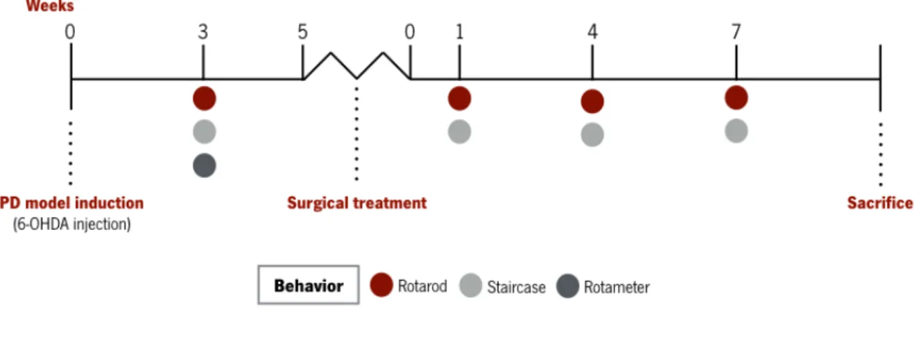
3.4. Behavioral assessment ... 29
3.4.1. Rotarod ... 29
3.4.2. Skilled paw reaching test (Staircase) ... 29
3.4.3. Apomorphine turning behavior (Rotameter) ... 30
3.5. Histological analysis ... 30
3.5.1. Tyrosine hydroxylase immunohistochemistry ... 30
3.5.2. Stereological analysis ... 31
3.5.3. Striatal fiber density measurement ... 31
3.5.4. BrdU administration and in vivo immunostaining ... 32
3.6. Proteomics - Mass Spectrometry and SWATH Acquisition ... 32
3.6.1. In gel digestion/Sample preparation ... 32
3.6.2. SWATH acquisition ... 34
3.7. Data analysis ... 36
Chapter 4 - Results ... 37
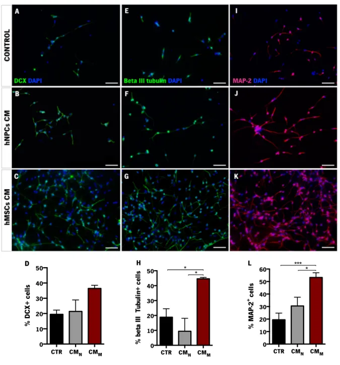
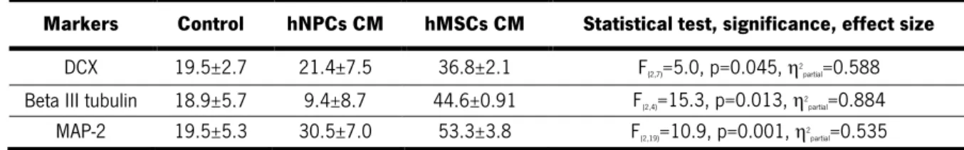
4.1. Neuronal differentiation of hNPCs induced by hMSCs and hNPCs conditioned medium ... 39
4.2. In vivo assay ... 41
4.2.1. Phenotypic characterization of 6-OHDA lesions ... 41
4.2.2. Transplantation of hMSCs, hNPCs and conditioned medium ... 43
4.2.2.1. Rotarod ... 43
4.2.2.2. Staircase ... 44
4.3. Assessment of the extension of the lesion ... 47
4.4. Secretome of hNPCs and hMSCs increased BrdU and TH-positive cells ... 50
4.5. hMSCs and hNPCs secretome proteomic analysis ... 51
Chapter 5 - Discussion ... 55
Chapter 6 - Concluding Remarks ... 65
Chapter 7 - References ... 69
xiii
LIST OF ABBREVIATIONS
# 6-OHDA - 6-hydroxydopamine A ACN - AcetonitrileALS - Amyotrophic lateral sclerosis ASCs - Adipose stem cells
B
BDNF - Brain-derived neurotrophic factor bFGF - Basic fibroblast growth factor
BM-MSCs - Bone marrow mesenchymal stem cells
BrdU - 5-bromo-2-deoxyuridine
C
CES - Collision energy spread cm - centimeter
CM - Conditioned medium CNS - Central nervous system CO2 - Carbone dioxide Cys C - Cystatin C D DA - Dopamine DAergic - Dopaminergic DAB - 3,3-diaminobenzidine DAPI - 4-6-diamidino-2-phenylindole-dhydrochloride
DAT - Dopamine transporter
DBS - Deep brain stimulation DCX - Doublecortin
DG - Dentate gyrus
DMEM - Dulbecco’s Modified Eagle Medium
E
EGF - Epidermal growth factor ESCs - Embryonic stem cells EVs - Extracellular vesicles
F
FA - Formic acid
FBS - Fetal bovine serum FDR - False discovery rate
G
g - g-force
GBA - Glucocerebrosidase GDN - Glia derived nexin
GDNF - Glial cell-derived neurotrophic factor GPi - Globus pallidus internus
H
h - hours
H2O2 - Hydrogen peroxidase
HCl - Hydrochloric acid HD - Huntington’s disease HGF - Hepatocyte growth factor
HUCPVCs - Human umbilical cord perivascular cells
xiv I
IDA - Information-dependent acquisition IGF-1 - Insulin-like growth factor 1 IL-10 - Interleukin 10
IL-6 - Interleukin 6 i.p. - Intraperitoneally
iPSCs - Induced pluripotent stem cells
ISCT - International Society for Cellular Therapy IV - Intravenous
K
kDA - kilo Daltons
L
LBs - Lewy bodies
LC - Liquid chromatography LNs - Lewy neurites
LRRK2 - Leucine-rich repeat kinase 2
M m - meter M - Molar
m/z - mass-to-charge ratio MAO - Monoamine oxidase MAO-B - Monoamine oxidase-B
MAP-2 - Microtubule associated protein-2 MFB - Medial forebrain bundle
mg - milligram
mg/kg - milligram per kilo mg/ml - milligram per milliliter
MHC-II - Major histocompatibility complex class II
MIF - Macrophage migration inhibitor factor min - minutes
miRNAs - micro RNAs ml - milliliter mm - millimeter MPTP - 1-methyl-4-phenyl-1,2,3,6-tetrahydropyridine MS - Mass spectrometry ms - millisecond
MS/MS - Tandem mass spectrometry MSCs - Mesenchymal stem cells
N
NaCl - Sodium chloride NBCS – Newborn calf serum NGF - Nerve growth factor NMS - Nonmotor symptoms NPCs - Neural progenitor cells NSCs - Neural stem cells
O
O.D. - Optical density
P
PBS - Phosphate buffered saline
PBS-T - Phosphate buffered saline-Triton PD - Parkinson’s disease
PEDF - Pigment epithelium-derived factor PFA - Paraformaldehyde
PINK1 - PTEN-induced putative kinase 1 ppm - parts per million
xv R
ROS - Reactive oxygen species rpm - Rotations per minute RT - Room temperature
S
s - seconds
SCF - Stem cell factor
SDF-1 - Stromal cell-derived factor 1 SEM - Standard error of the mean SEM7A - Semaphorin-7A
SEZ - Subependymal zone SGZ - Subgranular zone
SNpc - Substantia nigra pars compacta SODC - Superoxidase dismutase-cytoplasmatic SODM - Superoxidase dismutase-mitochondrial STN - Subthalamic nucleus
SVZ - Subventricular zone
SWATH - Sequential Windowed data independent Acquisition of the Total High-resolution Mass Spectra
T
TCA - Trichloroacetic acid
TGF-β -Transforming growth factor beta
TH - Tyrosine hydroxylase Tris-HCl - Tris-hydrochloride TrxR1- Thioredoxin reductase 1
U
UHCL1 - Ubiquitin carboxyl-terminal hydrolase L1
µl - microliter µm - micrometer
µl/min - microliter per minute
UPDRS – Unified Parkinson’s disease rating scale
UPS - Ubiquitin proteasome system
V V - Volts
VEGF - Vascular endothelial growth factor VTA - Ventral tegmental area
W
W - Watts
WJ-MSCS - Wharton jelly mesenchymal stem cells
xvii
LIST OF FIGURES
Figure 1. Neuropathology of Parkinson's disease. ... 5!
Figure 2. Key molecular mechanisms that contribute to the neurodegenerative process in dopaminergic neurons in Parkinson’s disease. ... 8!
Figure 3. Stem cells secretome-based therapy for Parkinson’s disease. ... 17!
Figure 4. Experimental design. ... 29!
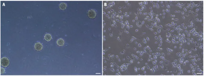
Figure 5. Expansion of hNPCs derived from telencephalon in vitro. ... 39!
Figure 6. In vitro differentiation of hNPCs. ... 40!
Figure 7. Behavioral characterization of 6-OHDA-induced lesions. ... 42!
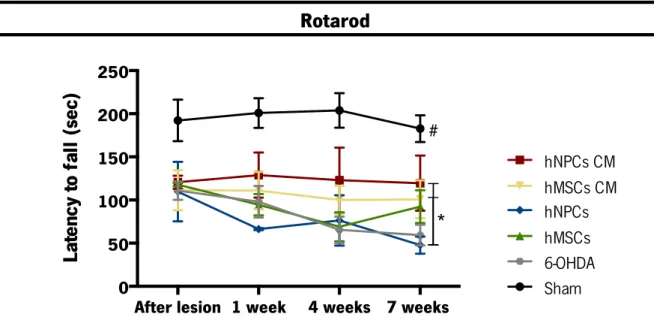
Figure 8. Motor coordination performance 1, 4 and 7 weeks after the transplantation of hMSCs, hNPCs and its CM (i.e. secretome) in the SNpc and striatum. ... 43!
Figure 9. Skilled motor performance 1, 4 and 7 weeks after the transplantation of hMSCs, hNPCs and its CM (i.e. secretome) in the SNpc and striatum. ... 45!
Figure 10. Representative micrographs of SNpc slices stained for TH. ... 48!
Figure 11. Representative micrographs of striatum slices stained for TH. ... 49!
Figure 12. Animals were injected daily with BrdU 5 days before sacrifice. ... 50!
Figure 13. Proteomics - Heatmap and Venn diagram. ... 51! Figure 14. Specific hMSCs and hNPCs CM proteins with neuroregulatory potential in CNS physiology. 53!
xix
LIST OF TABLES
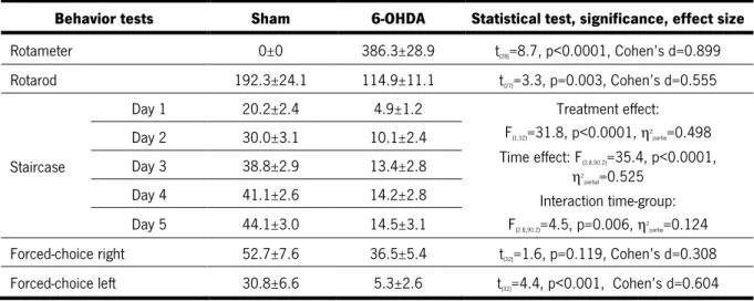
Table 1. Primary antibodies ... 27! Table 2. Secondary antibodies ... 27! Table 3. Statistical analysis of the in vitro assay (Data presented as mean±SEM) ... 41! Table 4. Statistical analysis of the phenotypic characterization of the 6-OHDA lesions (Data presented as mean±SEM) ... 42! Table 5. Statistical analysis of the rotarod test after treatments (Data presented as mean±SEM) ... 44! Table 6. Statistical analysis of the staircase test after treatments (Data presented as mean±SEM) ... 46! Table 7. Statistical analysis of the forced choice task for the left side after treatments (Data presented as mean±SEM) ... 46! Table 8. Statistical analysis of the forced choice task for the right side after treatments (Data presented as mean±SEM) ... 46! Table 9. Statistical analysis of the TH-positive cells in the SNpc (Data presented as mean±SEM) ... 47! Table 10. Statistical analysis of the TH-positive fibers in the striatum (Data presented as mean±SEM) 47! Table S1. List of proteins identified in both hMSCs and hNCPs secretome ………..87 Table S2. List of proteins identified in the hMSCs secretome ……….99 Table S3. List of proteins identified in the hNPCs secretome ………..100CHAPTER 1 - INTRODUCTION
CHAPTER 1
INTRODUCTION
3
1. INTRODUCTION
1.1. Parkinson’s DiseaseOriginally described by James Parkinson in 1817, Parkinson’s disease (PD) represents the second worldwide most common neurodegenerative disorder (de Lau and Breteler, 2006). Although an exact evaluation of PD epidemiological values is missing, studies have suggested that the prevalence in industrialized countries is generally estimated at 0.3% of the entire population, in which about 1% in people over 60 and 4% over 80 years of age are the most affected, demonstrating to be an aging-related disease (de Lau and Breteler, 2006; Dexter and Jenner, 2013; Pringsheim et al., 2014). Nowadays, the mean onset of PD has been established between 50-60 years. However, it has also been suggested that 10% of cases could occur between 20 and 50 years of age, being in this case classified as an young onset, which may represent a distinct disease group (Anisimov, 2009; Dexter and Jenner, 2013).
Clinically, PD is mainly characterized as a disease that affects the motor system. The diagnosis currently available depends on the identification of cardinal features namely, bradykinesia (slowness in the execution of voluntary movements), muscular rigidity (stiffness), postural instability (a tendency to fall even in the absence of weakness or cerebellar balance disturbance) and tremor at rest, with an asymmetric onset, which becomes bilateral with time (Gibb and Lees, 1988; Lees et al., 2009). Other motor signs such as akinesia (absence of normal unconscious movements like arm swing in walking), hypomimia (reduction of normal facial expression), speech and swallowing difficulties, decrease in size (micrographia) and speed of handwriting, as well as reduction of stride length during walking, have also been used in PD diagnosis (Dauer and Przedborski, 2003; Jankovic, 2008). Pathologically, these motor deficits are the result of the progressive loss of dopaminergic neurons (DAergic neurons) in the nigrostriatal pathway, particularly in the substantia nigra pars compacta (SNpc), leading, as consequence, to the reduction of dopamine (DA) levels in the striatum (i.e. putamen and caudate nucleus) (Figure 1) (Langston, 2006; Lees et al., 2009). In addition to DA, it has also been suggested that the content of norepinephrine and serotonin is also low. However, of the three biogenic amines, DA is the most drastically reduced (Shannak et al., 1994), being this loss the responsible mechanism that triggers the onset of the majority of motor signs (Chung et al., 2010; Kim et al., 2011). Another hallmark feature of PD is the presence of Lewy bodies (LBs) (Gibb and Lees, 1988), which are typically used as a post-mortem confirmation of PD (Olanow and Brundin, 2013). LBs are distinctive intracytoplasmatic inclusions, containing a variety of cellular proteins, being α–synuclein the most
4
abundant one (Benskey et al., 2016). The precise reason why LBs form and its role in pathogenesis of PD remains unclear (Dickson et al., 2009). However, in recent years, it has become clear that the initial sites displaying LBs are the dorsal motor nucleus of the vagus in the brainstem and the olfactory bulb (which is defined as stage I). This staging concept was firstly proposed by Braak and colleagues (Braak et al., 2003a), demonstrating that the disease most likely progresses in an upward direction via the pons (stage II) to the midbrain (stage III), followed by the basal prosencephalon and mesocortex (stage IV), and eventually reaching the temporal cortex and neocortex (stages V and VI) (Braak et al., 2003b). Therefore, it is only in stage III, when DAergic neuronal death exceeds a critical threshold (i.e. 70-80% of striatal nerve terminals and 50-60% of SNpc perikarya) that motor features of PD become evident, which means, that there is a substantive pre-symptomatic period of the disease that is hidden due to the existence of possible compensatory mechanisms (Bezard et al., 1999; Navntoft and Dreyer, 2016). Indeed, Zigmond and colleagues (Zigmond et al., 1990) proposed a model of compensatory changes, showing that the relationship between DAergic neuronal loss and functional impairments results from adaptive neurochemical changes that occur within the striatum. Recently, this dogma has been challenged, and several reports have shown that the classically accepted dopamine-mediated mechanisms are not the primarily involved in the initial compensation of DA depletion in PD, proposing a series of functional compensatory changes within and outside of the basal ganglia (Bezard et al., 2003; Obeso et al., 2004). In fact, it has been shown that the activation of the subthalamic nucleus (STN) increases the activity of SNpc DAergic neurons. Thus, the loss of DAergic projections and consequent decrease in DA concentration leads to an hyperactivity of the STN before the onset of functional changes in the putamen, suggesting that STN is implicated in the compensatory mechanisms in the initial phases of PD (Bezard et al., 1999; Hamani et al., 2004; Vila et al., 2000). Nevertheless, the precise nature of these compensatory mechanisms, and the reason for their ultimate failure has still been elusive.
To date, PD motor deficits are the main focus of the therapeutic interventions. However, there is a growing literature reporting that nonmotor symptoms (NMS) form an integral part of the clinical features of PD, suggesting that they could precede the manifestation of the characteristic motor symptoms, which could represent a new approach for its early prognosis (Khoo et al., 2013). Like motor signs, the NMS can be equally debilitating to PD patients, and include depression, anxiety, sensory abnormalities, autonomic dysfunctions and cognitive decline (Langston, 2006; Pantcheva et al., 2015). Moreover, although the real cause of NMS still remains poorly understood, the current strategies for treating PD are mainly effective against the motor symptoms but widely ineffective at addressing
5
NMS. Therefore, it is important to understand the molecular mechanisms that cause the appearance of motor signs, but also NMS, in order to establish the pathophysiology pattern of PD (Chaudhuri and Schapira, 2009).
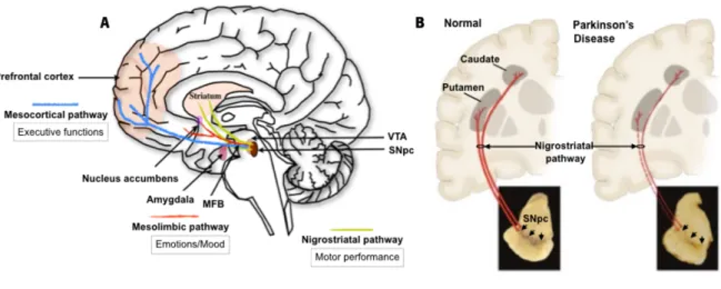
Figure 1. Neuropathology of Parkinson's disease.
A) DAergic network in the normal brain with main representative pathways: mesocortical, mesolimbic and nigrostriatal pathway. (B) When the disease occurs, the nigrostriatal pathway is the most affected, and is characterized by the progressive loss of DAergic neurons and consequent depigmentation of the SNpc. This neuronal loss leads to a DA deficiency in the striatum (i.e. putamen and caudate nucleus), which is responsible for characteristic motor symptoms. Adapted from (Dauer and Przedborski, 2003).
1.1.1. Etiology of Parkinson’s disease
The majority of the cases of PD appear to be sporadic, and these probably represent an interaction between genetic and environmental factors (Warner and Schapira, 2003). Age represents the main predisposing factor, however, it remains unknown if it is the chronological age or the aging process the responsible for PD susceptibility (Dexter and Jenner, 2013). Familial cases of PD are rare, but in recent years the role of genetic factors has been intensely explored, showing significant outcomes from the molecular point of view (Bras and Singleton, 2009; Pagano et al., 2016). Recently, Pagano and colleagues (Pagano et al., 2016) explored clinical characteristics of PD at different ages in diagnosed patients with untreated PD, and found that 25% of the patients had a familiar history of the disease, raising the possibility that some of them were carriers of a genetic mutation. In fact, different reports on familial PD have revealed at least 17 autosomal dominant and autosomal recessive gene mutations namely, α-synuclein duplications and triplications, parkin, leucine-rich repeat kinase 2 (LRRK2), ubiquitin carboxyl-terminal hydrolase L1 (UCH-L1), DJ-1 (PARK7), PTEN-induced putative kinase 1 (PINK1), among others (Bras and Singleton, 2009; Dexter and Jenner, 2013). In addition,
6
although it has been suggested that familial forms of PD have distinct clinical and pathological phenotypes, many of these neurodegeneration mechanisms overlap with the mechanisms involved in sporadic PD such as oxidative stress, mitochondrial dysfunction and abnormal protein aggregation (Bras and Singleton, 2009; Dexter and Jenner, 2013).
Concerning environmental factors, studies have proposed that farming occupation, rural living and the consequent exposure to pesticides such as rotenone and paraquat [structurally similar to 1-methyl-4-phenyl-1,2,3,6-tetrahydropyridine (MPTP)] may have an impact in the appearance of the disease. Nowadays, all these chemicals are being used to create animal models of PD, exploring its effects on the pathophysiology of the disease. However, Noyce and colleagues (Noyce et al., 2016) have recently suggested that environmental toxins most likely play a minor role in PD risk. Furthermore, there also exists a consistent association between PD and lifestyle factors such as smoking and coffee consumption (de Lau and Breteler, 2006; Noyce et al., 2016). Indeed, many epidemiological studies have shown a reduced risk of developing PD among cigarette smokers. The most probable explanation involves nicotine, as this component may stimulate dopamine release and acts as antioxidant (Quik, 2004). Some studies also related PD with coffee consumption, showing a significantly decreased PD risk for coffee drinkers (de Lau and Breteler, 2006). These observations were based in the effects of caffeine, a known inhibitor of the adenosine A2 receptor, which in turn has an important role in the
regulation of dopamine release (Chen et al., 2001).
1.1.2. Mechanisms of Neurodegeneration
Where does PD begins at the cellular level? This is the key question that still remains to be answered. Evidences from the literature have suggested that the degeneration of DAergic neurons in PD starts in the axonal and synaptic terminals, retrogradely progressing to the cells bodies in SNpc (Burke and O'Malley, 2013). In fact, at the time of the onset of motor deficits, more than 70% of DA (Bernheimer et al., 1973; Dauer and Przedborski, 2003), and more than 50% of the tyrosine hydroxylase (TH) and DA transporter (DAT) have been lost in the striatum (Beach et al., 2008; Dauer and Przedborski, 2003; Nandhagopal et al., 2008). On the other hand, the SNpc presents a decrease of around 30% of DAergic cells at this time (Cheng et al., 2010; Ross et al., 2004). Little is known about the mechanisms underlying the early deterioration of synapses and axons of DAergic neurons in PD, but available data indicate that the retrograde degeneration implicates a decline of the axonal trafficking of proteins and mitochondrias, followed by the aggregation of α-synuclein and the formation of axonal spheroids (Chu et al., 2012; Chung et al., 2009; Coleman, 2005; Kim-Han et al., 2011). Surrounding
7
astrocytes and microglial cells have also been described as potential modulators of these axonal impairments (Privat, 2003; Shokouhi et al., 2010). However, the real influence of these cells on the degeneration and deterioration progress of nigral or striatal DAergic pathways is still poorly understood (Halliday and Stevens, 2011). Despite these issues, nowadays, it is well accepted that oxidative stress, mitochondrial dysfunction and abnormal protein aggregation are the key molecular mechanisms involved in PD neurodegeneration process (Figure 2) (Dauer and Przedborski, 2003; Dexter and Jenner, 2013).
The central nervous system (CNS) is particularly sensitive to oxidative stress due to different reasons, including its high oxygen consumption even under basal conditions, high production of reactive species from specific neurochemical reactions as well as increased deposition of metal ions in the brain with aging (Chiurchiu et al., 2016). Throughout the whole lifespan, DAergic neurons are exposed to reactive oxygen species (ROS) as a result of the metabolism of DA itself. In DAergic cells, ROS generation occurs by deamination (auto-oxidation process) of DA by the monoamine oxidase (MAO) activity. This results in significant amounts of hydrogen peroxidase (H2O2) that can further interact with
metal ions (e.g. iron), leading to the origin of the reactive hydroxyl radical (!OH), which is highly toxic to the neurons (Bhat et al., 2015; Datta and Bhonde, 2012). Indeed, post-mortem analysis has shown that significantly higher concentrations of iron, in SNpc region, were found in brain tissue of PD patients when compared to healthy individuals (Griffiths et al., 1999). Indeed, DAergic neurons are more susceptible to oxidative damage when compared to other neuronal cells mainly because of the dual presence of DA and high levels of iron. Mitochondrial dysfunction is also another origin of oxidative stress that has been widely associated with the pathogenesis of PD. Neurons are metabolically very active, and as such, they greatly depend on mitochondria for energy production. Any pathological situation that leads to mitochondrial dysfunction can cause a higher increase in ROS, inducing (for instance) the release of cytochrome c in the cytosol, and consequently apoptosis (Bhat et al., 2015; Moon and Paek, 2015). In addition, the decrease of complex I (of the mitochondrial respiratory chain) enzyme activity was also observed to be an important player of degeneration in the SNpc of parkinsonian patients (Parker et al., 1989). In fact this process in known to cause excitotoxicity and axonal damage, leading to the progression of PD (Chiurchiu et al., 2016).
Finally,the abnormal deposition of proteins in brain tissue has also been an important feature in the pathophysiology of PD. Although it remains unclear how misfolded proteins could directly cause toxicity or damage the cells via the formation of protein aggregates, the prevailing hypothesis is that the formation of the latter triggers a cascade of neurodegenerative events (Diack et al., 2016). Oxidative
8
damage, linked to mitochondrial dysfunction and an abnormal DA metabolism, may also promote or predispose misfolded proteins conformations. Nonetheless, these events are not mutually exclusive, and one of the aims of the current PD research is to elucidate the sequence in which they act and understand the interaction between these pathways (Dauer and Przedborski, 2003).
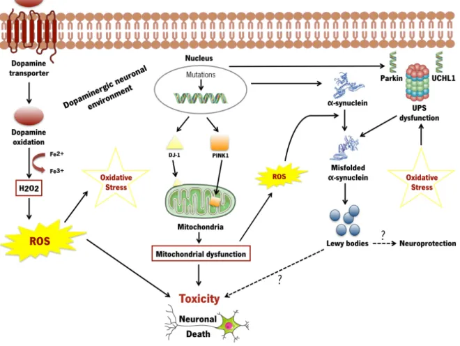
Figure 2. Key molecular mechanisms that contribute to the neurodegenerative process in dopaminergic neurons in Parkinson’s disease.
Cell death may be caused by oxidative stress, mitochondrial and UPS dysfunction, and α-synuclein aggregation. Pathogenic mutations may directly induce mitochondrial dysfunction (DJ-1, PINK-1), abnormal protein conformations (as believed to be the case with α -synuclein) or damage the ability of the cellular machinery to detect and degrade misfolded proteins (Parkin, UCHL1). Controversy exists regarding whether LBs promote toxicity or protect the cells from harmful effects of misfolded proteins. ROS generation occurs by the auto-oxidation process of DA resulting in significant amounts of H2O2 that can further interact with metal ions like iron. Oxidative damage, linked to mitochondrial dysfunction and abnormal dopamine metabolism, may also promote misfolded protein conformations. ROS: Reactive oxygen species. UPS: Ubiquitin proteasome system.
9
1.2. Current therapeutic approaches in Parkinson’s Disease: how far are we from the cure?
The treatment of PD has not significantly changed over the years, and the use of levodopa (L-DOPA) is still considered the gold standard treatment since its introduction in the early 1960s (LeWitt and Fahn, 2016). L-DOPA, is a naturally occurring aminoacid and is the immediate metabolic precursor for catecholamines like DA, and in contrast to DA, L-DOPA can readily cross the blood-brain barrier (BBB). This drug has revolutionized symptomatic treatment by providing improvement in activities of daily living and life quality. However, L-DOPA is just efficient during the first years of usage as its chronic administration has been associated with the appearance of undesirable side effects such as nausea, vomit, hypotension and long-term complications including motor fluctuations (loss of therapeutic effect benefit after each dose) and dyskinesias (excessive involuntary movements occurring at the peak of L-DOPA dosing) (Jankovic and Aguilar, 2008; Jimenez-Shahed, 2016; Rascol et al., 2003).
The use of DA agonists and enzyme inhibitors has been used as alternative to the above mentioned (Dexter and Jenner, 2013; Rascol et al., 2003). In the case of the DA agonists (e.g. pramipexole and ropinirole), studies have shown that they are efficient in controlling the cardinal motor symptoms of PD, particularly in early stages of the disease and in patients who have not been exposed to L-DOPA (Jankovic and Aguilar, 2008). For instance, the administration of pramipexole or ropinirole was found to significantly reduce the risk of motor complications compared to L-DOPA (Holloway et al., 2004; Rascol et al., 2000). However, the prolonged exposure to DA agonists also presents limiting features such as somnolence, sleep disturbances and impulse control disorders (Jankovic and Aguilar, 2008). On the other hand, the use of MAO-B inhibitors such as selegiline and rasagiline has attracted some attention (Dexter and Jenner, 2013). Safinamide, another MAO-B inhibitor compound, has recently been claimed as a promising agent for the treatment of PD. In Phase-III clinical trials, safinamide is a molecule with a dual mechanism of action based on the enhancement of the DAergic function and inhibition of the excessive release of glutamate. Indeed, safinamide was found to be a useful as a combinatory strategy to DA agonists in the early phases of PD, as well as to be able to reduce dyskinesias when used together with L-DOPA in patients with advanced PD (Kandadai et al., 2014; Onofrj et al., 2008). Despite these promising results, most of these treatments were not able to promote the total recovery of PD symptomatology, presenting long-term inefficiency as well as an inability to recover lost DAergic neurons or to protect the viability of the remaining ones.
Surgical treatments, such as deep brain stimulation (DBS) in the globus pallidus internus (GPi) or in the STN have been applied as an alternative in patients with significant motor complications where
10
the pharmacological treatment is no longer effective (Hariz et al., 2016; Hutchinson and Wick, 2016). Indeed, it has been reported that this surgical procedure is safe, leading to the lower consumption of anti-parkinsonian medications and dyskinesias (deSouza et al., 2013). However, DBS requires expertise in diagnosis, imaging and stereotaxic surgery, thereby limiting its widespread applicability (Rascol et al., 2003).
In addition to the conventional clinical treatments, some clinical trials based in the transplantation of human ventral mesencephalic tissues into the striatum of PD patients with advanced disease, were conducted in the late 1980s (Lindvall et al., 1990; Sawle et al., 1992). The results were quite promising, with patients displaying increased levels of DA, motor function amelioration as well as, reduction in the L-DOPA dosage requirement (Singh et al., 2007). Although these studies confirmed the relevance and feasibility of cell transplantation, the use of human tissue has some limitations associated with ethical and religion questions, as well as logistics of acquiring sufficient amount of fetal tissues (Kim et al., 2013; Suksuphew and Noisa, 2015).
In summary, all these interventions are not fully efficacious, and more importantly, the progression of the PD degenerative process is not avoided. Based on such limitations, cell-based strategies through the use of stem cells have been proposed as a possible therapeutic tool for the treatment of CNS disorders, including PD (Anisimov, 2009).
1.3. Stem cell-based therapeutic approaches
The low regeneration potential of CNS make it a challenge for the development of new protocols and strategies that could allow the generation of new functional neurons in response to injury (Williams, 2014). Endogenous stem cells are found in specific niches of human brain, which have the ability to differentiate and replace the damaged cells and secrete trophic factors required for tissue repair. However, this self-repair is not sufficient in most pathological processes, demanding external intervention (Buzhor et al., 2014). Recently, cell therapy has been proposed as an attractive option, and stem cells represent the most favorable cell source for such therapies, since these cells have the ability to renew themselves continuously, have high proliferation capability and are able to differentiate into different cell types (Kim et al., 2013; van der Kooy and Weiss, 2000). Over the years, several types of stem cells were investigated as potential agents for cellular therapy including embryonic stem cells (ESCs), neural stem cells (NSCs), mesenchymal stem cells (MSCs), or even induced pluripotent stem cells (iPSCs) from different sources, showing promising results in a wide panel of CNS disorders including PD (Goodarzi et al., 2015). From these, NSCs and MSCs have a number of interesting
11
properties that we will describe throughout this work. Indeed, it has been described that these cells display therapeutic effects of neuroprotection and immunomodulation, such as the capacity to protect and regenerate damaged DAergic neurons, as well as increase the motor function in PD animal models (Bonnamain et al., 2012; Drago et al., 2013; Kassem et al., 2004). Therefore, we focused our efforts in studying their potential use, as well as of its secretome, as a potential alternative therapy for PD.
1.3.1. Neural stem cells
NSCs are multipotent cells isolated from fetal and adult nervous system tissues, which have the ability to self-renew and differentiate into specialized functional neurons, astrocytes and oligodendrocytes (Buzhor et al., 2014; Fu et al., 2015), which makes them an interesting source of cells for neuronal repair after injury or disease (Bonnamain et al., 2012). It is known that NSCs exist not only in the developing brain but also in the adult brain (Palmer et al., 2001), particularly in the subgranular zone (SGZ) in the dentate gyrus (DG) of the hippocampus and the subependymal zone (SEZ) of the lateral ventricles (as reviewed by Salgado et al., 2015). These cells are commonly identified by the expression of the intermediate filament Nestin, GFAP, transcription factor Sox2, and the RNA binding protein Musashi1, together with absence of expression of the differentiated markers CD24, NeuN and O4 (Suksuphew and Noisa, 2015). NSCs are typically isolated from embryonic, fetal or adult nervous system tissue (Alvarez-Buylla and Garcia-Verdugo, 2002; Ogawa et al., 2009; Toma et al., 2001; Zhang et al., 2009), and can be cultured in vitro as neuroshperes, in the presence of growth factors such as basic fibroblast growth factor (FGF-2) and epidermal growth factor (EGF) (Bonnamain et al., 2012).
A growing number of studies have also highlighted NSCs as immunomodulatory agents and their capacity to reduce CNS inflammation (Ben-Hur, 2008; Kokaia et al., 2012). Although no reports have been presented regarding these effects of NSCs specifically in PD, it is known that inflammatory responses (e.g. T cell infiltration, increased expression cytokines, toxic mediators derived from activated glial cells) are prominent features of PD (Tufekci et al., 2012), and therefore, the immumodulation effects presented by these cells may be important for its use in cell transplantation.
Regarding the application of NSCs in PD, there are several examples of their impact on the reversion of the latter. For instance, Harrower et al. (Harrower et al., 2006) showed a reliable long-term survival and integration of transplanted NSCs in the striatum of rats lesioned with 6-hydroxydopamine (6-OHDA). According to the authors, an increase of DA fiber densities, as well as synapse formation was observed. Richardson and colleagues (Richardson et al., 2005) demonstrated that the transplantation of
12
adult NSCs (expanded from SEZ) in the striatum of 6-OHDA-lesioned rats led to a functional recovery in the animals, and the DAT immunoreactivity was restored in the host tissue. Using the same model (6-OHDA), Armstrong and colleagues (Armstrong et al., 2002) revealed that transplanted NPCs could differentiate into neurons, and indeed, a small number of TH-immunopositive neurons were present in both intrastriatal and intramesencephalic grafts. Yasuhara and co-workers (Yasuhara et al., 2006) also observed that by transplanting an immortalized NSC line (HB1.F3), functional improvements could be observed along with an evident preservation of TH immunoreactivity in the nigrostriatal pathway. Moreover, in other CNS related disorders, such as amyotrophic lateral sclerosis (ALS), Huntington’s disease (HD) and ischemic stroke, the transplantation of NSCs was also effective in delaying disease progression, exert neuronal protection and enhance motor function, and led to the increase of dendritic plasticity and axonal rewiring (Andres et al., 2011; Ryu et al., 2004; Xu et al., 2006).
Although promising, it is important to note that the application of NSCs for transplantation is still limited. Tissue availability, ethical and logistical concerns linked to the fact that it is also challenging to maintain and expand these cells for long periods of time, represent important issues to overcome in the future before resuming it for clinical applications (Bonnamain et al., 2012; Fu et al., 2015). Besides that, NSCs have been described as a potential stem cell source for the treatment of neurological disorders not only because they may provide a (tissue-specific) cellular reservoir for the replacement of lost or damaged cells, but also because of other capabilities, such as tissue trophic support (Ben-Hur, 2008; Drago et al., 2013).
1.3.2. Mesenchymal Stem Cells
MSCs represent a non-hematopoietic and multipotent stem cell population with self-renewal capacity and multiple differentiation potential (Wang et al., 2011). According to the International Society for Cellular Therapy (ISCT), there are three minimal criteria to define MSCs, namely: (1) the adherence to plastic surfaces when maintained in standard culture conditions; (2) the positive expression of specific surface markers such as CD105, CD90 e CD73, and negative expression of hematopoietic surface markers like CD14, CD34, CD45, HLA-DR, or CD11B, CD79α or CD19; and (3) in vitro differentiation into at least osteoblasts, adipocytes, and chondroblasts (Dominici et al., 2006). Friedenstein and colleagues (Friedenstein et al., 1974) were the first to isolate MSCs from bone marrow, describing them as fibroblastoid cells with clonogenic potential and plastic culture adherence. Following these early studies, countless reports have confirmed that in addition to bone marrow, MSCs can also be isolated from various adult and neonatal tissues such as adipose tissue, dental pulp,
13
amnion, placenta, Wharton jelly of the umbilical cord, and even the brain (Erices et al., 2000; Gronthos et al., 2000; Hass et al., 2011; Paul et al., 2012; Sarugaser et al., 2005; Wang et al., 2004; Zuk et al., 2002). The potential of MSCs has been attributed to their widespread availability throughout the human body, easy isolation and expanding, as well as maintenance of viability and regenerative capacity after cryopreservation (Uccelli et al., 2011a; Uccelli et al., 2011b; Wang et al., 2011). In addition to this, MSCs have also demonstrated low immunogenic properties due to the lack of the major histocompatibility complex class II (MHC-II), making them an attractive cell source for transplantation (Morandi et al., 2008). Furthermore, MSCs have been also described as immunomodulatory agents, being able to interact with different components of the immune system (Wang et al., 2011). For example, it has been described that MSCs are capable to regulate the proliferation, activation and maturation of T and B lymphocytes in vitro (Bartholomew et al., 2002), and to induce long-term survival in an allogeneic context (Aggarwal and Pittenger, 2005), which is an important concern for its use in transplantation.
Regarding PD, several reports have already shown that the transplantation of MSCs acts as a promoter of neuroprotection and/or neuronal function (Glavaski-Joksimovic and Bohn, 2013). Hellman and co-workers (Hellmann et al., 2006) demonstrated that after transplantation, bone marrow MSCs (BM-MSCs) were found to be viable and migrate in the brain parenchyma of a 6-OHDA PD rat model. Using the same PD model and the same MSCs population, Danielyan and colleagues (Danielyan et al., 2011) showed neuroprotective effects against nigrostriatal degeneration and improvements in the motor function of the 6-OHDA lesioned rats. Blandini et al. (Blandini et al., 2010) also achieved the same outcomes, verifying that although no differentiation of MSCs toward a neuronal (DAergic) phenotype was obtained in vivo, the animals that received the striatal MSCs grafts presented an increased survival of both cell bodies and terminals of DAergic neurons. With MSCs derived from adipose tissue (ASCs), Schwerk and colleagues (Schwerk et al., 2015) demonstrated a significant increase in TH-positive expression in transplanted animals when compared to the untransplanted group. The same results were also obtained by Xiong and colleagues (Xiong et al., 2010), which demonstrated neuroprotective and neuroregenerative effects in a rotenone-induced hemiparkinsonian rat model using MSCs isolated from umbilical cord. In patients, Venkataramana and co-workers (Venkataramana et al., 2010) observed that the transplantation of BM-MSCs led to a partial amelioration in the symptomatology and life quality of the patients [measured by Unified Parkinson’s disease rating scale (UPDRS)].
14
The specific mechanism by which MSCs are able to improve the motor performance either in animals or patients remains unclear. It is well studied that MSCs populations can be sub-passaged and differentiate into different cell lineages, however the differentiation into functional neuronal lineages is not likely to happen in such a relevant manner that could impact the recovery of PD animal models or patients (Maltman et al., 2011; Teixeira et al., 2013). Therefore, although some reports suggest the differentiation of MSCs into DAergic neurons or other neuronal lineages as the principal outcome of their therapeutical effects, recent evidences have proposed the secretome of these cells as the main responsible of their therapeutic action (Teixeira et al., 2013).
1.3.3. Stem cells secretome
The therapeutic effects of transplanted stem cells were initially attributed to their differentiation capacity. Indeed, most of the studies emphasize the ability of stem cells to migrate to the sites of injury, integrate the damaged tissue and differentiate into specialized cells (Drago et al., 2013). However, it has also been shown that only a small percentage of cells truly engraft and survive in the damaged host tissue, leading the current body of research to argue that the multipotent differentiation of stem cells, within the CNS, contributes minimally to the observed beneficial effects (Kupcova Skalnikova, 2013; Lavoie and Rosu-Myles, 2013). On the other hand, robust data has recently demonstrated that most of these potential effects, promoted by stem cells, are mainly mediated by the secretion of bioactive molecules (e.g., proteins, cytokines, vesicles), which is defined as secretome (Drago et al., 2013; Salgado et al., 2010; Salgado et al., 2015; Teixeira et al., 2013). The concept of secretome has been described as the proteins released by a cell, tissue or organism, being essential in the regulation of different cell processes (Teixeira et al., 2013). In fact, it has been demonstrated that these secreted molecules by stem cells act as modulators of cell survival, proliferation and differentiation, as well as regulators of inflammatory processes and promoters of angiogenesis (Teixeira et al., 2013). Although most of the secretome studies have been focused in their proteic soluble fraction (e.g. factors, growth factor and cytokines), nowadays it has also been described that stem cells are able to secrete a vesicular fraction that is constituted by microvesicles and exosomes, which involves the transference of proteins and genetic material to neighboring cells (Salgado et al., 2015; Yu et al., 2014).
In 2006, Crigler and colleagues (Crigler et al., 2006) were the first to show that BM-MSCs were able to induce neuronal cell survival and neurite outgrowth in a neuroblastoma cell line and in dorsal root ganglion explants, through the secretion of neurotrophic factors such as brain-derived neurotrophic factor (BDNF) and nerve growth factor (NGF). Further characterization studies have reported that,
15
indeed, these cells are able to secrete a wide panel of growth factors such as glial cell-derived neurotrophic factor (GDNF), FGF-2, insulin-like growth factor 1 (IGF-1), hepatocyte growth factor (HGF), vascular endothelial growth factor (VEGF) and EGF, as well as cytokines like interleukin 6 (IL-6), interleukin-10 (IL-10), transforming growth factor beta (TGF-β), stem cell factor (SCF) and stromal cell-derived factor 1 (SDF-1) (Baraniak and McDevitt, 2010; Meyerrose et al., 2010; Nakano et al., 2010; Ribeiro et al., 2012), which are described as important modulators of neuronal survival/differentiation, neurite outgrowth and glial cells. Cantinieaux and co-workers (Cantinieaux et al., 2013) showed in vitro that the BM-MSCs secretome was pro-angiogenic and was able to protect neurons from apoptosis. With ASCs secretome, Lu et al. (Lu et al., 2011) demonstrated its potential to protect a PC13 cell line model from glutamate excitotoxicity-induced apoptosis through the secretion of BDNF, VEGF and HGF. Salgado and colleagues (Salgado et al., 2010) revealed that the secretome of human umbilical cord perivascular cells (HUCPVCs) was also a modulator of neuronal viability and cell survival. In the context of PD, Kim and colleagues (Kim et al., 2009), using co-cultures of microglia and mesencephalic neurons together with BM-MSCs, observed that there was a decrease in the microglia activation due to the release of anti-inflammatory molecules such as IL-6 and TGF-β, thereby protecting dopaminergic neurons from death. Similar results were also presented by Wang and colleagues (Wang et al., 2010), which showed that BM-MSCs could exert neuroprotection in 6-OHDA-exposed dopaminergic neurons in vitro, through anti-apoptotic mechanisms promoted by the expression of SDF-1. In vivo, the secretome of MSCs also plays an important role, either by the active secretion in situ (after MSCs transplantation) or by the injection of the secretome itself in the form of conditioned media (CM) (Teixeira et al., 2013). Previous studies from Salgado’s lab showed that the injection of MSCs secretome was able to revert the parkinsonian phenotype from both histological and functional outcomes (data not published). This is in line with what has been described in the literature regarding the trophic capability of MSCs in PD. For instance, Sadan and colleagues (Sadan et al., 2009), using human BM-MSCs as neurotrophic factors secreting cells (NTF-SC), observed a remarkable attenuation in the amphetamine-induced rotation and other abnormal behavior, as wells as in the loss of TH immunoreactive nerve terminals when compared to the untreated MSCs group, attributing these effects to the secretion of BDNF and GDNF. Similar observations were also claimed by Cova and co-workers (Cova et al., 2010), which observed that after intrastriatal transplantation of MSCs there was an increase in the preservation of spared DAergic neurons, which was correlated with an increased expression of BDNF by MSCs. Interestingly, in a comparative study reported by Teixeira and colleagues (Teixeira et al., 2015), animals injected just with
16
the secretome of HUCPVCs into the DG of adult rats, disclosed levels of neuronal survival and differentiation very similar to those observed in cell-transplanted groups.
Neurotrophic growth factors such as NGF, BDNF, GDNF have also been found to be increased after NSCs transplantation (Drago et al., 2013). Likewise MSCs, currently there are no studies regarding the application of NSCs secretome alone in animal models of PD. However, studies have suggested NSCs as neurotrophic-factor secreting cells (Drago et al., 2013). For instance, Yasuhara and co-workers (Yasuhara et al., 2006) showed that after intrastriatal transplantation of NSCs in 6-OHDA PD model, there was an improvement in the behavioral performance of the animals, which was correlated with the increase of TH innervation due to an active expression of SCF. Similar outcomes were also presented by Ourednik and colleagues (Ourednik et al., 2002), which demonstrated, using a MPTP PD model, that after the transplantation of NSCs there was an increased recovery of TH and DAT activity due to an in situ expression of GDNF. Moreover, the authors suggest that the NSCs have the capacity to create host environments rich in trophic and neuroprotective support to rescue imperiled host cells (Ourednik et al., 2002). Ebert and colleagues (Ebert et al., 2008) demonstrated that NSCs overexpressing either IGF-1 or GNDF were able to significantly reduce amphetamine-induce rotational behavior and DA neuronal loss in 6-OHDA PD animals, when compared to the untransduced NSCs. Behrstock et al. (Behrstock et al., 2006) demonstrated, in vitro, that the number of primary neurons staining for TH significantly increased after the addition of NSCs CM. In vivo, using NSCs genetically modified to release GDNF, the same authors showed that more TH positive neurons were present in the transplanted rats (partial lesioned with 6-OHDA), verifying fewer rotations compared to the untransplanted group (Behrstock et al., 2006).
Besides the paracrine soluble factors released by stem cells to the extracellular space, intensive research has also been investigating the role of secreted extracellular vesicles (EVs) in the therapeutic potential of stem cells (Drago et al., 2013). Exosomes and microvesicles are the most well studied classes of EVs. These vesicles are involved in cell-to-cell communication, with the ability to transfer proteins and functional genetic material such as micro-RNAs (miRNAs) to other cells, which are implicated in important physiological processes such as antigen presentation, genetic exchange, immune responses and angiogenesis (Lener et al., 2015; Lopez-Verrilli et al., 2016; Xin et al., 2013; Yu et al., 2014). A more detailed characterization of EVs secreted by both MSCs and NSCs is required, in an effort to select and identify the molecules responsible for the therapeutic effects of the secretome (Drago et al., 2013; Lai et al., 2010).
17
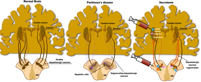
Altogether, these findings strongly suggest that stem cells’ secreted factors are the key players on stem cells’ mediated effects in models of injury and disease in the CNS. Therefore, the use of secretome as a possible replacement of cell transplantation is of enormous interest and may be a new and important tool for the treatment of PD (Figure 3).
Figure 3. Stem cells secretome-based therapy for Parkinson’s disease.
The trophic action of stem cells has been increasingly accepted nowadays as a new concept for the regeneration of the CNS, including PD. The ability to secrete growth factors, cytokines and chemokines seems to be one of the reasons to the contribution to the protection/survival of the preexisting DAergic neurons in lesioned areas, leading to functional amelioration and improvement of motor function. Adapted from (Teixeira et al., 2013).
CHAPTER 2 - RESEARCH OBJECTIVES
CHAPTER 2
RESEARCH OBJECTIVES
21
2. RESEARCH OBJECTIVES
Stem cells have been on the forefront of new possible therapeutic strategies for CNS regeneration. Recent evidences have indicated that most of the beneficial actions caused by these cells are related with their secretome and its trophic capability. This is extremely important as it should minimize biological variability, allowing precise dosing, and overcome several stem cells related issues including the number of available cells for transplantation and its survival after this procedure. Therefore, the main goal of the present project is to determine the role of secretome as a potential cell-free therapy for PD, when compared to cell-based transplantation approaches. Therefore, the main objectives of the present thesis are:1. Characterize the secretome of human MSCs (hMSCs) and human NPCs (hNPCs), performing a comparative study through proteomic-based approaches.
2. Determine the impact of hMSCs and hNPCs secretome on the neuronal survival and differentiation of hNPCs in vitro.
3. Establish the therapeutic potential of hMSCs secretome in an in vivo model of PD (6-OHDA), comparing it to the outputs obtained from animals transplanted with hMSCs, hNPCs and their secretome.
CHAPTER 3 - MATERIALS AND METHODS
CHAPTER 3
MATERIALS AND METHODS
25
3. MATERIALS AND METHODS
3.1. Cell Culture3.1.1. Expansion of hMSCs and collection of conditioned medium
hMSCs derived from bone marrow (Lonza, Switzerland) were thawed and plated into T-75 gelatin (0.1%, Sigma, USA)-coated culture flasks (SPL Life Sciences, Korea) with 12 mL of serum-free growth medium (PPRF-msc6). The formulation and preparation of PPRF-msc6 has previously been described in detail (Jung et al., 2010). The medium was renewed every 3 days and the culture maintained at 37°C in a humidified atmosphere containing 5% CO2. When the cells reached 80-90% of
confluence, they were enzymatically dissociated using 0.05% trypsin-EDTA (Life Technologies, USA) during 5 min at 37°C. Dulbecco’s Modified Eagle Medium (DMEM; Life Technologies, USA) supplemented with Fetal bovine serum (FBS, Biochrom, Germany) was then added to stop the reaction. After that, cells were centrifuged at 1200 rpm (4°C) for 5 min. The supernatant was removed and the pellet was resuspended in fresh growth medium, in which a small volume of cells was diluted in Trypan Blue (Life Technologies, USA) to perform cell counts. At last, the cells were plated into new gelatin-coated culture flasks at a density of 5000 cells/cm2 for experiments, and 12 000 cells/cm2 for
proteomic procedures. At passage 5 (P5), after 72 hours of growth, the medium was removed and the cells were washed twice with Neurobasal A medium (Life Technologies, USA). Following this, Neurobasal-A medium supplemented with 1% kanamycin (Life Technologies, USA) was added to the cells, which were placed at 37°C in a humidified atmosphere containing 5% CO2. After 24 h, this
medium, containing the factors secreted by hMSCs (called conditioned medium (CM)) was collected and centrifuged at 1200 rpm for 10 min to remove any cell debris, and then stored at -80°C until it was required for further experiments.
3.1.2. Expansion of hNPCs and collection of conditioned medium
hNPCs were a kind gift from Prof. Leo A. Behie (University of Calgary, Canada). Cells were isolated from the telencephalon region of a 10 week post-conception fetus according with the protocols and strict ethical guidelines previously established and approved by the Conjoint Health Research Ethics Board (CHREB, University of Calgary, Canada; ID:E-18786) (Baghbaderani et al., 2010; Mendez et al., 2002; Mendez et al., 2005). hNPCs were thawed and the content placed in T-75 culture flasks containing 15 mL of serum-free medium PPRF-h2 (Baghbaderani et al., 2010). After 3 days, the cells were mechanically triturated using a P1000 Pipetman set to 850µL (25-30 times) into a single cell
26
suspension, being then cultured in fresh growth medium (PPRF-h2). Every 3 days, 40% of spent medium was replaced with fresh growth medium and the culture was maintained at 37°C in a humidified atmosphere containing 5% CO2. After 10-12 days of growth, hNPCs were centrifuged at 1000
rpm during 6 min and then enzymatically dissociated using 0.05% Trypsin-EDTA (1 mL) during 10 min at 37°C. Afterwards, was added 5 mL of growth medium to the cell suspension to stop trypsin activity. Then, the content were centrifuged at 1000 rpm for 10 min. The supernatant was discard and the pellet was resuspended 25-30 times in fresh growth medium. A small volume of cells was then diluted in Trypan Blue to perform cell counts. Finally, the cells were plated into new tissue culture flasks at a density of 5000 cells/cm2 for experiments, and 12 000 cells/cm2 for proteomic procedures. At P5, after
10-12 days of growth, the cells were centrifuged at 1000 rpm for 5 min. The supernatant was discard and then Neurobasal A medium supplemented with 1% kanamycin was added to the cells, and these were placed in a humidified incubator, operating at 37°C and 5% CO2. After 24 h, the medium was
removed, centrifuged at 1200 rpm for 10 min to remove any cell debris and then stored at -80°C until it was required for further experiments.
3.2. In vitro assay
3.2.1. Growth of hNPCs and incubation with hMSCs and hNPCs conditioned medium Pre-isolated and cryopreserved hNPCs were thawed at 37ºC and placed in T-75 culture flasks with 15 mL of serum-free medium PPRF-h2. After 3 days, the cells were mechanically dissociated into a single cell suspension and cultured in fresh growth medium. Every 4 days, the 40% of the spent medium was replaced with fresh growth medium and the culture as maintained at 37°C in a humidified atmosphere containing 5% CO2. After 10-15 days of growth, hNPCs were passaged and plated on
pre-coated [poly-D-lysine (100 µg/mL, Sigma, USA) and laminin (10 µg/mL, Sigma, USA)] 24-well plates at a density of 50 000 cells per well, for 5 days,with the hMSCs and hNPCs CM and placed at 37°C in a humidified atmosphere containing 5% CO2. Neurobasal-A medium supplemented with 1% kanamycin
was used as control group.
3.2.2. In vitro immunostaining of hNPCs
hNPCs were fixed in 4% paraformaldehyde (PFA, Merck, Portugal) for 30 min at room temperature (RT), to retain the antigenicity of the target molecules and preserve cells morphology. Cells were permeabilized in 1X phosphate buffered saline (PBS) with 0.1% Triton X-100 (Sigma, USA) (PBS-T) for 5 min at RT and washed three times with 1X PBS. Blockage of non-specific binding sites was
27
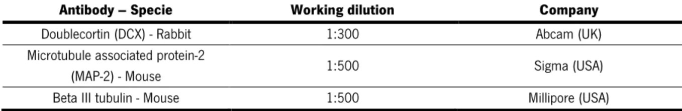
performed using 1X PBS with 10% newborn calf serum (NBCS; Biochrom, Germany) for 1 h at RT. hNPCs were then incubated with the primary antibodies (Table 1) diluted in 1X PBS with 10% NBCS for 1h at RT, after which they were washed with 1X PBS with 0.5% NBCS and incubated with the secondary antibodies (Table 2) diluted in 1X PBS with 10% NBCS for 1 h at RT. The cells were then incubated with the nuclear counterstain 4-6-diamidino-2-phenylindole-dhydrochloride (DAPI, 1:1000; Life Technologies, USA) for 10 min at RT. Afterwards, coverslips were mounted on glass slides using immu-mount (Thermo Scientific, UK). Finally, for quantification analysis, samples were observed under blind conditions using a fluorescence microscope (BX61, Olympus, Japan). For this purpose, four coverslips per condition and ten representative fields were chosen and analyzed. In order to normalize the data between the different sets, the results are presented in percentage of cells. This was calculated by counting the positive cells for the respective markers (Table 1), dividing this value by the total number of cells/field (DAPI-positive cells).
Table 1. Primary antibodies
Antibody – Specie Working dilution Company Doublecortin (DCX) - Rabbit 1:300 Abcam (UK) Microtubule associated protein-2
(MAP-2) - Mouse 1:500 Sigma (USA)
Beta III tubulin - Mouse 1:500 Millipore (USA) Table 2. Secondary antibodies
Antibody – Antigenicity Working dilution Company Alexa Fluor 488 - Goat anti-rabbit
1:1000 Life Technologies (USA) Alexa Fluor 488 - Goat anti-mouse
Alexa Fluor 594 - Goat anti-mouse
3.3. Stereotaxic surgeries 3.3.1. 6-OHDA lesions
All the experiments were done after the consent from the Portuguese national authority for animal research, Direcção Geral de Alimentação e Veterinária (ID: DGAV28421) and Ethical Subcommittee in Life and Health Sciences (SECVS; ID: SECVS-008/2013, University of Minho), conducted in accordance with the local regulations on animal care and experimentation (European Union Directive 2010/63/EU). Eight-weeks old Wistar-Han male rats (Charles River, Barcelona) were housed in pairs, in appropriate cages, under standard controlled conditions (12 h light/12 h dark cycles; RT at 22-24°C and 55% humidity; food and water ad libitum). Animals were handled for 1 week