Cell-based technologies for
Huntington’s disease
Mônica Santoro Haddad1, Cristiane Valverde Wenceslau2, Celine Pompeia3, Irina Kerkis4
ABSTRACT. Huntington’s disease (HD) is a fatal genetic disorder, which causes the progressive breakdown of neurons in the human brain. HD deteriorates human physical and mental abilities over time and has no cure. Stem cell-based technologies are promising novel treatments, and in HD, they aim to replace lost neurons and/or to prevent neural cell death. Herein we discuss the use of human fetal tissue (hFT), neural stem cells (NSCs) of hFT origin or embryonic stem cells (ESCs) and induced pluripotent stem cells (IPSCs), in clinical and pre-clinical studies. The in vivo use of mesenchymal stem cells (MSCs), which are derived from non-neural tissues, will also be discussed. All these studies prove the potential of stem cells for transplantation therapy in HD, demonstrating cell grafting and the ability to differentiate into mature neurons, resulting in behavioral improvements. We claim that there are still many problems to overcome before these technologies become available for HD patient treatment, such as: a) safety regarding the use of NSCs and pluripotent stem cells, which are potentially teratogenic; b) safety regarding the transplantation procedure itself, which represents a risk and needs to be better studied; and finally c) technical and ethical issues regarding cells of fetal and embryonic origin. Key words: Huntington’s disease, stem cells, safety issues, cell therapy.
TECNOLOGIAS CELULARES NO DOENÇA DE HUNTINGTON
RESUMO. A doença de Huntington (DH) é uma desordem genética que provoca a destruição progressiva dos neurônios no cérebro humano. A DH deteriora progressivamente as habilidades físicas e mentais humanas, e é incurável. Tecnologias terapêuticas baseadas em células representam novas alternativas para diversas doenças neurodegenerativas, pois visam substituir neurônios e/ou prevenir a morte neuronal. Nesta revisão discutirmos o uso de tecido fetal humano, células tronco neurais (CTN) de origem fetal ou de células tronco embrionárias ou células tronco pluripotentes induzidas, em estudos pré-clínicos e clínicos. Além disso, o uso terapêutico de células derivadas de tecidos não-neurais, como células tronco mesenquimais, também será discutido. Todos estes estudos provam o potencial do transplante celular na DH, demonstrando a sua habilidade em enxertar no encéfalo e diferenciar em neurônios in vivo, resultando em melhorias comportamentais e motoras em modelos animais da DH. Nós também discutimos que há muitos problemas a serem resolvidos quanto à terapia celular na DH, tais como: a) questões associadas à segurança do uso de CTNs, as quais são consideradas potencialmente teratogênicas; b) segurança do procedimento de transplante intracerebral, que representa um risco ao paciente; c) e, finalmente, questões técnicas e éticas associadas ao uso de células de origem fetal e embrionária.
Palavras-chave: doença de Huntington, células tronco, aspectos de segurança de células tronco, terapia celular.
INTRODUCTION
A
merican physician George Huntington irst described the disease in 1872. Hun-tington’s disease, which is also denominated Huntington’s syndrome or Huntington’s chorea, is a chronic, progressive and fatal neuropsychiatric disorder. HD is a hereditary autosomal dominant disease – 50% ofchil-dren born from disease carriers inherit HD. his disease induces destruction of neurons in the subcortical parts of the brain hemi-spheres (mainly – striped body): mostly the caudate and lenticular nuclei. HD leads to the expansion of the anterior horns of the lateral ventricles - a sign used to identify HD during neuroimaging.1,2
This study was conducted at the Instituto Butantan – Genética, São Paulo, SP, Brazil.
1MD. Faculdade de Medicina da Universidade de São Paulo – Neurologia São Paulo, São Paulo, SP, Brazil. 2 MD. Instituto Butantan - Genética, São Paulo, SP, Brazil. 3MD. Instituto Butantan - Genética, São Paulo, SP, Brazil. 4 MD, PhD. Instituto Butantan - Genética, São Paulo, São Paulo, Brazil.
Irina Kerkis. Instituto Butantan - Genética. Avenida Vital Brasil, 1500 – 05503-900 São Paulo SP – Brazil. E-mail: [email protected]
Disclosure: The authors report no conflicts of interest.
Received October 07, 2016. Accepted in final form November 10, 2016.
Neuronal death in HD occurs as a result of abnormal synthesis of the Huntingtin protein (HTT), which is par-ticularly prone to misfold and accumulates, leading to axonal and synaptic dysfunction. his accumulation of the misfolded mutant Huntingtin protein (mHTT) may afect intracellular transport of the protein or impair pro-tein degradation by the proteasome, leading to autoph-agy.3,4 However, it is not clear how mHTT accumulates in
the neuronal processes and induces an early pathological event in the brains of HD carriers.
here are two types of disease: adult-onset HD or juvenile HD, deined by the manifestations of the irst signs of the disease. In adult-onset HD, the irst symp-toms usually appear after age 30, but possibly may start earlier.3HD is characterized by a combination of motor
and neuropsychiatric disorders that can occur simulta-neously or in succession. Movement disorders include involuntary grimace, excessive gestures and others. Neu-ropsychiatric disorders, which usually appear after the motor disorders, may afect both cognitive and intellec-tual functions, as well as emotional status. Cognitive dys-function is most often expressed in problems of visual-spatial orientation.4,5Memory can also sufer, especially
in the ability to retain the information necessary for worklow. Patients may not be able to plan and organize their activities. With time, symptom severity increases, reaching that of dementia. Emotional and behavioral dis-orders primarily present themselves by the emergence of unmotivated aggression and irritability (observed in more than half of patients).6,4Approximately one third of
patients sufer from depression. Remarkably, the cause of death among HD carriers is not the disease itself, but accompanying illnesses, such as pneumonia and heart failure; approximately 30% of patients die from suicide. Juvenile HD is a less common, early-onset form of HD that starts in childhood or adolescence. Juvenile HD evolves very rapidly and presents severe symptoms that quickly lead to disability.7
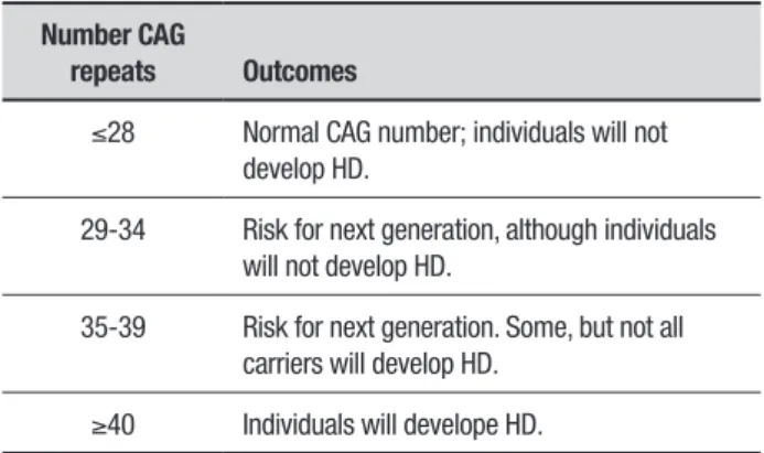
Currently, the diagnosis of HD is based on clinical (see above), genotypic and imaging indings. Since the discovery of the HD gene in 1993, a genetic test was developed that analyzes the HD mutation by counting the number of CAG repeats in the HD gene. his analysis enables direct conirmation of the diagnosis of HD in patients that exhibit HD-like symptoms. Using a blood sample, it is possible to demonstrate that an individual who does not have HD usually has 28 or fewer CAG repeats. HD carriers have 40 or more repeats, while a small proportion of individuals show a number of repeats that fall within a borderline region (Table 1).8
Magnetic Resonance Imaging (MRI) is a
neuroim-aging modality that provides the greatest spatial and contrast resolution for assessing the type of lesion asso-ciated with HD and allows the most precise diagnosis. Magnetic resonance spectroscopy (MRS) can also be used to show impaired brain energy metabolism associated with, for example, increased regional brain lactate that plays an important role in the pathogenesis of HD. In addition, computerized tomography (CT) scan, which determines the damaged areas of the brain, is also use-ful in HD diagnosis.9-14
In conclusion, neuroimaging, particularly MRI, remains a keystone in diagnosing and assessing the severity of HD. Genetic testing is extremely important to conirm the diagnosis, especially if the family history is not forthcoming and, moreover, genetic counseling of HD patients may provide guidance on the implications of the disease.
No cure is currently available. All HD treatment focuses on symptom relief, thus achieving only tempo-rary improvement of the patient’s neurological status. Although such treatment improves patient quality of life, the progress of the disease is unchanged and the neuro-nal loss persists.
Neuronal cells and diferent types of stem cells are a promising raw material for the development of new therapeutic strategies in HD: they may prevent neuronal loss and consequently delay the disease progress.
CELL-BASED THERAPIES
he main goal of cell-based technologies is to repair the mechanisms underlying disease initiation and progres-sion, achieved by replacement of dead or defective cells and through the trophic efect, which some cell types may confer after their transplantation into the injured site.15-17 Diferent cell types can be utilized in these
ther-apies, including fetal cells and tissues, progenitor cells or primary stem cells isolated from diferent tissues of
Table 1. Number of CAG repeats and HD outcomes that lead to HD development.
Number CAG
repeats Outcomes
≤28 Normal CAG number; individuals will not develop HD.
29-34 Risk for next generation, although individuals will not develop HD.
35-39 Risk for next generation. Some, but not all carriers will develop HD.
an adult organism.15,18-21 his review covers the current
state of cell-based therapies designed for HD. Some of these therapies are still at an initial stage of develop-ment and must be tested in animal models (preclinical studies) before they can be approved by regulatory agencies for use in clinical trials (involving humans). On the other hand, only a few studies have already reached clinical trials as will be discussed later.
HUMAN FETAL TISSUE TRANSPLANTS
IN HD PATIENTS
Human fetal tissue (hFT) has been used in basic research for decades.18 Clinical studies involving hFT aim to
rebuild brain structures and neural circuitries by trans-plantation of hFT into the damaged central nervous system (CNS) in HD patients. he hFT is derived from elective surgical terminations of pregnancy in fetuses at between 6 and 12 weeks of gestation. In the case of the hFT used for clinical transplantation, the tissue consists of the whole ganglionic eminence, corresponding to the striatal primordium that ultimately develops into the caudate and putamen.22-26 HD patients may receive
unilateral or bilateral hFT cell transplantation from several donated embryos (between ive and nine).
Generally, the surgery and procedure of hFT trans-plantation into the brain of HD patients is considered to be safe.22,26 However, one study reported that three out
of seven HD patients that underwent bilateral stereo-tactic transplantation developed subdural haemorrhages and two others required surgical drainage.24 No patients
had adverse efects to the associated cyclosporin immu-nosuppression, nor did any patients exhibit deteriora-tion following the procedure.26 he immunosuppressive
protocol adopted in these studies eiciently prevents immune rejection of the graft. Histological evidence of immune rejection, including the appearance of microglia and macrophages in the hFT transplantation sites, has never been observed.25,26
All these studies show the ability of hFT cells to delay disease progression and provide stabilization or improve-ment in several neurological indices of cognitive func-tioning, although these changes were not uniform across HD patients.22,24-26
MRI analyses showed hFT graft survival and even growth without damage to the surrounding tissue.
Another key concern that needs to be considered is the safety of hFT transplantation regarding tumor for-mation.27 Although the majority of the studies do not
show tumor formation after hFT transplantation,26
recently, one study emphasized the instability and risk of hFT grafts: the patient enrolled in a NIH-funded study
did not show any behavioral improvement after hFT transplantation into the brain, yet presented a growth of tissue mass, termed “graft overgrowth”.28 Such graft
overgrowth may be explained by the presence of imma-ture neuroepithelial cells actively dividing cells that express NSC markers such as Sox2, which are occasion-ally preserved in the hFT-derived cell suspension used for transplantation.28 Both these studies26,28 compromised
the safety of hFT transplantation and indicate the need for further in-depth studies using chemical and trans-genic preclinical models and a large number of animals. Additionally, it has been shown that not only hFT trans-plantation, but also fetal stem cell transtrans-plantation, may sporadically lead to brain tumor formation at the site of transplantation.29 Although these cells are subject
to restrictions regarding safety, they are able to recover medium spiny neurons (MSN).
A postmortem analysis of a brain carried out six months after a patient underwent transplantation demonstrated that hFT-derived cells were able to difer-entiate into MSNs which expressed dopamine-receptor related phosphoprotein (32 kDa) (DARPP-32), the neu-ronal nuclear antigen (NeuN), calretinin that can protect the MSNs against neurodegeneration in HD, and soma-tostatin, which is increased in the basal ganglia in HD.20,28
hese results are important given that HD is considered a disease of the striatum, characterized by vulnerability to degeneration and death of MSNs.1,2 Also, hFT
graft-derived astrocytes have also been observed after trans-plantation.20 Astrocytes are a sub-type of glial cells in the
CNS that are protective neurons against excitotoxicity by removing excessive glutamate from the extracellular space. Astrocyte recovery after hFT transplantation is a highly beneicial efect of the procedure, since in the HD brain, where mHTT accumulates in glial nuclei, there is a decrease in the expression of glutamate transporters in neurons and astroglial cells.30
Misfolded mHTT protein aggregates, which accumu-late as cytoplasmic aggregates and nuclear inclusions, are a hallmark feature of HD.3,4 As we mentioned above,
mHTT aggregates clump together and damage neurons. Recent research suggests that these mHTT proteins may also be transmitted from neuron to neuron. Inter-estingly, this hypothesis of pathogenic protein spread during neurodegeneration has been critically evalu-ated recently by Walsh,31 who suggests experimental
after the treatment did not show the presence of neuro-nal protein aggregates of mHTT within the hFT graft.32
In this context, all experimental data that support or detract from this hypothesis are of great importance.
ANIMAL MODELS OF HD
To date, of the possible transplant options that can beneit HD patients, only hFT has been used in clin-ical HD studies. hus, for the purposes of the present review, we have provided a short explanation about animal HD models, which are most commonly used in preclinical studies for other treatment approaches. Preferably, the HD animal model should provide similar genotypes and phenotypes as human HD.15,33 Chemical
HD animal models are commonly induced by treatment with quinolinic acid (QA)19,34,35 or 3 nitropropionic acid
(3-NP)36-39 Both of these chemical agents induce rapid
development of progressive HD, early disease onset, abnormal behavior, and neuropathological features. hese models are useful for rapid evaluation of the ther-apeutic hypothesis (acute models).15 In contrast,
trans-genic models are providing more precise systems for disease replication that allow evaluation of the therapy of interest in slow progressive degeneration.40
Transgenic models result from the random insertion of a portion of the human HTT gene containing the
cod-ing region for the polyglutamine repeat into the mouse genome, under control of several promoters. he most commonly used mouse HD models are R6/1, R6/2 and N171-Q82, which express truncated N-terminal frag-ments.41 Moreover, in contrast to the chemical models,
the transgenic HD models are powerful research tools because they recapitulate speciic HD features such as the accumulation of intracellular aggregates of mHTT in the cytoplasm or nucleus of neurons, known as inclusion bodies (IBs) (Figure 1).
Rodent HD models such as rats and mice are widely used to investigate cell-based therapy technologies.40 More recently, a transgenic nonhuman primate model in rhesus monkeys (Macaca mulatta) of HD was
devel-oped.42 Also, transgenic HD models were established
in large farm animals such as sheep (Ovis aries)43 and
Tibetan miniature pigs.44 However, transgenic HD
mod-els in large animals are high cost and technically diicult to use in routine cell-based therapy testing.
NEURAL STEM CELL TRANSPLANTATION
IN HD ANIMAL MODELS
It was long thought that neural cells could not undergo regeneration in injured or diseased human brain. However, advanced studies with NSCs in injured animal
A
B
Figure 1. Aggregate formation by HTT mutation in neuron cell derives inclusion bodies: [A] Inclusion body accumulation at axons and dendrites (cytoplasm); and [B] nucleus. Black arrows indicate inclusion bodies.
NSCs
Self-renewal Unlimited proliferative ability
Multipotent Differentiate into: neurons, astrocytes and
oligodendrocytes
NPGCs
Limited proliferative ability Unipotent
Bipotent Multipotent Neural precursor cells (NPCs)
Neural stem cells (NPCs) + Neural progenitor cells (NPGCs)
Figure 2. Hierarchical relationship between types of brain-derived stem cells.
models have shown the regenerative potential of these cells.15,45,46 NSCs are self-renewing cells able to generate
neurons, glial cells and astrocytes. NSCs can be derived from embryonic, fetal or adult tissues.47,48
Embryonic-fetal neural precursor cells (NPCs) are composed of a mixed population of multipotent NSCs and transit amplifying/intermediate progenitor cells (NPGCs) (Figure 2).49
Discrete populations of NSCs can be found in speciic areas of the postnatal human brain and seem to play an important role in postnatal growth as well as in recovery of neural tissue from injury, anoxia, or disease.50 NSCs
can be isolated at diferent stages of development and cultivated in vitro under appropriate culture conditions
NSCs (Figure 2). Neurospheres, when allowed to adhere to the substrate, start to produce neural rosettes (Figure 2), which are long-term self-renewing neuroblast-like cells. Murine or human NSCs have been used in preclin-ical studies to show the potential therapeutic efect of NSC-based technologies on neurogenesis in HD animal models.34,51,52 hese studies showed that transplantation
of NSCs improves motor function, extends life span and even lessens mHTT intracellular aggregate formation. In these studies, the cells were injected mainly by the intrastriatal route due to the inability of NSCs to pass though the blood brain barrier (BBB). NSCs exhibit robust engraftment at the site of injury. However, the diferentiation of these cells into neural cells is still controversial.34,37,38,51-53
he evaluation of teratogenicity of embryo-fetal derived NSCs is still lacking, although it is essential for the further translation of these studies into clinical tri-als. Due to the commitment of NSCs to neural fate, they may be considered an ideal cell type for the treatment of neurological diseases. However, NSCs are diicult to obtain in therapeutically signiicant quantities and pose serious ethical and religious limitations given that they are obtained from aborted fetuses.15
HFT AND NSC FROM HFT TRANSPLANTATION
AND ETHICAL CONCERNS
here is no doubt that hFT and NSCs are important in many research areas, especially in studies of human neurodegenerative diseases. HFT and NSCs help scien-tists investigate many aspects of basic science that cannot be studied in any other way. However, isolation of hFT and NSCs from hFT raises major ethical, political and religious controversies.21,54 We believe that these
ethical issues, associated with the limited number of experiments carried out so far, only minor success in these experimental therapies, as well as the potential teratogenicity of fetal and embryonic cells, are reasons why the use of ethically-accepted alternatives is starting to dominate the ield, especially in Brazil.
PLURIPOTENT STEM CELL TRANSPLANTATION
IN HD ANIMAL MODELS
Embryonic stem cells (ESCs) are pluripotent cells isolated from the inner cell mass of early embryos. hese cells can generate a whole organism when re- introduced back into the embryonic environment (e.g. blastocysts) (Figure 3). ESCs are also able to form gametes – repro-ductive cells. Additionally, ESCs form noncancerous tumors called teratomas, which is one of their funda-mental traits.55,56
As mentioned before, tumor formation is a major safety concern for those who hope to use the cells thera-peutically.57 Due to this feature of ESCs, they cannot be
used unspecialized in preclinical and clinical studies. To analyze the therapeutic potential of human ESCs in HD animal models, they have been reversed to NPCs that have been transplanted directly into the striatum of the animal models. Diferent types of NPCs may be obtained, which depends on the protocol of human ESC diferentia-tion.58-60 Rosette-forming ESC–derived NPCs (Figure 3)
are unable to diferentiate in vivo into medial spiny
neu-rons (MSN), which is a principal target of HD therapies.59
However, they have the capacity to diferentiate into other neuron types and into astrocytes. Preclinical short-term studies show efective recovery of motor deicit after hESC-derived NSC transplantation in the QA rat model.58-60 he main problem in using ESC-derived NPCs
in the clinic is the need to control their proliferation in order to avoid neural cell graft overgrowth.59,60
Laboratory-grown induced pluripotent stem cells (iPSCs) are a type of pluripotent cell that can be gener-ated directly from adult cells (Figure 3). Currently, practi-cally any type of somatic adult cell can be reprogrammed back into an embryonic-like pluripotent state. he iPSCs share all principal characteristics of ESCs, having the abil-ity of long-term self-renewal, maintaining an unspecial-ized state; and giving rise to (under speciic treatments) specialized cell types and teratomas when injected in
vivo.61,62 However, the pluripotential ability of the iPSCs
may be inluenced by the adult donor cell source.63,64 he
therapeutic potential of hiPSCs, as well as that of hiPSC-derived NPCs, has also been investigated using HD ani-mal models.65-67 hese cells have been transplanted into
both chemical65 and transgenic HD rodent models via the
ipsilateral ventricular route.67 hese cells graft into
recipi-ent brains and diferrecipi-entiate into GABAergic MSNs65,67,68
and astrocytes.68 After transplantation, a modest
reduc-tion in striatal neuronal atrophy, a hallmark of HD dis-ease that appears before the onset of motor symptoms, can be observed.68,69
After transplantation of NSCs derived from iPSCs, short- and medium-term functional motor improve-ments have been documented in comparison with sham group animals.65,67,68 However, long-term motor
func-tional recovery from HD followingESC- and iPSC-derived NSC transplantation still needs to be further evaluated.
MESENCHYMAL STEM CELLS
IN HD ANIMAL MODELS
placenta), bone marrow and adipose tissue. Isolation of these cells is associated with fewer ethical concerns when compared with ESCs. MSCs have a ibroblast-like morphology (Figure 3), express a set of speciic markers and adhere to plastic. hey are of exceptional interest because they are unspecialized cells capable of self-renewal and diferentiation into speciic cell types, especially into mesoderm derivatives.70-73 However,
a growing number of studies has demonstrated their capacity to also diferentiate into neural cells.74
Further-more, the number of clinical trials using MSCs is rising every year and, at least, the clinical safety of these cells has been conirmed.75
he biological function of MSCs is to replace tissues in response to normal cellular turnover or trauma.76 It
has been also suggested that these cells can be recruited to nascent microvascular walls during development and postnatal growth.77 Accordingly, MSCs in their
anatomi-cal sites – stem cell niches – have their growth arrested until they are triggered to restart proliferation and even diferentiation in response to physiological cues, such as tissue turnover or repair, or experimentally, when iso-lated and cultivated in vitro. In bone marrow, for
exam-ple, subendothelial osteoprogenitors become stem cells acting as pericytes in diferent postnatal tissues. here-fore, as an alternative to diferentiation, tissue-speciic MSCs may function to support the regeneration of other local cell types. Such support is carried out by MSCs after transplantation into the unhealthy organism via the secretion of a variety of bioactive molecules such as cyto-kines, which have ‘trophic activities’ that can promote a regenerative microenvironment.16,17 MSCs also beneit
tissue reconstruction by stimulating angiogenesis, the production of immunomodulatory mediators and even by delivering other molecules to the injured site. MSCs also act by reducing chronic inlammation, inhibiting apoptosis and decreasing scar formation. hey are able to stimulate mitosis of tissue-intrinsic stem cells and reduce the destructive efects of oxidative stress.16,17,78
MSCs express very low levels of MHC class I proteins and lack MHC class II proteins, and can therefore be trans-planted into other organisms of the same or diferent species without rejection.73,79
MSCs isolated from bone, adipose tissue and umbili-cal cord have been used in diferent chemiumbili-cal (QA and 3-NP) and genetic animal models (R6/2-J2, N171-82Q,
R6/2) of HD, which were transplanted using the intra-cerebral route (directly into the striatum). hese stud-ies demonstrate that MSC transplantation leads to behavioral and memory improvements, reduced brain damage, improvement of striatal degeneration, and enhanced expression of several striatal growth factors, which are attributed to the neuroprotective efect of MSCs. MSC transplantation shows robust cell engraft-ment at the site of lesion, as well as the ability of these cells to migrate to adjacent areas.80-83 Although the actual
diferentiation of MSCs into neurons is controversial, it is not required to justify the beneicial activity of these cells in HD. Furthermore, MSC transplantation has never been shown to lead to teratoma or graft overgrowth for-mation, indicating the safety of these cells, at least in animal models.
NEUROTROPHIC FACTORS
Neurotrophic factors, such as brain-derived neuro-trophic factor (BDNF), are essential contributors of CNS neuron function. Studies demonstrate their reduced availability in diseased brains, thus suggesting that they play an important role in neurological disorders and, in particular, in HD.84,85 Under non-pathologic conditions,
BDNF is synthesized in the cortex, the substantia nigra pars compacta, the amygdala, and in the thalamus. All these regions supply the striatum with BDNF.86,87 In
HD, the deicit of BDNF in the striatum may be due to reduced BDNF gene transcription in the cerebral cortex or reduced BDNF vesicle transport (or both).88,89 he
decrease in BDNF expression observed in HD impairs dopaminergic neuronal function, which may be asso-ciated with HD motor disturbances. As a result, many studies have been carried out to examine whether increasing BDNF levels may help treat HD.37,83,88
NPCs and MSCs, besides their diferentiation ability to produce neurons, have been extensively investigated with respect to neurotrophic factor secretion.37,90
Stud-ies have shown that MSCs derived from adipose tissue and bone marrow are able to secrete BDNF in vitro.83,91-93
However, MSCs secrete BDNF at low levels, which are dependent on tissue source used for MSC isolation and on donor characteristics.92,94,95
herefore, recent studies have generated NSCs derived from ESCs that overexpress BDNF in order to evaluate whether they have enhanced therapeutic abili-ties in HD. Neurons derived from BDNF-GFP-expressing ESCs harbor a more complex dendritic morphology and diferentiate into the GABAergic lineage.96
CONCLUSION
Great advances have been made in cell-based therapies over recent decades and it is expected that, in the future, such therapies will be provided alone or in combination with traditional medication, new small molecule drugs and biological drugs to patients who sufer from neuro-logical conditions such as HD. Figure 2 summarizes the major advances that have been made in cell-based technologies and their use for the treatment of HD in clinical and preclinical studies.
Notably, all these cell-based technologies may be roughly divided into two: NSC- and MSC-based, since none of the pluripotent cells (such as ESCs or IPSCs) can be used intact/unspecialized due to the risk of terato-genicity after transplantation into the lesion site. NSCs derived from iPSCs look set to substitute hFT, fetal- as well as ESC-derived NSCs, thus avoiding ethical and reli-gious considerations.
Another important conclusion is that these difer-ent cell types produce very similar clinical outcomes and therefore can be used interchangeably. he major advantage of MSCs over NSC derived from diferent cell types is that they do not produce teratomas or graft over-growth. Additionally, they express neurotrophic factors, albeit at low levels, which are apparently important for HD improvement.
he major problem of cell-based technologies in HD concerns the route of administration, which is usually intracerebral. his will be a great obstacle for the use of cell-based technologies in HD patients, which is a genetic disease and may require more than one cell transplanta-tion. None of the clinical and preclinical HD studies have yet used the systemic intravenous route,21,24,26,27 probably
due to the fear that these cells will not penetrate the BBR in signiicant numbers. However, we believe that MSCs can be systemically administrated and have tropism for sites of injury, even the brain, as in the case of HD. However, further studies are necessary to better under-stand this administration route and prove its eicacy and safety in HD.
REFERENCES
1. Davies S, Ramsden DB. Huntington’s disease. Mol Pathol. 2001;54(6): 409-13.
2. Vonsattel JP, DiFiglia M. Huntington disease. J Neuropathol Exp Neurol. 1998;57(5):369-84.
3. The Huntington’s Disease Collaborative Research Group. A novel gene containing a trinucleotide repeat that is expanded and unstable on Huntington’s disease chromosomes. Cell. 1993;72(6):971-83. 4. Ross CA, Tabrizi SJ. Huntington’s disease: from molecular pathogenesis
to clinical treatment. Lancet Neurol. 2011;10:83-98. 5. Hayden MR. Huntington’s chorea. London: Springer; 1981.
6. Folstein SE. The psychopathology of Huntington’s disease. Res Publ Assoc Res Nerv Ment Dis. 1991;69:181-91.
7. Ribaï P, Nguyen K, Hahn-Barma V, Gourfinkel-An I, Vidailhet M, Legout A, et al. Psychiatric and cognitive difficulties as indicators of juvenile huntington disease onset in 29 patients. Arch Neurol. 2007;64(6):813-9. 8. Agostinho LA, dos Santos SR, Alvarenga RMP, Paiva CLA. A systematic
review of the intergenerational aspects and the diverse genetic profiles of Huntington’s disease. Genet Mol Res. 2013;2(2):1974-81.
9. Ho VB, Chuang HS, Rovira MJ, Koo B. Juvenile Huntington disease: CT and MR features. AJNR Am J Neuroradiol. 1995;16(7):1405-12. 10. Paulsen JS, Zimbelman JL, Hinton SC, Langbehn DR, Leveroni CL,
Benjamin ML, et al. fMRI biomarker of early neuronal dysfunction in presymptomatic Huntington’s Disease. AJNR Am J Neuroradiol. 2004;25(10):1715-21.
11. Dormont D, Seidenwurm DJ. Expert Panel on Neurologic Imaging; Amer-ican College of Radiology. Dementia and movement disorders. AJNR Am J Neuroradiol. 2008;29(1):204-6.
12. Maddock RJ, Buonocore MH. MR spectroscopic studies of the brain in psychiatric disorders. Curr Top Behav Neurosci. 2012;11:199-251. 13. Ahmad R, Bourgeois S, Postnov A, Schmidt ME, Bormans G, Van Laere
K, et al. PET imaging shows loss of striatal PDE10A in patients with Huntington disease. Neurology. 2014;82(3):279-81.
14. Macerollo A, Perry R, Stamelou M, Batla A, Mazumder AA, Adams ME, et al. Susceptibility-weighted imaging changes suggesting brain iron accumulation in Huntington’s disease: an epiphenomenon which causes diagnostic difficulty. Eur J Neurol. 2014;21(2):e16-7.
15. Kerkis I, Haddad MS, Valverde CW, Glosman S. Neural and mesen-chymal stem cells in animal models of Huntington’s disease: past experi-ences and future challenges.Stem Cell Res Ther. 2015;6:232. 16. Caplan AI, Dennis JE. Mesenchymal stem cells as trophic mediators. J
Cell Biochem. 2006;98(5):1076-84.
17. Caplan AI. MSCs: the new medicine. In: Vertes AA, Qureshi N, Caplan AI, Babiss LE, editors. Stem Cells in Regenerative Medicine: Science, Regulation and Business Strategies. John Wiley & Sons; 2013. 18. Fetal Research and Applications: A Conference Summary. Conference
Committee on Fetal Research and Applications, Institute of Medicine. National Academy Press, Washington, D.C. 1994.
19. Bantubungi K, Blum D, Cuvelier L, Wislet-Gendebien S, Rogister B, Brouillet E, et al. Stem cell factor and mesenchymal and neural stem cell transplantation in a rat model of Huntington’s disease. Mol Cell Neurosci. 2008;37:454-70.
20. Capetian P, Knoth R, Maciaczyk J, Pantazis G, Ditter M, Bokla L, et al. Histological findings on fetal striatal grafts in a Huntington’s disease patient early after transplantation. Neuroscience. 2009;160(3):661-75. 21. Freeman TB, Cicchetti F, Bachoud-Levi AC, Dunnett SB. Technical
factors that influence neural transplant safety in Huntington’s disease. Exp Neurol. 2011;227:1-9.
22. Kopyov OV, Jacques S, Lieberman A, Duma CM, Eagle KS. Safety of intrastriatal neurotransplantation for Huntington’s disease patients. Exp Neurol. 1998;149(1):97-108.
23. Philpott LM, Kopyov OV, Lee AJ, Jacques S, Duma CM, Caine S, et al. Neuropsychological functioning following fetal striatal transplanta-tion in Huntington’s chorea: three case presentatransplanta-tions. Cell Transplant. 1997;6(3):203-12.
24. Hauser RA, Furtado S, Cimino CR, Delgado H, Eichler S, Schwartz S, et al. Bilateral human fetal striatal transplantation in Huntington’s disease. Neurology. 2002;58:687-95.
25. Gallina P, Paganini M, Lombardini L, Saccardi R, Marini M, De Cristofaro MT, et al. Development of human striatal anlagen after transplantation in a patient with Huntington’s disease. Exp Neurol. 2008;213(1):241-4. 26. Gallina P, Paganini M, Lombardini L, Giordano G, Mascalchi M, Romoli
AM, et al. Progress in restorative neurosurgery: human fetal striatal trans-plantation in Huntington’s disease. J Neurosurg Sci. 2011;55(4):371-81. 27. Jeong JO, Han JW, Kim JM, Cho HJ, Park C, Lee N, et al. Malignant
tumor formation after transplantation of short-term cultured bone marrow
mesenchymal stem cells in experimental myocardial infarction and diabetic neuropathy. Circ Res. 2011;108:1340-7.
28. Keene CD, Chang RC, Leverenz JB, Kopyov O, Perlman S, Hevner RF, et al. A patient with Huntington’s disease and long-surviving fetal neural transplants that developed mass lesions. Acta Neuropathol. 2009;117(3):329-38.
29. Amariglio N, Hirshberg A, Scheithauer BW, Cohen Y, Loewenthal R, Trakhtenbrot L, et al. Donor-derived brain tumor following neural stem cell transplantation in an ataxia telangiectasia patient. PLoS Med. 2009; 6(2):e1000029.
30. Maragakis NJ, Rothstein JD. Glutamate transporters in neurologic disease. Arch Neurol. 2001;58(3):365-70.
31. Walsh DM, Selkoe DJ.A critical appraisal of the pathogenic protein spread hypothesis of neurodegeneration. Nat Rev Neurosci. 2016;17(4): 251-60.
32. Freeman TB, Cicchetti F, Hauser RA, Deacon TW, Li XJ, Hersch SM, et al. Transplanted fetal striatum in Huntington’s disease: pheno-typic development and lack of pathology. Proc Natl Acad Sci U S A. 2000;97(25):13877-82.
33. Ramaswamy S, McBride JL, Kordower JH. Animal models of Hunting-ton’s disease. ILAR J. 2007;48(4):356-73.
34. Lee ST, Chu K, Park JE, Lee K, Kang L, Kim SU, et al. Intravenous administration of human neural stem cells induces functional recovery in Huntington’s disease rat model. Neurosci Res. 2005;52:243-9. 35. Pierozan P, Fernandes CG, Dutra MF, Pandolfo P, Ferreira F, de Lima
BO, Porciúncula L, Wajner M, Pessoa-Pureur R.Biochemical, histopatho-logical and behavioral alterations caused by intrastriatal administration of quinolic acid to young rats. FEBS J. 2014;281(8):2061-73.
36. Brouillet E, Jenkins BG, Hyman BT, Ferrante RJ, Kowall NW, Srivastava R, et al. Age-dependent vulnerability of the striatum to the mitochondrial toxin 3- nitropropionic acid. J Neurochem. 1993;60(1):356-9. 37. Ryu JK, Kim J, Cho SJ, Hatori K, Nagai A, Choi HB, et al. Proactive
transplantation of human neural stem cells prevents degeneration of striatal neurons in a rat model of Huntington disease. Neurobiol Dis. 2004;16:68-77.
38. Rosenstock TR, Carvalho AC, Jurkiewicz A, Frussa-Filho R, Smaili SS. Mitochondrial calcium, oxidative stress and apoptosis in a neurodegen-erative disease model induced by 3-nitropropionic acid. J Neurochem. 2004;88(5):1220-8.
39. Roberts TJ, Price J, Williams SC, Modo M. Preservation of striatal tissue and behavioral function after neural stem cell transplantation in a rat model of Huntington’s disease. Neuroscience. 2006;139:1187-99. 40. Pouladi MA, Morton AJ, Hayden MR. Choosing an animal model for the
study of Huntington’s disease. Nat Rev Neurosci. 2013;14(10):708-21. 41. Mangiarini L, Sathasivam K, Seller M, Cozens B, Harper A, Hetherington
C, et al. Exon 1 of the HD gene with an expanded CAG repeat is suffi-cient to cause a progressive neurological phenotype in transgenic mice. Cell. 1996;87(3):493-506
42. Yang SH, Cheng PH, Banta H, Piotrowska-Nitsche K, Yang JJ, Cheng EC, et al. Towards a transgenic model of Huntington’s disease in a non-human primate. Nature. 2008;453:921-4.
43. Jacobsen JC, Bawden CS, Rudiger SR, McLaughlan CJ, Reid SJ, Wald-vogel HJ, et al. An ovine transgenic Huntington’s disease model. Hum Mol Genet. 2010;19(10):1873-82.
44. Yang D, Wang CE, Zhao B, Li W, Ouyang Z, Liu Z, et al. Expression of Huntington’s disease protein results in apoptotic neurons in the brains of cloned transgenic pigs. Hum Mol Genet. 2010;19(20):3983-94. 45. Armstrong RJ, Watts C, Svendsen CN, Dunnett SB, Rosser AE. Survival,
neuronal differentiation, and fiber outgrowth of propagated human neural precursor grafts in an animal model of Huntington’s disease. Cell Trans-plant. 2000;9(1):55-64.
46. Doeppner TR, Kaltwasser B, Bähr M, Hermann DM. Effects of neural progenitor cells on post-stroke neurological impairment-a detailed and comprehensive analysis of behavioral tests. Front Cell Neurosci. 2014;8:338.
47. Doe CQ. Neural stem cells: balancing self-renewal with differentiation. Development. 2008;135:1575-87
48. Brustle O, Jones KN, Learish RD, Karram K, Choudhary K, Wiestler OD, et al. Embryonic stem cell-derived glial precursors: a source of myelina-ting transplants. Science. 1999;285(5428):754-6.
49. Calof AL, Bonnin A, Crocker C, Kawauchi S, Murray RC, Shou J, et al. Progenitor cells of the olfactory receptor neuron lineage. Microsc Res Tech. 2002;58(3):176-88.
51. McBride JL, Behrstock SP, Chen EY, Jakel RJ, Siegel I, Svendsen CN, et al. Human neural stem cell transplants improve motor function in a rat model of Huntington’s disease. J Comp Neurol. 2004;475:211-9. 52. Johann V, Schiefer J, Sass C, Mey J, Brook G, Kruttgen A, et al. Time
of transplantation and cell preparation determine neural stem cell survival in a mouse model of Huntington’s disease. Exp Brain Res. 2007;177:458-70.
53. Vazey EM, Chen K, Hughes SM, Connor B. Transplanted adult neural progenitor cells survive, differentiate and reduce motor function impair-ment in a rodent model of Huntington’s disease. Exp Neurol. 2006;199: 384-96.
54. Lunn JS, Sakowski SA, Hur J, Feldman EL. Stem cell technology for neurodegenerative diseases. Ann Neurol. 2011;70(3):353-61. 55. Thomson JA, Itskovitz-Eldor J, Shapiro SS, Waknitz MA, Swiergiel JJ,
Marshall VS, et al. Embryonic stem cell lines derived from human blas-tocysts. Science. 1998;282(5391):1145-7.
56. Wu J, Yamauchi T, Izpisua Belmonte JC. An overview of mammalian pluripotency. Development. 2016;143(10):1644-8.
57. Blum B, Bar-Nur O, Golan-Lev T, Benvenisty N. The anti-apoptotic gene surviving contributes to teratoma formation by human embryonic stem cells. Nat Biotechnol. 2009;27(3):281-7.
58. Song J, Lee ST, Kang W, Park JE, Chu K, Lee SE, et al. Human embry-onic stem cell-derived neural precursor transplants attenuate apomor-phine-induced rotational behavior in rats with unilateral quinolinic acid lesions. Neurosci Lett. 2007;423(1):58-61.
59. Aubry L, Bugi A, Lefort N, Rousseau F, Peschanski M, Perrier AL. Striatal progenitors derived from human ES cells mature into DARPP32 neurons in vitro and in quinolinic acid-lesioned rats. Proc Natl Acad Sci USA. 2008;105(43):16707-12.
60. Vazey EM, Dottori M, Jamshidi P, Tomas D, Pera MF, Horne M, et al. Comparison of transplant efficiency between spontaneously derived and noggin- primed human embryonic stem cell neural precursors in the quinolinic acid rat model of Huntington’s disease. Cell Transplant. 2010;19(8):1055-62.
61. Takahashi K, Yamanaka S. Induction of pluripotent stem cells from mouse embryonic and adult fibroblast cultures by defined factors. Cell. 2006;126(4):663-76.
62. Takahashi K, Tanabe K, Ohnuki M, Narita M, Ichisaka T, Tomoda K, et al. Induction of pluripotent stem cells from adult human fibroblasts by defined factors. Cell. 2007;131(5):861-72.
63. Beltrão-Braga PC, Pignatari GC, Maiorka PC, OliveiraNA, Lizier NF, Wenceslau CV, et al. Feeder-free derivation of inducedpluripotent stem cells from human immature dental pulp stem cells. Cell Transplant. 2011;20(11-12):1707-19.
64. Quattrocelli M, Giacomazzi G, Broeckx SY, Ceelen L, Bolca S, Spaas JH, et al. Equine-Induced Pluripotent Stem Cells Retain Lineage Commit-ment Toward Myogenic and Chondrogenic Fates. Stem Cell Reports. 2016;6(1):55-63.
65. Jeon I, Lee N, Li JY, Park IH, Park KS, Moon J, et al. Neuronal proper-ties, in vivo effects, and pathology of a Huntington’s disease patient-derived induced pluripotent stem cells. Stem Cells. 2012;30(9):2054-62. 66. Juopperi TA, Kim WR, Chiang CH, Yu H, Margolis RL, Ross CA, et al.
Astrocytes generated from patient induced pluripotent stem cells recapit-ulate features of Huntington’s disease patient cells. Mol Brain. 2012;5:17. 67. Jeon I, Choi C, Lee N, Im W, Kim M, Oh SH, et al. In Vivo Roles of a Patient- Derived Induced Pluripotent Stem Cell Line (HD72-iPSC) in the YAC128 Model of Huntington’s Disease. Int J Stem Cells. 2014;7(1): 43-47. 68. Mu S, Wang J, Zhou G, Peng W, He Z, Zhao Z, et al. Transplantation of induced pluripotent stem cells improves functional recovery in Hunting-ton’s disease rat model. PLoS One 2014;9(7):e101185.
69. Montserrat N, Nivet E, Sancho-Martinez I, Hishida T, Kumar S, Miquel L, et al. Reprogramming of human fibroblasts to pluripotency with lineage specifiers. Cell Stem Cell. 2013;13(3):341-50.
70. Friedenstein AJ. Osteogenic stem cells in bone marrow. In: Heersche JNM, Kanis JA, editors. Bone and Mineral Research. Amsterdam: Else-vier; 1990:243-72.
71. Caplan AI. Mesenchymal stem cells. J Orthop Res. 1991;9(5):641-50 72. Caplan AI. Review: mesenchymal stem cells: cell-based reconstructive
therapy in orthopedics. Tissue Eng. 2005;11(7-8):1198-211. 73. Dominici M, Le Blanc K, Mueller I, Slaper-Cortenbach I, Marini F, Krause
D, et al. Minimal criteria for defining multipotent mesenchymal stromal cells. The International Society for Cellular Therapy position statement. Cytotherapy. 2006;8:315-7.
74. Bianco P, Robey PG, Simmons PJ. Mesenchymal Stem Cells: Revisiting History, Concepts and Assays. Cell Stem Cell. 2008;2(4):313-9. 75. Gratwohl A, Pasquini MC, Aljurf M, Atsuta Y, Baldomero H, Foeken L, et
al. Worldwide Network for Blood and Marrow Transplantation (WBMT). One million haemopoietic stem-cell transplants: a retrospective obser-vational study. Lancet Haematol. 2015;2(3):e91-100.
76. Kørbling M, Estrov Z. Adult stem cells for tissue repair - a new thera-peutic concept? N Engl J Med. 2003;349(6):570-82
77. Jain RK. Molecular regulation of vessel maturation. Nat Med. 2003;9: 685-93.
78. Bernardo ME, Fibbe WE. Mesenchymal stromal cells: sensors and switchers of inflammation. Cell Stem Cell. 2013;13(4):392-402. 79. Ankrum JA, Ong JF, Karp JM. Mesenchymal stem cells: immune evasive,
not immune privileged. Nat Biotechnol. 2014;32(3):252-60.
80. Snyder BR, Chiu AM, Prockop DJ, Chan AW. Human multipotent stromal cells (MSCs) increase neurogenesis and decrease atrophy of the stri-atum in a transgenic mouse model for Huntington’s disease. PLoS One. 2010;5:e9347.
81. Lee ST, Chu K, Jung KH, Im WS, Park JE, Lim HC, et al. Slowed progression in models of Huntington disease by adipose stem cell trans-plantation. Ann Neurol. 2009;66:671-81.
82. Moraes L, Vasconcelos-dos-Santos A, Santana FC, Godoy MA, Rosado-de-Castro PH, Jasmin, et al. Neuroprotective effects and magnetic reso-nance imaging of mesenchymal stem cells labeled with SPION in a rat model of Huntington’s disease. Stem Cell Res. 2012;9:143-55. 83. Rossignol J, Boyer C, Leveque X, Fink KD, Thinard R, Blanchard F, et
al. Mesenchymal stem cell transplantation and DMEM administration in a 3NP rat model of Huntington’s disease: morphological and behavioral outcomes. Behav Brain Res. 2011;217:369-78.
84. Zuccato C, Ciammola A, Rigamonti D, Leavitt BR, Goffredo D, Conti L, et al. Loss of huntingtin-mediated BDNF gene transcription in Huntington’s disease. Science. 2001;293(5529):493-8.
85. Zuccato C, Cattaneo E. Brain-derived neurotrophic factor in neurode-generative diseases. Nat Rev Neurol. 2009;5(6):311-22.
86. Altar CA, Cai N, Bliven T, Juhasz M, Conner JM, Acheson AL, et al. Anterograde transport of brain-derived neurotrophic factor and its role in the brain. Nature. 1997;389(6653):856-60.
87. Baquet ZC, Gorski JA, Jones KR. Early striatal dendrite deficits followed by neuron loss with advanced age in the absence of anterograde corti-calbrain-derived neurotrophic factor. J Neurosci. 2004;24(17):4250-8. 88. Zuccato C, Cattaneo E. Role of brain-derived neurotrophic factor in
Huntington’s disease. Prog Neurobiol. 2007;81(5-6):294-330. 89. Baydyuk M, Xu B. BDNF signaling and survival of striatal neurons. Front
Cell Neurosci. 2014;8:254.
90. Lu P, Jones LL, Snyder EY, Tuszynski MH. Neural stem cells constitu-tively secrete neurotrophic factors and promote extensive host axonal growth after spinal cord injury. Exp Neurol. 2003;181(2):115-29. 91. Sadan O, Shemesh N, Cohen Y, Melamed E, Offen D. Adult neurotrophic
factor- secreting stem cells: a potential novel therapy for neurodegenera-tive diseases. Isr Med Assoc J. 2009;11(4):201-4.
92. Zemelkov VI, Kozhukharova IV, Kovaleva ZV, Domnina AP, Pugovkina NA, Fridlianskaia II, et al. BDNF secretion in human mesenchymal stem cells isolated from bone marrow, endometrium and adipose tissue. Tsitologiia. 2014;56(3):204-11.
93. Kurozumi K, Nakamura K, Tamiya T, Kawano Y, Ishii K, Kobune M, et al. Mesenchymal stem cells that produce neurotrophic factors reduce ischemic damage in the rat middle cerebral artery occlusion model. Mol Ther. 2005;11(1):96-104
94. Razavi S, Razavi MR, Zarkesh Esfahani H, Kazemi M, Mostafavi FS. Comparing brain-derived neurotrophic factor and ciliary neurotrophic factor secretion of induced neurotrophic factor secreting cells from human adipose and bone marrow-derived stem cells. Dev Growth Differ. 2013;55(6):648-55
95. Kim SJ, Moon GJ, Chang WH, Kim YH, Bang OY; STARTING-2 (STem cell Application Researches and Trials In NeuroloGy-2) collaborators. Intravenous transplantation of mesenchymal stem cells preconditioned with early phase stroke serum: current evidence and study protocol for a randomized trialal. Trials. 2013;14:317.
![Figure 1. Aggregate formation by HTT mutation in neuron cell derives inclusion bodies: [A] Inclusion body accumulation at axons and dendrites (cytoplasm); and [B] nucleus](https://thumb-eu.123doks.com/thumbv2/123dok_br/15189156.527167/4.892.447.781.108.365/aggregate-formation-mutation-inclusion-inclusion-accumulation-dendrites-cytoplasm.webp)
