Stem cells and bioengineering in the current context of dentistry and
general health
Células-tronco e bioengenharia no contexto atual da odontologia e saúde
geral
DOI:10.34117/bjdv6n11-570
Recebimento dos originais: 25/10/2020 Aceitação para publicação: 26/11/2020
Marcella Vieira Ambrosio
Health Sciences Center - Dentistry, State University of Northern Paraná – UENP, Jacarezinho, PR, Brazil.
João Lopes Toledo Neto
Health Sciences Center - Dentistry, State University of Northern Paraná – UENP, Jacarezinho, PR, Brazil.
Augusto Alberto Foggiato
Health Sciences Center - Dentistry, State University of Northern Paraná – UENP, Jacarezinho, PR, Brasil.
Research Institute in Dentistry, Medicine and Phototherapy Foggiato, Jacarezinho, PR, Brazil [email protected]
Douglas Fernandes da Silva
Health Sciences Center - Dentistry, State University of Northern Paraná – UENP, Jacarezinho, PR, Brazil.
Research Institute in Dentistry, Medicine and Phototherapy Foggiato, Jacarezinho, PR, Brazil [email protected]
ABSTRACT
Mesenchymal stem cells (MSC) are undifferentiated cell structures with the ability to both renew themselves and differentiate into different specialized cell types. These can be found in some structures of the dental organ, such as in the dental pulp, in the periodontal ligament, in the dental follicle and in the apical papilla, changing to being called dental stem cells (DSC). When compared with other types of MSC, DSC have the benefit of being obtained in a minimally invasive and painless way. These particularities make them the ideal tool for applicability in dental and non-dental therapies. In this review was emphasize that, in an appropriate culture medium, DSC can be used for the restoration of dental structures, in the repair of nervous and bone tissue and in the treatment of neurodegenerative, hepatic and ocular deficits. In addition, in the current context of COVID-19, MSC are being investigated and applied to treat complications caused by the new coronavirus, which has been extended to DSC. In summary, this review article concludes that understanding the functioning of these cells and their properties may allow the development of more effective treatments or even the cure of several pathologies.
Keywords: mesenchymal stem cells, odontology, culture medium, bioengineering, SARS-CoV-2.
RESUMO
As células-tronco mesenquimais (MSC) são estruturas celulares indiferenciadas com a capacidade de se renovar e se diferenciar em diferentes tipos de células especializadas. Estas podem ser encontradas em algumas estruturas do órgão dentário, como na polpa dentária, no ligamento periodontal, no folículo dentário e na papila apical, passando a ser chamadas de células-tronco dentárias (DSC). Quando comparado com outros tipos de MSC, o DSC tem o benefício de ser obtido de forma minimamente invasiva e indolor. Essas particularidades os tornam a ferramenta ideal para aplicabilidade em terapias odontológicas e não odontológicas. Nesta revisão foi enfatizado que, em meio de cultura apropriado, o DSC pode ser utilizado na restauração de estruturas dentais, na reparação de tecido nervoso e ósseo e no tratamento de déficits neurodegenerativos, hepáticos e oculares. Além disso, no contexto atual do COVID-19, os MSC estão sendo investigados e aplicados no tratamento de complicações causadas pelo novo coronavírus, que foi estendido ao DSC. Em suma, este artigo de revisão conclui que a compreensão do funcionamento dessas células e de suas propriedades pode permitir o desenvolvimento de tratamentos mais eficazes ou mesmo a cura de diversas patologias.
Palavras-chave: células-tronco mesenquimais, odontologia, meio de cultura, bioengenharia, SARS-CoV-2.
1 INTRODUCTION
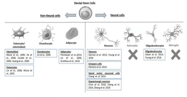
Mesenchymal stem cells (MSC) are defined as multipotent cells that have the ability to differentiate into different cell lines, such as osteoblasts, chondrocytes, adipocytes (GARCIA; ROQUE; SILVA, 2017) and neurocytes (JI et al., 2019) (figure 1) under the presence of extracellular matrix and growth factors (SOARES et al., 2007).
Figure 1: Summary of differentiation of dental stem cells (DSC) into non-neural cells (eg, osteocytes, chondrocytes and adipocytes) or neural cells (eg, neurons and oligodendrocytes). The X indicates that there is no data on the differentiation of DSCs into astrocytes or microglia (RELAÑO-GINÉS et al., 2019).
Among the various types of MSC, such as bone marrow (BM-MSCs) represent the most popular source used in clinical applications. However, they have some negative points, such as invasive collection and a low rate of cell yield (ISOBE et al., 2016). Thus, it is notable that there is a need for alternative sources to obtain MSC.
In the current scenario of bioengineering, teeth are promising options in the acquisition of these cells. According to the various studies present in this article, dental stem cells (DSC) can be used both in oral rehabilitation (YANG et al., 2017) and to treat problems in nervous and bone tissue (GARCIA; ROQUE; SILVA, 2017). In addition, they are being investigated for the treatment of complications caused by the coronavirus of severe acute respiratory syndrome 2 (SARS-Cov-2), the etiological agent of COVID-19.
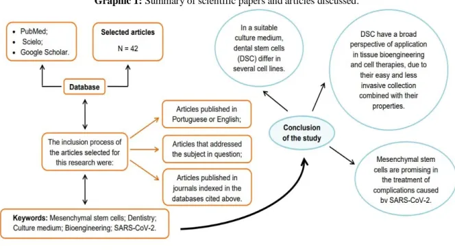
In this article, a literature review was made on the application of stem cells in dental and non-dental therapies, highlighting their properties and the need for an adequate culture medium for cell differentiation to occur. This review article is summarized in the graphical abstract (Graphic 1).
Graphic 1: Summary of scientific papers and articles discussed.
2 MESENCHYMAL STEM CELLS DERIVED FROM DENTAL PULP, PERIODONTAL LIGAMENT, DENTAL FOLLICLE AND APICAL PAPILLA
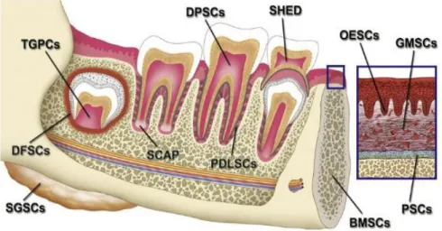
Dental pulp, as well as other tooth structures, are alternative sources for obtaining MSC (RELAÑO-GINÉS et al., 2019). These, coming from dental structures, also known as dental stem cells (DSC), can be divided into different types, according to their origin and as shown in figure 2 (EGUSA et al., 2012). In embryonic development, they originate from the neural crest
(RELAÑO-GINÉS et al., 2019), which gives them more potent neurogenic properties when compared to other types of MSC (JARMALAVIČIŪTĖ et al., 2015).
According to Garcia et al. (GARCIA; ROQUE; SILVA, 2017), DSC are found in several regions of the dental organ and attached structures, such as the dental pulp, the periodontal ligament, the apical papilla and the dental follicle. According to the same authors, these cells represent new approaches in dental and non-dental therapies, since they have a high capacity for proliferation and differentiation, combined with their minimally invasive and painless collection. In view of the premises and the various works in the literature (GARCIA; ROQUE; SILVA, 2017; RELAÑO-GINÉS et al., 2019; SOARES et al., 2007), the dental organ presents itself as a promising source of cells in question.
Gronthos et al. (GRONTHOS et al., 2000), at the beginning of the study of stem cells derived from teeth, they used impacted third molars to identify for the first time a population of stem cells from dental pulp of permanent teeth (DPSC). After three years, Miura et al. (MIURA et al., 2003) discovered stem cells from the pulp of human exfoliated primary teeth (SHED). These cells, as demonstrated in the current literature, have a higher proliferation rate compared to DPCS (GARCIA; ROQUE; SILVA, 2017).
In a later study, Seo et al. (SEO et al., 2004) isolated stem cells from the periodontal ligament (PDLSC). After one year, stem cells from the dental follicle (DFSC) were identified (MORSCZECK et al., 2005). Today, the recently discovered DCS population is the apical papilla stem cells (SCAP) (SONOYAMA et al., 2006) which, when contrasted with DPSC, have a superior mineralization capacity (BAKOPOULOU et al., 2011).
Thus, DSC are a type of MSC that has great potential for several therapeutic applications, due to their less invasive acquisition associated with their high capacity for differentiation.
Figure 2: Sources of stem cells in the oral and maxillofacial region, in particular: stem cells from dental pulp of permanent teeth (DPSC); pulp stem cells from human exfoliated primary teeth (SHED); stem cells of the periodontal ligament (PDLSC); dental follicle stem cells (DFSC); stem cells of the apical papilla (SCAP) (EGUSA et al., 2012).
3 CULTURE MEDIUM COMPOSITION
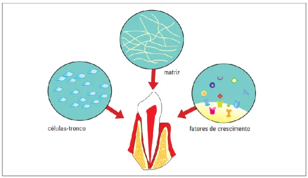
For the DSC to demonstrate its therapeutic therapies in clinics, cell differentiation is essential (GARCIA; ROQUE; SILVA, 2017). Thus, with the purpose that tissue bioengineering takes place effectively and with greater use of cells, the culture medium must be formed by a triad composed of stem cells, matrix and growth factors (SOARES et al., 2007) (Figure 3).
According to Soares et al. (SOARES et al., 2007), the matrix functions as a framework, which is rich in nutritional substances and favors the transport of nutrients and metabolic waste to cells. In DSC, the matrix components activate the morphogens, which are secreted extracellularly and function as growth and/or differentiation (NAKASHIMA; AKAMINE, 2005). During this bioprocess, the matrix is gradually destroyed and replaced by regenerated tissue. The studies by Deng et al. (DENG et al., 2005) using stem cells from the first gill arch of mice to the stimulus transforming growth factor-human β1 (TGF-β1) and dentin non-collagen proteins (DNCP), suggested that proteins derived from the matrix an essential function in odontoblastic differentiation.
Growth factors are important proteins for the differentiation of stem cells in culture media, as they induce the desired potentiality in a specific tissue (GARCIA; ROQUE; SILVA, 2017) and govern morphogenesis during epithelial-mesenchymal interactions (NAKASHIMA; AKAMINE, 2005). Bone morphogenetic proteins (BMPs) are examples of growth factors used in the differentiation of DSC (GARCIA; ROQUE; SILVA, 2017). Although these comprise five protein families involved in tooth development, the studies by Nakashima et al.
(NAKASHIMA; REDDI, 2003) demonstrated that BMPs are sufficient for the formation of tertiary dentin.
Therefore, it is extremely important that the culture media are composed of the triad presented so that, in this way, there is cell differentiation and a greater use of the stem cells fed.
Figure 3: Factors needed for bioengineering in Dentistry. The differentiation of stem cells depends directly on the culture medium, which must be composed of stem cells, matrix and growth factors (SOARES et al., 2007).
4 APPLICATION IN ODONTOLOGY
Classical dentistry, based on the use of synthetic materials in the treatment of oral diseases, gained prominence after the Second World War, when it was noticed that improvements needed to be made in the oral hygiene of soldiers in order for the army to remain healthy (YELICK; SHARPE, 2019). Since then, the community has made efforts to create increasingly improved methods in dental replacement therapies and recovery of lost tissues.
Possible dental treatments using stem cells have advantages when compared to current prosthetic methodologies, since a DSC therapy maintains the vitality, elasticity and hydration of dental tissues (GARCIA; ROQUE; SILVA, 2017), in addition to renewing well-being and recovering the patient's oral health. Thus, dental tissue bioengineering is based on the use of DSC and has stood out to provide better adaptation and functionality to previously damaged dental structures (YANG et al., 2017).
In the current context of regenerative therapies, among all DSC, those with the greatest therapeutic potential and which are most used for research purposes are stem cells derived from dental pulp (FUJII et al., 2015; SAEZ et al., 2019; TANIKAWA et al., 2020), as they have a
great regenerative potential and a wide capacity to differentiate into different cell lines (AURREKOETXEA et al., 2015), as in odontoblasts, osteoblasts, adipocytes and nerve cells (RELAÑO-GINÉS et al., 2019).
Piva et al. (PIVA et al., 2017), used third molars obtained from dental extractions for the isolation and expansion of DPSC in standard culture conditions containing fetal bovine serum (DPSCs-FBS) or human serum (DPSCs-HS). In this study was demonstrated that the DPSC maintain their pulp regenerative properties even in environments devoid of animal serum. In addition, experiments showed that transplantation of DPSC into a tooth slice produced a robust angiogenic response and further induced the formation of pre-dentin.
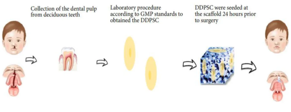
In a recent study, the authors Tanikawa et al. (TANIKAWA et al., 2020) reported for the first time the use of deciduous dental pulp stem cells (DDPSC) associated with a hydroxyapatite-collagen sponge (Bio-Oss Colagen® 250 mg, Geistlich) for maxillary alveolar reconstruction in patients with cleft lip and palate, as shown in figure 4.
Figure 4: Schematic representation of autologous bone tissue engineering using DDPSC associated with 250 mg of Bio-Oss Collagen® (TANIKAWA et al., 2020).
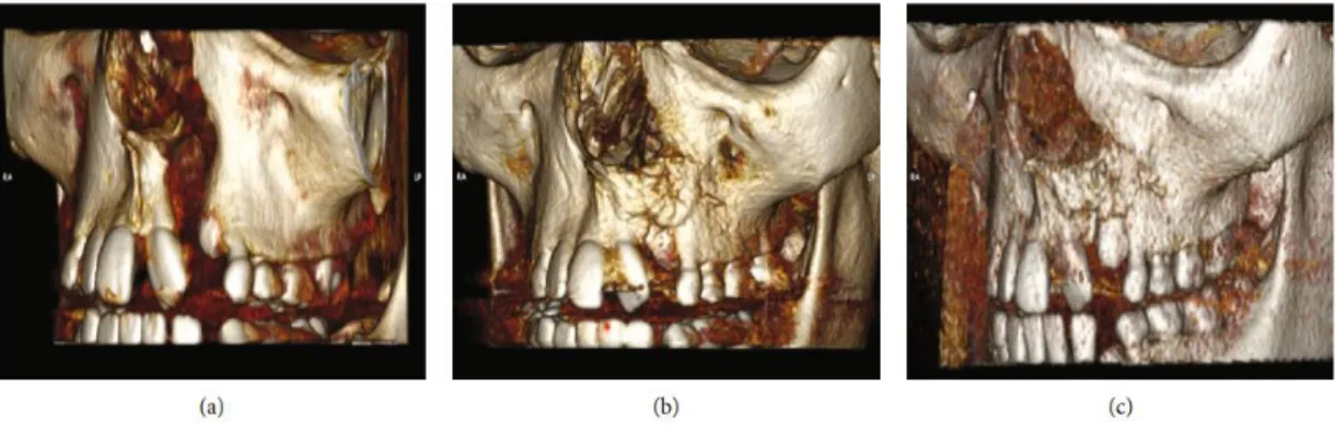
Tanikawa et al. (TANIKAWA et al., 2020) selected six patients aged 8 to 12 years, and as an inclusion factors, unilateral alveolar cleft defects. In the experimental group, individuals were subjected to the use of the sponge associated with DDPSC. In historical controls, rhBMP-2 (recombinant human bone morphogenetic protein-rhBMP-2) was used in group one and traditional bone graft from the iliac crest for upper alveolar reconstruction in group two. As a result, the authors reported that stem cell therapy resulted in satisfactory bone regeneration with tooth eruption and reduced morbidity (Figure 5) when compared to traditional iliac crest bone grafting and rhBMP-2.
Figure 5: Computed tomography images of the same patient, showing the filling of the alveolar cleft by the bone between 6 and 12 months after the use of bone tissue engineering procedures (DDPSC associated with Bio-Oss Collagen®) and the rash of the canine tooth after 12 months. (a) Preoperative: presence of alveolar cleft; (b) 6 months postoperatively: alveolar cleft filled with new bone; (c) 12 months postoperatively: the canine tooth erupted in the new bone formed using bone tissue engineering (TANIKAWA et al., 2020).
In addition to the dental pulp, there is the periodontal ligament, which consists of fibers that are inserted in the cementum and bone (KATCHBURIAN; ARANA, 2017). In the study by Shi et al. (SHI et al., 2018), the combination of periodontal ligament stem cells (PDLSC) and porous improved biphasic calcium phosphate (BCP) has shown promise in periodontal regeneration.
Around the developing tooth there is the dental follicle, which is responsible for forming the cementum, periodontal ligament and alveolar bone (KATCHBURIAN; ARANA, 2017). Through experiments carried out on mice, it was observed that epithelial stem cells from the human dental follicle (hDF-EpiSCs), when cultured under three-dimensional induction conditions in vitro, are able to regenerate the dysfunction of salivary glands (XU et al., 2017). The apical papilla, which is only present in the root development, is responsible for forming dentin and pulp (KATCHBURIAN; ARANA, 2017). When compared to DPSC, apical papilla stem cells (SCAP) are more effective in forming mineralized tissues (BAKOPOULOU et al., 2011). Through guinea pigs was possible to identify the influence of SCAP and PDLSC on the recovery of strength and appearance of the teeth, because the cells contributed to the formation of a complete root able to support a crown and perform the functions of the tooth (SONOYAMA et al., 2006).
DSC are very useful in the field of Dentistry, as in the fields of aesthetic dentistry and periodontics. In this first, stem cells are used to repair enamel and dentin. From PDLSC, specifically the epithelial remains, the authors Shinmura et al. (SHINMURA et al., 2008) showed that there was the formation of cells similar to ameloblasts, which can develop enamel. In a study published in “Scientific Reports” (NEVES et al., 2017), researchers stimulated the natural formation of dentin in experiments with mice. The idea of the study was to stimulate
the formation of restorative dentin and reestablish lost dentin by activating the process of differentiation of dental pulp stem cells into odontoblasts.
In the field of periodontics, advances in research are also observed in the literature and are of great relevance, as periodontitis causes loss of the periodontal ligament and underlying bone tissue (LINDHE, JAN; LANG, 2018). In this scenario, studies manipulating PDLSC obtained complete root (SONOYAMA et al., 2006) and regeneration of the periodontal bone defect (TSUMANUMA et al., 2011).
In short, tissue bioengineering applied to dentistry is gaining more and more space, since it promotes the well-being of patients through better adjustment and functionality to deteriorated tissues.
5 APPLICATION IN NON-DENTAL THERAPIES
A few years ago, the therapeutic applications of dental stem cells (DSC) were limited only to oral rehabilitation, as for example, in the regeneration of the dentin-pulp complex (GARCIA; ROQUE; SILVA, 2017) and the periodontal ligament (SHI et al., 2018). However, over time, many studies have shown that stem cells found mainly in the pulp of permanent and deciduous teeth could be a new alternative for regenerative therapies in various areas of the body, from bone to nervous tissue (GARCIA; ROQUE; SILVA, 2017).
Facial nerve regeneration, as a practical and successful example, occurs after the application of immature stem cells from the human dental pulp (iDPSC) at the injury site (SAEZ et al., 2019). In this study, Saez et al. used 70 Winstar rats submitted to a facial nerve crush injury, which were separated into two groups: group 1 (animals subjected to injury) and group 2 (animals subjected to injury and treatment with iDPSC). Using as a parameter the symmetrical or asymmetric movement and the position of the vibrissae, the results showed that group 2 showed complete functional recovery at 14 days, while group 1 recovered only after 42 days. These findings showed that iDPCS, in a favorable microenvironment, directly contributed to facial nerve regeneration in rats.
In the context of neurological deficits, stem cells from dental pulp of exfoliated primary teeth (SHED) have shown promise in the therapy of a parkinsonian rat model induced by 6-hydroxy-dopamine (6-OHDA) (FUJII et al., 2015). From SHED, there was the induction of DAergic neuron cells (dSHEDs), which contributed to the neuroprotection against 6-ODHA-induced neurodegeneration and to the restoration of the nigrostriatal tract. The results of this study suggested that the grafted dSHEDs promoted the recovery of neurological deficits in parkinsonian rats mainly through their paracrine and neuroprotective activities.
Mita et al. (MITA et al., 2015) revealed that the multifaceted capacity of SHED can considerably improve cognitive function in mice with Alzheimer's disease, thus evidencing the therapeutic efficacy of stem cells in neural injuries and neurodegenerative diseases.
SHED, in addition to being used in nervous tissue therapies, were able to form cells similar to hepatocytes and to regenerate liver tissue. Thus, this type of DSC has been shown to be a new alternative in the treatment of liver disease (ISHKITIEV et al., 2012).
Soares et al. (SOARES et al., 2019) evaluated the inflammatory effect and bone formation in non-critical defects in the tibia of rats after implantation of a Hemospon® collagen sponge with stem cells from the human dental pulp and a species of succulent plant of the genus Aloe (Aloe vera). The rats were separated into five experimental groups, according to the treatment, and were subsequently evaluated for bone repair capacity. Thus, the results suggested that in the group treated with the combination of Hemospon®, Aloe vera and stem cells from the dental pulp, bone failure was repaired and the effects of the inflammatory cascade were reduced.
In another experiment with animal guinea pigs (SYED-PICARD et al., 2015), adult dental pulp stem cells demonstrated their ability to differentiate into keratocytes in vitro and in
vivo, subsequently generating tissue similar to the corneal stroma. In this study, Syed-Picard et al. revealed that stem cell therapy may be a new form of clinical treatment for corneal recovery,
since keratoplasty with allogeneic cadaveric tissue, the most common method of treatment, presents several difficulties, such as the scarcity of donors and the possibility of rejection of the transplanted tissue.
These studies, as well as the present work, present promising data on the use of dental stem cells not only in dental therapies, but also in regenerative therapies of nervous and bone tissue and in treatments of neurodegenerative, hepatic and ocular deficits.
6 MESENCHYMAL STEM CELLS AND A PERSPECTIVE IN THE TREATMENT OF COVID-19 AND SEVERE ACUTE RESPIRATORY SYNDROME
The COVID-19 pandemic, a disease caused by the new coronavirus (SARS-CoV-2) (LI et al., 2020), is a major concern worldwide, as while treatment options are scarce, the numbers of serious cases and deaths rise dramatically every day.
Today, several drugs, including antivirals (ELFIKY, 2020) and antimalarials (WANG et al., 2020), are under investigation for the treatment of patients with SARS-CoV-2. Although these therapeutic strategies provide better recovery and survival for patients, they do not lead
to the restoration of lung damage caused by the new coronavirus (CHRZANOWSKI; KIM; MCCLEMENTS, 2020).
Since the principle of therapies for COVID-19 is related to the management of the cytokine storm in the lungs, mesenchymal stem cells (MSC) are promising, since their main mechanism of action is through immunomodulatory, anti -inflammatory, antioxidant and regenerative (CHRZANOWSKI; KIM; MCCLEMENTS, 2020; SOARES et al., 2019; TANIKAWA et al., 2020). These cells appear as new strategies for the treatment of complications caused by this new disease.
Recently, MSC were investigated in acute respiratory distress syndrome (ARDS) both in the preclinical context (CURLEY et al., 2013) and in the clinical context (WILSON et al., 2015), which demonstrated its ability to promote the pulmonary epithelial repair. In another study (BHATTACHARYA; MATTHAY, 2013), researchers noted that these cells stabilized the leakage of endothelial fluid and maintained a capillary barrier function, attenuating the development of interstitial pulmonary edema.
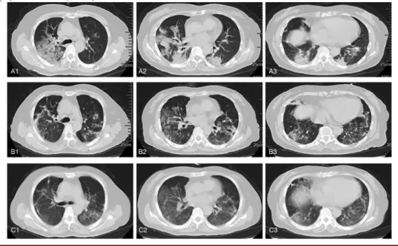
In order to determine whether the infusion of human umbilical cord mesenchymal stem cells (hUC-MSC) could be effective and safe for the treatment of severe COVID-19, experiments on critically ill patients demonstrated that the management of these cells in treatments decreased the mortality rate and disease progression from severe to critical (SHU et al., 2020), in addition to attenuating the pulmonary inflammatory reaction (LIANG et al., 2020), as shown in figure 6.
Figure 6: Typical computed tomography (CT) images of the lung. A1 - A3: CT images on day 2 indicate that there are lesions and an increasing shadow of mass density in the right and left lungs; B1 - B3: CT images on day 20 indicate relief in the right and left lungs; C1 - C3: CT images on day 25 indicate additional relief in the right and left lungs (LIANG et al., 2020).
To date, therapies based on dental stem cells (DSC) have not been performed in patients with COVID-19. However, a study report for a randomized controlled trial was developed to assess the safety and therapeutic effects of dental pulp stem cells (DPSC) in the treatment of severe COVID-19 pneumonia (YE et al., 2020). In this study scheduled for 2021, twenty serious cases of the disease will be selected, which will be separated into two groups: experimental (venous injection of DPSC suspension in 30 ml of saline) and control (saline that will work as a placebo). The results will be evaluated according to six ordered grades (corresponding to grade 1 for patient discharge and grade 6 for death).
Although the number of clinical trials based on MSC is limited, their prospects for use in the clinical environment to treat complications caused by SARS-CoV-2 appear to be promising, since, as reported in studies, these cells have immunomodulatory and anti-inflammatory properties, combined with the great potential for regeneration. These studies open countless perspectives for the application of the most diverse stem cells found in the structures of dental organs (DPSC, SHED, PDLSC, DFSC and SCAP), as already discussed and presented in this work.
7 CONCLUSION
This literature review focused on mesenchymal stem cells - in particular DPSC, SHED, PDLSC, DFSC and SCAP - with regard to their contribution and effectiveness in dental treatments or not. Based on the studies presented, these cells have, in fact, a broad perspective of application in tissue bioengineering and cell therapies, due to their easy and less invasive collection combined with their regenerative, anti-inflammatory and immunomodulatory properties. In addition, despite the reduced number of studies, MSC are also promising in the treatment of complications caused by SARS-CoV-2. In a culture medium composed of the presented triad, DSC demonstrates their ability to differentiate into multiple cell lines. In this context, understanding the functioning of these cells and their properties allows the development of more effective therapies or even the cure of diseases, such as Severe Acute Respiratory Syndrome.
ACKNOWLEDGEMENTS
The authors wish to thank Universidade Estadual do Norte do Paraná - UENP, Jacarezinho, Parana.
REFERENCES
AURREKOETXEA, M.; GARCIA-GALLASTEGUI, P.; IRASTORZA, I.; LUZURIAGA, J.; URIBE-ETXEBARRIA, V.; UNDA, F.; IBARRETXE, G. Dental pulp stem cells as a multifaceted tool for bioengineering and the regeneration of craniomaxillofacial tissues. Frontiers in Physiology, [s. l.], v. 6, n. OCT, p. 1–10, 2015. Disponível em: <http://journal.frontiersin.org/Article/10.3389/fphys.2015.00289/abstract>
BAKOPOULOU, A.; LEYHAUSEN, G.; VOLK, J.; TSIFTSOGLOU, A.; GAREFIS, P.; KOIDIS, P.; GEURTSEN, W. Comparative analysis of in vitro osteo/odontogenic differentiation potential of human dental pulp stem cells (DPSCs) and stem cells from the apical papilla (SCAP). Archives of Oral Biology, [s. l.], v. 56, n. 7, p. 709–721, 2011. Disponível em: <http://dx.doi.org/10.1016/j.archoralbio.2010.12.008>
BHATTACHARYA, J.; MATTHAY, M. A. Regulation and Repair of the Alveolar-Capillary Barrier in Acute Lung Injury. Annual Review of Physiology, [s. l.], v. 75, n. 1, p. 593–615, 2013. Disponível em: <http://www.annualreviews.org/doi/10.1146/annurev-physiol-030212-183756>
CHRZANOWSKI, W.; KIM, S. Y.; MCCLEMENTS, L. Can Stem Cells Beat COVID-19: Advancing Stem Cells and Extracellular Vesicles Toward Mainstream Medicine for Lung Injuries Associated With SARS-CoV-2 Infections. Frontiers in Bioengineering and Biotechnology, [s. l.], v. 8, 2020. Disponível em: <https://www.frontiersin.org/article/10.3389/fbioe.2020.00554/full>
CURLEY, G. F.; ANSARI, B.; HAYES, M.; DEVANEY, J.; MASTERSON, C.; RYAN, A.; BARRY, F.; O’BRIEN, T.; TOOLE, D. O.; LAFFEY, J. G. Effects of Intratracheal Mesenchymal Stromal Cell Therapy during Recovery and Resolution after Ventilator-induced Lung Injury. Anesthesiology, [s. l.], v. 118, n. 4, p. 924–932, 2013. Disponível em: <http://anesthesiology.pubs.asahq.org/Article.aspx?doi=10.1097/ALN.0b013e318287ba08>
DENG, M.; SHI, J.; SMITH, A. J.; JIN, Y. Effects of transforming growth factor β1 (TGFβ-1) and dentin non-collagenous proteins (DNCP) on human embryonic ectomesenchymal cells in a three-dimensional culture system. Archives of Oral Biology, [s. l.], v. 50, n. 11, p. 937–945, 2005. Disponível em: <https://linkinghub.elsevier.com/retrieve/pii/S0003996905000932>
EGUSA, H.; SONOYAMA, W.; NISHIMURA, M.; ATSUTA, I.; AKIYAMA, K. Stem cells in dentistry – Part I: Stem cell sources. Journal of Prosthodontic Research, [s. l.], v. 56, n. 3, p. 151–165, 2012. Disponível em: <http://dx.doi.org/10.1016/j.jpor.2012.06.001>
ELFIKY, A. A. Anti-HCV, nucleotide inhibitors, repurposing against COVID-19. Life Sciences, [s. l.], v. 248, n. January, p. 117477, 2020. Disponível em: <https://linkinghub.elsevier.com/retrieve/pii/S0024320520302253>
FUJII, H.; MATSUBARA, K.; SAKAI, K.; ITO, M.; OHNO, K.; UEDA, M.; YAMAMOTO, A. Dopaminergic differentiation of stem cells from human deciduous teeth and their therapeutic benefits for Parkinsonian rats. Brain Research, [s. l.], v. 1613, p. 59–72, 2015. Disponível em: <http://dx.doi.org/10.1016/j.brainres.2015.04.001>
GARCIA, T.; ROQUE, J. S.; SILVA, D. F. CÉLULAS-TRONCO: BIOENGENHARIA APLICADA À ODONTOLOGIA. Nanocell News, [s. l.], v. 4, n. 6, p. NA-NA, 2017. Disponível em: <http://www.nanocell.org.br/celulas-tronco-bioengenharia-aplicada-a-odontologia/>
GRONTHOS, S.; MANKANI, M.; BRAHIM, J.; ROBEY, P. G.; SHI, S. Postnatal human dental pulp stem cells (DPSCs) in vitro and invivo. Proceedings of the National Academy of Sciences, [s. l.], v. 97, n. 25, p. 13625–13630, 2000. Disponível em: <http://www.pnas.org/cgi/doi/10.1073/pnas.240309797>
ISHKITIEV, N.; YAEGAKI, K.; IMAI, T.; TANAKA, T.; NAKAHARA, T.; ISHIKAWA, H.; MITEV, V.; HAAPASALO, M. High-purity hepatic lineage differentiated from dental pulp stem cells in serum-free medium. Journal of Endodontics, [s. l.], v. 38, n. 4, p. 475–480, 2012. Disponível em: <http://dx.doi.org/10.1016/j.joen.2011.12.011>
ISOBE, Y.; KOYAMA, N.; NAKAO, K.; OSAWA, K.; IKENO, M.; YAMANAKA, S.; OKUBO, Y.; FUJIMURA, K.; BESSHO, K. Comparison of human mesenchymal stem cells derived from bone marrow, synovial fluid, adult dental pulp, and exfoliated deciduous tooth pulp. International Journal of Oral and Maxillofacial Surgery, [s. l.], v. 45, n. 1, p. 124– 131, 2016. Disponível em: <http://dx.doi.org/10.1016/j.ijom.2015.06.022>
JARMALAVIČIŪTĖ, A.; TUNAITIS, V.; PIVORAITĖ, U.; VENALIS, A.; PIVORIŪNAS, A. Exosomes from dental pulp stem cells rescue human dopaminergic neurons from 6-hydroxy-dopamine–induced apoptosis. Cytotherapy, [s. l.], v. 17, n. 7, p. 932–939, 2015. Disponível em: <https://linkinghub.elsevier.com/retrieve/pii/S1465324915008828>
JI, L.; BAO, L.; GU, Z.; ZHOU, Q.; LIANG, Y.; ZHENG, Y.; XU, Y.; ZHANG, X.; FENG, X. Comparison of immunomodulatory properties of exosomes derived from bone marrow mesenchymal stem cells and dental pulp stem cells. Immunologic Research, [s. l.], v. 67, n. 4–5, p. 432–442, 2019. Disponível em:
<http://link.springer.com/10.1007/s12026-019-09088-6>
KATCHBURIAN, E.; ARANA, V. Histologia e Embriologia Oral. 4. ed. [s.l: s.n.].
LI, H.; LIU, S.-M.; YU, X.-H.; TANG, S.-L.; TANG, C.-K. Coronavirus disease 2019 (COVID-19): current status and future perspectives. International Journal of Antimicrobial Agents, [s. l.], v. 55, n. 5, p. 105951, 2020. Disponível em: <https://doi.org/10.1016/j.ijantimicag.2020.105951>
LIANG, B.; CHEN, J.; LI, T.; WU, H.; YANG, W.; LI, Y.; LI, J.; YU, C.; NIE, F.; MA, Z.; YANG, M.; XIAO, M.; NIE, P.; GAO, Y.; QIAN, C.; HU, M. Clinical remission of a critically ill COVID-19 patient treated by human umbilical cord mesenchymal stem cells. Medicine, [s.
l.], v. 99, n. 31, p. e21429, 2020. Disponível em:
<https://journals.lww.com/10.1097/MD.0000000000021429>
LINDHE, JAN; LANG, N. P. Tratado de Periodontia Clínica e Implantologia Oral. 6a
Edição ed. [s.l: s.n.].
MITA, T.; FURUKAWA-HIBI, Y.; TAKEUCHI, H.; HATTORI, H.; YAMADA, K.; HIBI, H.; UEDA, M.; YAMAMOTO, A. Conditioned medium from the stem cells of human dental pulp improves cognitive function in a mouse model of Alzheimer’s disease. Behavioural Brain Research, [s. l.], v. 293, p. 189–197, 2015. Disponível em: <http://dx.doi.org/10.1016/j.bbr.2015.07.043>
MIURA, M.; GRONTHOS, S.; ZHAO, M.; LU, B.; FISHER, L. W.; ROBEY, P. G.; SHI, S. SHED: Stem cells from human exfoliated deciduous teeth. Proceedings of the National Academy of Sciences, [s. l.], v. 100, n. 10, p. 5807–5812, 2003. Disponível em: <http://www.pnas.org/cgi/doi/10.1073/pnas.0937635100>
MORSCZECK, C.; GÖTZ, W.; SCHIERHOLZ, J.; ZEILHOFER, F.; KÜHN, U.; MÖHL, C.; SIPPEL, C.; HOFFMANN, K. H. Isolation of precursor cells (PCs) from human dental follicle of wisdom teeth. Matrix Biology, [s. l.], v. 24, n. 2, p. 155–165, 2005. Disponível em: <https://linkinghub.elsevier.com/retrieve/pii/S0945053X04001593>
NAKASHIMA, M.; AKAMINE, A. The Application of Tissue Engineering to Regeneration of Pulp and Dentin in Endodontics. Journal of Endodontics, [s. l.], v. 31, n. 10, p. 711–718, 2005. Disponível em: <https://linkinghub.elsevier.com/retrieve/pii/S0099239906610911>
NAKASHIMA, M.; REDDI, A. H. The application of bone morphogenetic proteins to dental tissue engineering. Nature Biotechnology, [s. l.], v. 21, n. 9, p. 1025–1032, 2003. Disponível
em: <http://www.nature.com/articles/nbt864>
NEVES, V. C. M.; BABB, R.; CHANDRASEKARAN, D.; SHARPE, P. T. Promotion of natural tooth repair by small molecule GSK3 antagonists. Scientific Reports, [s. l.], v. 7, n. 1, p. 39654, 2017. Disponível em: <http://dx.doi.org/10.1038/srep39654>
PIVA, E.; TARLÉ, S. A.; NÖR, J. E.; ZOU, D.; HATFIELD, E.; GUINN, T.; EUBANKS, E. J.; KAIGLER, D. Dental Pulp Tissue Regeneration Using Dental Pulp Stem Cells Isolated and Expanded in Human Serum. Journal of Endodontics, [s. l.], v. 43, n. 4, p. 568–574, 2017. Disponível em: <https://linkinghub.elsevier.com/retrieve/pii/S0099239916309682>
RELAÑO-GINÉS, A.; LEHMANN, S.; DEVILLE DE PÉRIÈRE, D.; HIRTZ, C. Dental stem cells as a promising source for cell therapies in neurological diseases. Critical Reviews in Clinical Laboratory Sciences, [s. l.], v. 56, n. 3, p. 170–181, 2019. Disponível em: <https://doi.org/10.1080/10408363.2019.1571478>
SAEZ, D. M.; SASAKI, R. T.; MARTINS, D. de O.; CHACUR, M.; KERKIS, I.; DA SILVA, M. C. P. Rat Facial Nerve Regeneration with Human Immature Dental Pulp Stem Cells. Cell Transplantation, [s. l.], v. 28, n. 12, p. 1573–1584, 2019. Disponível em: <http://journals.sagepub.com/doi/10.1177/0963689719854446>
SEO, B.-M.; MIURA, M.; GRONTHOS, S.; MARK BARTOLD, P.; BATOULI, S.; BRAHIM, J.; YOUNG, M.; GEHRON ROBEY, P.; WANG, C. Y.; SHI, S. Investigation of multipotent postnatal stem cells from human periodontal ligament. The Lancet, [s. l.], v. 364,
n. 9429, p. 149–155, 2004. Disponível em:
<https://linkinghub.elsevier.com/retrieve/pii/S0140673604166270>
SHI, H.; ZONG, W.; XU, X.; CHEN, J. Improved biphasic calcium phosphate combined with periodontal ligament stem cells may serve as a promising method for periodontal regeneration. American Journal of Translational Research, [s. l.], v. 10, n. 12, p. 4030–4041, 2018. SHINMURA, Y.; TSUCHIYA, S.; HATA, K.; HONDA, M. J. Quiescent epithelial cell rests of Malassez can differentiate into ameloblast-like cells. Journal of Cellular Physiology, [s. l.], v. 217, n. 3, p. 728–738, 2008. Disponível em: <http://doi.wiley.com/10.1002/jcp.21546>
SHU, L.; NIU, C.; LI, R.; HUANG, T.; WANG, Y.; HUANG, M.; JI, N.; ZHENG, Y.; CHEN, X.; SHI, L.; WU, M.; DENG, K.; WEI, J.; WANG, X.; CAO, Y.; YAN, J.; FENG, G. Treatment of severe COVID-19 with human umbilical cord mesenchymal stem cells. Stem cell research & therapy, [s. l.], v. 11, n. 1, p. 361, 2020. Disponível em:
<http://www.ncbi.nlm.nih.gov/pubmed/32811531>
SOARES, A. P.; KNOP, L. A. H.; JESUS, A. A. De; ARAÚJO, T. M. De. Células-tronco em odontologia. Revista Dental Press de Ortodontia e Ortopedia Facial, [s. l.], v. 12, n. 1, p. 33–40, 2007. Disponível em: <https://www.scielo.br/pdf/dpress/v12n1/a06v12n1.pdf>
SOARES, I. M. V.; FERNANDES, G. V. de O.; CAVALCANTE, L. C.; LEITE, Y. K. P. de C.; BEZERRA, D. de O.; CARVALHO, M. A. M. De; CARVALHO, C. M. R. S. The influence of Aloe vera with mesenchymal stem cells from dental pulp on bone regeneration: characterization and treatment of non-critical defects of the tibia in rats. Journal of Applied Oral Science, [s. l.], v. 27, p. 1–11, 2019. Disponível em:
<http://www.scielo.br/scielo.php?script=sci_arttext&pid=S1678-77572019000100440&tlng=en>
SONOYAMA, W.; LIU, Y.; FANG, D.; YAMAZA, T.; SEO, B.-M.; ZHANG, C.; LIU, H.; GRONTHOS, S.; WANG, C.-Y.; SHI, S.; WANG, S. Mesenchymal Stem Cell-Mediated Functional Tooth Regeneration in Swine. PLoS ONE, [s. l.], v. 1, n. 1, p. e79, 2006. Disponível em: <https://dx.plos.org/10.1371/journal.pone.0000079>
SYED-PICARD, F. N.; DU, Y.; LATHROP, K. L.; MANN, M. M.; FUNDERBURGH, M. L.; FUNDERBURGH, J. L. Dental Pulp Stem Cells: A New Cellular Resource for Corneal Stromal Regeneration. STEM CELLS Translational Medicine, [s. l.], v. 4, n. 3, p. 276–285, 2015.
TANIKAWA, D. Y. S.; PINHEIRO, C. C. G.; ALMEIDA, M. C. A.; OLIVEIRA, C. R. G. C. M.; COUDRY, R. D. A.; ROCHA, D. L.; BUENO, D. F. Deciduous Dental Pulp Stem Cells for Maxillary Alveolar Reconstruction in Cleft Lip and Palate Patients. Stem Cells International, [s. l.], v. 2020, n. Figure 1, 2020.
TSUMANUMA, Y.; IWATA, T.; WASHIO, K.; YOSHIDA, T.; YAMADA, A.; TAKAGI, R.; OHNO, T.; LIN, K.; YAMATO, M.; ISHIKAWA, I.; OKANO, T.; IZUMI, Y. Comparison of different tissue-derived stem cell sheets for periodontal regeneration in a canine 1-wall defect model. Biomaterials, [s. l.], v. 32, n. 25, p. 5819–5825, 2011. Disponível em: <http://dx.doi.org/10.1016/j.biomaterials.2011.04.071>
WANG, M.; CAO, R.; ZHANG, L.; YANG, X.; LIU, J.; XU, M.; SHI, Z.; HU, Z.; ZHONG, W.; XIAO, G. Remdesivir and chloroquine effectively inhibit the recently emerged novel coronavirus (2019-nCoV) in vitro. Cell Research, [s. l.], v. 30, n. 3, p. 269–271, 2020. Disponível em: <http://www.nature.com/articles/s41422-020-0282-0>
WILSON, J. G.; LIU, K. D.; ZHUO, H.; CABALLERO, L.; MCMILLAN, M.; FANG, X.; COSGROVE, K.; VOJNIK, R.; CALFEE, C. S.; LEE, J.-W.; ROGERS, A. J.; LEVITT, J.; WIENER-KRONISH, J.; BAJWA, E. K.; LEAVITT, A.; MCKENNA, D.; THOMPSON, B. T.; MATTHAY, M. A. Mesenchymal stem (stromal) cells for treatment of ARDS: a phase 1 clinical trial. The Lancet Respiratory Medicine, [s. l.], v. 3, n. 1, p. 24–32, 2015. Disponível em: <http://dx.doi.org/10.1016/S2213-2600(14)70291-7>
XU, Q. L.; FURUHASHI, A.; ZHANG, Q. Z.; JIANG, C. M.; CHANG, T. H.; LE, A. D. Induction of Salivary Gland-Like Cells from Dental Follicle Epithelial Cells. Journal of Dental Research, [s. l.], v. 96, n. 9, p. 1035–1043, 2017.
YANG, L.; ANGELOVA VOLPONI, A.; PANG, Y.; SHARPE, P. T. Mesenchymal Cell Community Effect in Whole Tooth Bioengineering. Journal of Dental Research, [s. l.], v. 96,
n. 2, p. 186–191, 2017. Disponível em:
<http://journals.sagepub.com/doi/10.1177/0022034516682001>
YE, Q.; WANG, H.; XIA, X.; ZHOU, C.; LIU, Z.; XIA, Z.; ZHANG, Z.; ZHAO, Y.; YEHENALA, J.; WANG, S.; ZHOU, G.; HU, K.; WU, B.; WU, C.-T.; WANG, S.; HE, Y. Safety and efficacy assessment of allogeneic human dental pulp stem cells to treat patients with severe COVID-19: structured summary of a study protocol for a randomized controlled trial (Phase I / II). Trials, [s. l.], v. 21, n. 1, p. 520, 2020. Disponível em: <https://trialsjournal.biomedcentral.com/articles/10.1186/s13063-020-04380-5>
YELICK, P. C.; SHARPE, P. T. Tooth Bioengineering and Regenerative Dentistry. Journal of Dental Research, [s. l.], v. 98, n. 11, p. 1173–1182, 2019. Disponível em: <http://journals.sagepub.com/doi/10.1177/0022034519861903>