RESUMO.- [Efeito do sobrenadante de isolados de Lac-tobacillus sp. sobre Escherichiacoli O157:H7 reforça o papel da produção de ácidos orgânicos como fator de controle patogênico.] Muitas tentativas têm sido feitas para se estabelecer o controle de patógenos de origem ali-mentar através do uso de estirpes de Lactobacillus e dos seus produtos de metabolismo, com sucesso sendo sucedi-do em várias situações. O objetivo deste trabalho foi investi-gar o efeito antagônico do sobrenadante de culturas de oito
isolados de Lactobacillus, incluindo L. casei subsp. pseudo-plantarum, L. plantarumL. reuteri e L. delbrueckii subsp. del-brueckii, sobre Escherichiacoli amostra O157:H7. Os efeitos
inibidores de culturas puras e de dois “pools” de cultura de
Lactobacillus sobre o crescimento da bactéria foram avalia-dos através do método de inibição em ágar e através do mo-nitoramento da turbidez da cultura bacteriana. A atividade
antimicrobiana foi confirmada para Lactobacillus reuteri e Lactobacillus delbrueckii subsp. delbrueckii e para o “pool” de bactérias acido-láctica. O sobrenadante neutralizado do “pool” de Lactobacillus exerceu uma atividade
antimicro-biana mais elevada do que aquela das estirpes individuais. Além disso, ácido D-láctico e ácido acético foram produzi-dos durante o crescimento produzi-dos Lactobacillus estudados. TERMOS DE INDEXAÇÃO: Lactobacillus, antagonismo, Escherichia coli O157:H7, ácido orgânico.
INTRODUCTION
Escherichia coli O157:H7 represents one of the most
impor-tant enteropathogenic bacteria; it is responsible for nume-rous reports of diarrhea is transmitted through food, water
Effect of
Lactobacillus
sp. isolates supernatant on
Escherichia
coli
O157:H7 enhances the role of organic
acids production as a factor for pathogen control
1Larissa B. Poppi2, Javier D. Rivaldi2,4, Thais S. Coutinho2, Claudete S. Astolfi-Ferreira3, Antonio J. Piantino Ferreira3* and Ismael M. Mancilha2,5
ABSTRACT.- Poppi L.B., Rivaldi J.D., Coutinho T.S., Astolfi-Ferreira C.S., Ferreira A.J.P. & Mancilha I.M. 2015. Effect of Lactobacillus sp. isolates supernatant onEscherichia coli O157:H7 enhances the role of organic acids production as a factor for pathogen con-trol. Pesquisa Veterinária Brasileira 35(4):353-359. Departamento de Patologia, Faculdade
de Medicina Veterinária e Zootecnia, Universidade de São Paulo, Av. Prof. Dr. Orlando Mar-ques de Paiva 87, São Paulo, SP 05508-270, Brazil. E-mail: [email protected]
Many attempts have been made to establish the control of foodborne pathogens through Lactobacillus isolates and their metabolism products with success being obtained in sev-eral situations. The aim of this study was to investigate the antagonistic effect of eight Lac-tobacillus isolates, including L. casei subsp. pseudoplantarum,L. plantarum, L. reuteri and
L. delbrueckii subsp. delbrueckii, on the pathogenic Escherichia coli strain O157:H7. The inhibitory effect of pure cultures and two pooled cultures supernatants of Lactobacillus on
the growth of pathogenic bacteria was evaluated by the spot agar method and by
monitor-ing turbidity. Antimicrobial activity was confirmed for L. reuteri and L. delbrueckii subsp. delbrueckii and for a pool of lactic acid bacteria. The neutralized supernatant of the pool ex-erted a higher antimicrobial activity than that of the individual strains. Furthermore, D-lac -tic acid and ace-tic acid were produced during growth of the Lactobacillus isolates studied. INDEX TERMS: Lactobacillus, antagonism, Escherichiacoli O157:H7, organic acid.
1 Received on January 13, 2015.
Accepted for publication on April 21, 2015.
2 Departamento de Biotecnologia, Faculdade de Engenharia, Universida -de -de São Paulo (USP), Área I, Estrada Municipal do Campinho s/n, Lorena, SP 12.602-810, Brazil
3 Departamento de Patologia, FMVZ-USP, Avenida Prof. Dr. Orlando Mar -ques de Paiva 87, São Paulo, SP 05508-270, Brazil. *Corresponding author: [email protected]
4 Facultad de Quimica, Universidad National de Asunción, Barrio Villa Universitaria, San Lorenzo, Paraguay.
and the environment, affecting mainly infants and immu-nosuppressed adults, all over the world (Monteiro-Neto et
al. 1997, Fábrega et al. 2002). The pathogenicity of E. coli
O157:H7 is attributed to the production of potent entero-toxins, the Shiga-like toxins (Stx1 and Stx2, also called ve-rocytotoxins), that directly affect the activity of cells in the intestinal wall, resulting in hemorrhage and thousands of deaths annually (Kudva et al. 1996, Cantarelli et al. 2000, Garcia et al. 2010). Many different types of food were
iden-tified as potential sources of Shiga-toxin-producing Escheri-chia coli (STEC), including raw and undercooked foodstuffs (Garcia et al. 2010). The natural hosts of Escherichia coli
O157:H7 are wildlife and farm ruminants, mainly cattle and swine. In the state of Rio de Janeiro (Brazil), studies con-ducted by Cerqueira et al (1999) reported the presence of STECin 71 % of the fecal samples of healthy cattle from
dai-ry farms, beef farms and slaughterhouses. This was the first
report concerning the isolation of STEC from the intestines of dairy and beef cattle in Brazil, although several studies had already reported the presence of other enteropathoge-nic strains in the food industry (Cerqueira et al. 1999).
One approach that led to the reduction and, in a number of cases, the elimination of intestinal pathogenic bacteria in humans and animals includes the ingestion of probiotics
in the diary diet (Guarner & Schaafsma 1998, Gopal et al.
2001). Probiotics are live microorganisms that, when
ad-ministered in adequate amounts, confer beneficial effects
on the host by altering indigenous microbiota and
pre-venting infections (FAO/WHO 2001). Lactic acid bacteria
(LAB) with probiotic properties, such as Bifidobacterium
spp. and Lactobacillus spp. were used to prevent some in-testinal pathogenic infections and to stimulate the host’s immune system in both humans and animals (Perdigón et
al. 1999, Fang et al. 2000, Nakazato et al. 2011). It is well
documented that Lactobacillus spp. with probiotic proper-ties prevent the growth and toxin production of bacteria such as Campylobacter jejuni, Listeria monocytogenes, He-licobacter pylori, Salmonella, Shigella and Escherichia coli (Jin et al. 1996, Kalantzopoulos 1997, Servin & Coconnier
2003, Poppi et al. 2008, Scapin et al. 2013).
The antagonist activity of probiotics on pathogenic bac-teria could be associated with the competition for nutrients and sites of adhesion in the mucosa of the small intestine and the production of carbon dioxide, hydrogen peroxide
and diacetyl (Gopal et al. 2001). Furthermore, the inhibitory
effect on the growth of several enteropathogenic bacteria is likely associated with the antimicrobial compounds produ-ced by lactic acid bacteria, such as bacteriocin and lactic, ace-tic and other short-chain organic acids, which are
responsi-ble for a reduction in the intestinal pH (Servin & Coconnier
2003, Cheng et al. 2003, Varalakshmi et al. 2013). Lactic acid represents the main antimicrobial compound present in cul-tures of lactic acid bacteria (Earnshaw 1992, Navarro et al.
2000, Todorov & Dicks 2005, Rossland et al. 2005, Moraes et al. 2013). Weak acids possess higher antimicrobial activi -ty than strong acids, which ionize completely in an aqueous
solution (Axe & Bailey 1995). The non-dissociated forms of
organic acids are able to function as protonophores,
indu-cing the acidification of the cytoplasm and the accumulation
of toxic anions. The decrease in the cell’s internal pH affects
the influx of protons through the cell membrane, which dis -sipates the proton-motive force, reducing cellular energy
(ATP) and affecting substrate uptake in the cell (Axe & Bai
-ley 1995, Diez-Gonzalez & Russell 1997).
Several in vitro and in vivo studies demonstrated the
an-tagonism of numerous strains of Lactobacillus, including L. delbrueckii var delbrueckii, L. plantarum, L. acidophilus, L. reuteri and L. casei, against a variety of pathogens (Poppi et
al. 2008, Servin & Coconnier 2003, Kalantzopoulos 1997,
Jin et al. 1996). In spite of many detailed studies concer-ning the antagonistic effects of these bacteria on patho-gens, there is still a need for new bacterial strains with
an-timicrobial properties for clinical and commercial benefits (Chaucheyras-Durand & Durand 2010). In vitro screening
methods such as agar spotting and the monitoring of tur-bidity represent a fast and effective tool for this purpose. Therefore, it is desirableto use these methods to select pro-mising strains of Lactobacillus for the development of new probiotic preparations at the industrial scale.
The main objective of this work was to evaluate the in vitro performance of eight strains of Lactobacillus isolated from poultry litter with respect to their inhibitory effect on the growth of Escherichia coli O157:H7.
MATERIALS AND METHODS
Lactobacillus isolates and Escherichia coli strain O157:H7
Eight isolates of Lactobacillus, previously isolated from poul-try litter (Paço et al. 2003), were selected to study their antago-nistic effects on an Escherichia coli O157:H7 strain that was kin-dly provided by Dr. Isabel Scaletsky (Department of Microbiology, Immunology and Parasitology, Federal University of São Paulo, São Paulo, Brazil). The bacterial isolates L.casei subsp. pseudo-plantarum (isolates 30b and 30c), L. plantarum (isolates 11fb, 22c and 41b), L. reuteri (isolates 18fa and 19fa) and L. delbrueckii subsp. delbrueckii (isolate 17fb) were obtained from the Labora -tory of Avian Diseases at the School of Veterinary Medicine and Animal Science of the University of São Paulo, Brazil. The Lacto-bacillus isolates were maintained at 4°C in MRS (Manosa-Rogosa and Sharpe) agar slant. The pathogenic strain of Escherichia coli 0157:H7 was grown in BHI broth (Difco, Sparks, MD, USA) under aerobic conditions at 37°C for 18h.
The physiological characteristics and purity of the Lactoba-cillus isolates were analyzed by observation of their morphological characteristics using a light microscope, Gram staining, a catalase test, a motility test and their ability to ferment various substra-tes. Sugar and sugar- alcohol assimilation tests were performed in test tubes by inoculation of the isolates into sterilized media, as described. The following substrates were used: peptone (10 g.L-1),
yeast extract (5 g.L-1) , potassium phosphate (5 g.L-1), ammonium
citrate (2 g.L-1)sodium acetate (5 g.L-1), manganese sulfate (0.5
g.L-1), magnesium sulfate (0.01 g.L-1), Tween 80 (0.05 g.L-1),
phe-nol red (0.5 g.L-1). All products have been purchased from Merck,
Darmstadt, Germany. The carbohydrate sources used: arabinose, cellobiose, galactose, glucose, lactose, maltose, mannitol, manno-se, melezitomanno-se, melibiomanno-se, raffinomanno-se, rhamnomanno-se, salicin, sucromanno-se, trehalose and xylose (Sigma, St. Louis, MO, USA) at concentration 10g.L-1 for each sugar type. The stock solutions were prepared from a 20% (w/v) solution previously filtered through a 0.45 µm
membrane filter (Millipore Corp., Billerica, MA, USA). The tubes were inoculated and incubated at 37oC for 7 days to observe red
Growth conditions
The strains of Lactobacillus were activated by transferring a full loop from the stock culture to a 125mL Erlenmeyer flask con -taining 45mL of MRS broth, followed by incubation at 37°C for 18 h. After three successive propagations in the same conditions, strains were grown independently in test tubes containing 5.0mL of MRS broth at 37°C for 16h.
Two different pools of Lactobacillus were prepared, which were named pool A (PA) and pool B (PB). PA was prepared by mi-xing 4.0-mL of each pre-activated culture (32-mL total) in a 500-mL flask containing 268-500-mL sterile MRS broth and incubating the mixture at 37°C for 18 h. PB consisted of the same mixture as PA, but without the subsequent incubation. Cells of individual and pooled cultures were collected by centrifugation (10,000xg at 4°C for 10 min), washed twice with 0.1 M PBS (phosphate sa-line buffer, pH 7.4), suspended in 20 % sterile sucrose solution and frozen at -80°C for later use. The supernatants from these cultures were used to study their inhibitory capability against Es-cherichia coli O157:H7. All of the experiments were carried out at least in duplicate. Replicates differ by less than 10%, and typically by less than 5%. The statistical significance was evaluated by the Student’s t test.
Antibacterial activity against Escherichia coli O157:H7 - agar
spot test
The antimicrobial activity of Lactobacillus strains against Es-cherichia coli O157:H7 was analyzed by a spot agar test as
descri-bed by Schillinger & Lüke (1989). Aliquots of 2μL of concentrated cell suspensions of pure cultures and pool A of Lactobacillus were spotted on the surface of MRS agar plates and MRS agar plates bu-ffered with 35 mM sodium bicarbonate. The plates were dried out at room temperature for 30 min and incubated microaerobically at 37°C for 24 h. Afterward, the plates were covered with 10mL of soft BHI (Brain Heart Infusion) agar containing 108 CFU/mL of an
overnight culture of Escherichia coli O157:H7 suspension. Then, the plates were incubated under anaerobic condition at 37°C for an additional 24h. The formation of clear zones of growth inhibi-tion around Lactobacillus colonies and their diameters were re-corded. Inhibition was considered positive if the diameter of the clear zone that formed around colonies was 5 mm or larger.
Inhibitory effect of Lactobacillus supernatants on Escherichia coli O157:H7
The supernatants from each culture and pool of Lactobacil-lus were separated into two groups, a neutralized fraction and an acidic fraction. One fraction was adjusted to pH 7.0 with sodium bicarbonate, while the pH of the other fraction remained unal-tered. Both fractions were filtered using a 0.45 μm membrane filter (Millipore Corp., Billerica, MA, USA) and stored at -80°C for further use.
The direct antagonism of compounds contained in Lactobacil-lus cell-free supernatants against E. coli O157:H7 was monitored by turbidimetry. Aliquots (300 μL) of the neutralized fraction and the acidic fraction of supernatants from each pure culture and PA and PB were transferred to sterile tubes containing 300μLof an E. coli O157:H7 cell suspension (108 CFU/mL) and incubated at
37°C for 7h. The blank used for standardization consisted of a mixture of 300μL each of sterile MRS and BHI broth. As a control run, 300μL of sterile MRS broth was added to 300 μL of the patho -genic cell suspension, followed by incubation as indicated above. Samples were taken at intervals of 1 hour, and their OD was de-termined using a spectrophotometer Biomate 3 (Thermo Fisher Scientific, Waltham, MA, USA) at 600 nm. After each absorbance measurement, cell viability was confirmed using a BHI agar plate incubated in the same conditions.
Effect of pH on Escherichia coli O157:H7
To investigate the survival of Escherichia coli O157:H7 in the culture media at different pH values. Afterwards, pH was adjusted to 3.6 and 4.2, respectively, with 1.0 N HCl. The bacterial cultures were inoculated with an aliquot of Escherichia coli O157:H7 cell suspension (108 CFU/mL) and incubated at 37°C for 7h. The cell
growth was monitored following the procedures described above.
Lactic acid and acetic acid assays
The D-lactic acid and acetic acid concentrations were determi-ned by HPLC Waters 786 (Spectralab Scientific, Ontario, Canada) with a refractive index (IR) detector and Bio-Rad HPX-87-H (300 x 7.8mm) column at 45°C using 5 mM sulfuric acid as the eluent, a flow rate of 0.6ml.min-1 and a sample volume of 20 µl. All samples
were conveniently diluted and filtered using a Sep Pak C18 colu -mn (Millipore Corp., Billerica, MA, USA).
RESULTS AND DISCUSSION
The physiological characteristics of Lactobacillus strains previously isolated from poultry litter (Paço et al. 2003) were analyzed with regard to their morphology, physiology
and biochemical characteristics. All strains were confirmed
as Lactobacillus using characteristics and properties expec-ted to be on this bacterium, such as a rod shape, positive Gram staining, lack of motility, catalase-negativity, absence of endospores, fermentation of most of the sugars and su-gar alcohols tested and resistance to the phenol compound.
The strains exhibited a carbohydrate fermentation profile
similar to L. plantarum, L. reuteri, L. delbruecki subsp. del-bruecki and L. casei subsp. pseudoplantarum (Kandler & Weiss 1986).
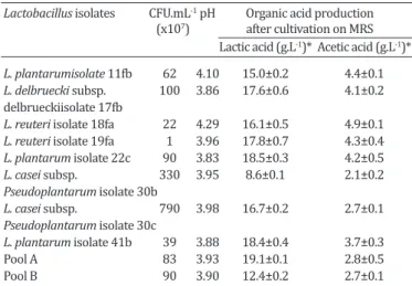
Table 1 shows the total viable cells, final pH, lactic and
acetic acid concentrations of cell-free supernatants obtai-ned from pure and pooled cultures of Lactobacillus. The
total number of colony formation units (CFU) of the strains
were generally high, ranging from 1.107 to 1.109 CFU mL-1.
These results were similar to those obtained by Avonts &
De Vuyst (2001) using seven commercial strains of lactoba-cilli grown in MRS broth under the same conditions, also as shown in Table 2.
Table 1. Results of colony-forming unit, pH values and organic acid concentrations from Lactobacillus isolates after
24 h of cultivation
Lactobacillus isolates CFU.mL-1 pH Organic acid production (x107) after cultivation on MRS Lactic acid (g.L-1)* Acetic acid (g.L-1)*
L. plantarumisolate 11fb 62 4.10 15.0±0.2 4.4±0.1
L. delbruecki subsp. 100 3.86 17.6±0.6 4.1±0.2
delbrueckiisolate 17fb
L. reuteri isolate 18fa 22 4.29 16.1±0.5 4.9±0.1
L. reuteri isolate 19fa 1 3.96 17.8±0.7 4.3±0.4
L. plantarum isolate 22c 90 3.83 18.5±0.3 4.2±0.5
L. casei subsp. 330 3.95 8.6±0.1 2.1±0.2
Pseudoplantarum isolate 30b
L. casei subsp. 790 3.98 16.7±0.2 2.7±0.1
Pseudoplantarum isolate 30c
L. plantarum isolate 41b 39 3.88 18.4±0.4 3.7±0.3
All strains, except L. casei subsp. pseudoplantaum
iso-late 30b, were able to produce lactic acid at concentra-tions higher than 12.4g. L-1. The highest concentration was attained with pool A (19.1g.L-1), followed by L.
plan-tarum isolate 41b (18.4g.L-1) and L. plantarum isolate 22c (18.5g.L-1). In addition, all lactobacilli produced acetic acid at concentrations higher than 2.0g.L-1. These short-chain organic acids are known to exhibit high antimicrobial ac-tivity against microorganisms due to the easy diffusion of the non-dissociated form through the cell membranes of
pathogens (Axe & Bailey 1995). Cheng et al. (2003) stu
-died the influence of organic acids, including acetic acid,
propionic acid and lactic acid on the growth and survival of E. coli O157:H7. The authors reported that lactic acid exerts greater inhibitory effects on the growth of the pa-thogenic bacteria than acetic acid and propionic acid; ho-wever, the combination of the organic acids exhibited an important synergic effect in the inhibition of this pathogen (Cheng et al. 2003, Varalakshmi et al. 2013). According to
Diez-Gonzalez & Russell (1997) small amounts of sodium
acetate (1.6g. L-1) can inhibit the growth of E. coli O157:H7. This level of acetate is similar to the levels observed in the present work.
The antagonistic effects of pure and pooled cultures of
Lactobacillus strains on E. coli O157:H7 were studied using the agar spot method. Based on the data in Figure 1, in non --neutralized media, all of the strains and the pool of Lacto-bacillus strains were antagonistic towards E.coli O157:H7
(Table 2). The strains L. delbrueckii isolate17fb and L. casei
subsp. pseudoplantarum isolate 30c displayed significantly
greater (p<0.05) inhibitory effects on the pathogen than the other studied strains, with zones of inhibition larger than
20mm (Fig.1). Pool A and L. plantarum isolate 41b
exhi-bited a similar pattern (19mm of inhibition halo). Among the strains studied, differences between 10 and 77% of the diameter of clear zone formation were observed. The total
colony count in 2 μL of cell suspension obtained by parallel
culture on an MRS plate ranged from 1.105 to 1.108 CFU -mL-1, while the inoculum concentration of E. coli O157:H7 was 108 CFU in 10 mL of liquefied BHI agar. The lower level of inhibition by L. reuteri isolate 18fa, L. reuteri isolate 19fa
and L. plantarum isolate 22c in MRS media was likely rela-ted to their slower growth capacity (Table 2).
Furthermore, when the same strains and pools of Lac-tobacillus were tested on neutralized MRS medium (with
sodium bicarbonate, 3g.L-1), the mixed strains (Pool A) exhibited higher inhibitory activity (19mm halo diameter) than the pure cultures (0 to 15mm). Differences in the halo diameter of between 30 and 100% were observed for
cul-tures in MRS and MRS-neutralized agar medium (Fig.1). Si -milar results have been described in a study that examined the inhibitory effects of different strains of Bifidobacterium against E. coli; halo diameters in the range of 13 to 30mm
were reported for the un-neutralized agar media associa-ted with a considerable reduction in halo size during gro-wth in media containing sodium bicarbonate (Gagnon et al. 2004).
In our study, the E. coli O157:H7inhibition by the Lacto-bacillus isolates in the presence of sodium bicarbonate may be associated with non-acidic substances, such as
hydro-gen peroxide and bacteriocins. Furthermore, it has been confirmed that the failure of L. plantarum isolate 22c and L. reuteri isolate 19fa to grow in the media containing sodium bicarbonate was due to an unfavorable pH for the growth of these microorganism.
The combination of strains in pool A displayed greater inhibitory activity toward E. coli O157:H7 than the indivi-dual cultures in almost all cases, regardless of the
presen-ce or absenpresen-ce of sodium bicarbonate (Fig.1). These results
demonstrate variation in the inhibitory strength of pure cultures of Lactobacillus, as well as in the synergistic effect of co-cultures against E. coli O157:H7, emphasizing the
im-portance of the precise selection of the strains for probiotic applications.
Figure 2A shows the optical density of E. coli O157:H7 grown in media containing the supernatants from different cultures of Lactobacillus. The absorbance values
demons-trate that E. coli was able to grow in media containing the neutralized fraction of supernatants from pure and pooled cultures of Lactobacillus. However, the pathogenic strain exhibited slower growth rate and reached lower final
OD values in neutralized media compared to the control
(Fig.2A).
The results suggest that pH adjustment results in the loss of the antibacterial properties of the supernatants. Ho-wever, inhibitory activity was observed in all of the media containing the acidic fractions of the supernatants, as
con-Fig.1. Inhibitory effects of Lactobacillus isolates supernatant on Escherichia coli O157:H7 by the spot agar method using MRS agar and MRS after treatment with sodium bicarbonate.
Table 2. Results of optical density of Escherichia coli O157:H7
after 7 h of incubation in acidified BHI and MRS media
Broth Optical density* (600 nm)
Initial OD Final OD
MRS, pH 3.6 0.063±0.005 0.076±0.004 MRS, pH 4,2 0.103±0.008 0.117±0.003 MRS, pH 6,3 0.083±0.003 0.212±0.010 BHI, pH 3,6 0.090±0.004 0.113±0.005 BHI, pH 4,2 0.094±0.002 0.120±0.008 BHI, pH 7,4 0.111±0.010 1.015±0.016
firmed by optical density values that were near zero. Inde -ed, it is well known that most of the antagonistic effects of
Lactobacillus on E. coli growth could be due to the presence
of organic acids that are produced during cell growth (Axe
& Bailey 1995, Diez-Gonzalez & Russell 1997).
According to the results shown in Figure 2B, substan -ces present in the supernatants exerted bacteriostatic or bactericidal effects on the pathogenic Escherichia coli. The
supernatants from L. delbruecki subsp. Delbrueckii isolate
17fb, L. reuteri isolate 19fa and pool B cultures exhibited bactericidal activity on E. coli; after 7 h of exposure to these
supernatants, no colony formation was observed on
spe-cific agar media (data not shown). Some strains, such as L. reuteri, can produce reuterin, a non-proteic substance that is soluble in water and possesses antibacterial, antifungal and antiprotozoan properties (Sung et al. 2003).
Lash et al. (2005) studied the inhibitory effects and sta-bility of L. plantarum supernatants at different
temperatu-res, pHs and after treatment with proteolytic enzymes. The results revealed that the supernatant from L. plantarum
lost its antimicrobial activity when the pH was adjusted to values higher than 5.0 and lower that 4.0, suggesting that the compound responsible for the inhibition was active only in this pH range. In the present work, a similar effect was detected with the supernatants from Lactobacillus
iso-lates 17fb and 19fa, and pool B, which exhibited antimicro -bial activity toward E. coli O157:H7 except at pH 7.0, where
the pathogenic bacteria grew normally.
Ogawa et al. (2001) reported that L. casei strain Shirota and L. acidophilus strain YIT 0070 exerted inhibitory effects
on E. coli growth. Their antimicrobial activity was assayed
in co-culture, suggesting that the antimicrobial effect of
Lactobacillus depends on the pH, due to accumulation of lactic acid in the culture medium. The authors also tested the antagonistic effect of L. brevis; however, this organism
was unable to affect E. coli growth. The lack of antagonis-tic effects indicates that, as observed in the present work, the antimicrobial activity is dependent on the Lactobacillus
strain. On the other hand, Aricia et al. (2004) observed a decrease in the antibacterial activity of supernatants of
Lactobacillus that were adjusted to pH 6.5 and contained catalase, suggesting that hydrogen peroxide and organic acids promoted the inhibition of the growth of pathogens, including E. coli.
The pH exerts an important negative effect on bacterial metabolism and growth. In this study, the E. coli O157:H7
was subjected to a moderately acidic environment at 37°C for 7 h. Table 2 shows the optical density (OD 600 nm) of the pathogenic E. coli inoculated in BHI and MRS broth whi-ch had been adjusted to different pH values. The results de-monstrate that E. coli O157:H7 was unable to grow in both
media at pH 3.6 and 4.2; the effect on pathogen growth was
bacteriostatic. However, the acidified media did not exert a
bactericidal effect on E. coli; after 7h of exposure, the
patho-genic strains were able to grow on BHI agar plates (data not shown). Previous studies showed that E. coli and other pa-thogenic bacteria are sensitive to pH 3.5 and that exposure to low pH can result in adaptive resistance to acidic media
(Koutsoumanis & Sofos 2004; Cheng et al. 2003). E.coli and
others pathogens bacteria can survive under conditions of extreme acids stress thanks to an acidic-resistance (AR) systems, which is a group of amino acid decarboxylases and antiporters amino-acid dependent. Cells that have
gro-wn in amino-acid deficient medium (minimal medium) can
succumb at pH 2.5 given the lack of glutamate or arginine
(Foster 2004).
Cheng et al. (2003) reported that the percentage of E. coli O157:H7 cells that survived in MRS broth at pH 3.0 were 4.2%. However, the authors mentioned that the bac-tericidal effect was not observed after 120 min of exposure to these conditions and that by increasing the pH of the cul-ture broth to 4.0, the percentage of survival grew to 70% of the control growth (pH 7.0). Previous researches showed that many Lactobacillus isolates may prevent the binding
of pathogens bacteria in the human and animal intestinal cells. After the banning of growth promoters antibiotics (GPA) in worldwide, many alternatives were launched, but
still the Lactobacillus strains are the best choice for many
situations, like the use as probiotics for chicken, cattle, swi-ne and others production animals, such as meat-type or egg-type. Nevertheless, we must consider the metabolism product of these bacteria for use in animal production fee-ding. The Lactobacillus studied could prove useful as dieta-ry supplements as well as antimicrobial agents in food and packaging applications.
CONCLUSIONS
Almost all strains of Lactobacillus investigated in this study exhibited bactericidal effects on Escherichia coli
O157:H7.
The highest inhibitory activity corresponded to Lac-tobacillus delbrueckii subsp. delbrueckii 17fb and L. casei
subsp. pseudoplantarum 30c and, suggesting that antimi-crobial compounds production, such as hydrogen peroxide or bacteriocins may be responsible by bacterial inhibition.
All strains tested produced lactic and acetic acids. The amount of lactic acid produced has no
demonstra-ble influence on the inhibition; in pool A, the supernatants
with the highest lactic acid concentrations were unable to exert a more pronounced bactericidal effect on the patho-genic bacteria than the other strains.
Thus, the strains of Lactobacillus studied could be used
as antimicrobial agents in packaging or as dietary supple-ments to control pathogenic microorganisms such as Es-cherichia coli O157:H7.
Acknowledgements.- The authors gratefully acknowledge the financial support of CAPES (Coordenação de Aperfeiçoamento de Pessoal de Nível Superior), FAPESP (Fundação de Amparo à Pesquisa do Estado de São Paulo), and CNPq (Conselho Nacional de Pesquisa e Desenvolvimento Tec-nológico). A.J. Piantino Ferreira is recipient of CNPq fellowship.
REFERENCES
Aricia M., Bilgin B., Sadgic O. & Ozdemir C. 2004. Some characteristic of
Lactobacillus isolates from infant faeces. Food Microbiol. 21:19-24.
Avonts L. & De Vuyst L. 2001. Antimicrobial potential of probiotic lactic acid bacteria. Meded Rijksuniv. Gent., Fak. Landbouwkd. Toegep. Biol. Wet. 66:543-550.
Axe D.D. & Bailey J.E. 1995. Transport of lactate and acetate through the cytoplasmic membrane of Escherichia coli. Biotechnol. Bioeng. 47:8-19. Cantarelli V., Nagayama K., Takahashi A., Honda T., Cauduro P., Dias G.A.G.,
Mezzari A. & Brodt T. 2000. Isolation of Shiga toxin-producing Escher-ichia coli (STEC) serotype O91:H21 from a child with diarrhea in Porto Alegre city, RS, Brazil. Braz. J. Microbiol. 31:266-270.
Cerqueira A.M.F., Guth B.E.C., Joaquim R.M. & Andrade J.R.C. 1999. High occurrence of Shiga toxin-producing Escherichia coli (STEC) in healthy cattle in Rio de Janeiro State, Brazil. Vet. Microbiol. 70:111-121. Chaucheyras-Durand F. & Durand H. 2010. Probiotics in animal nutrition
and health. Beneficial Microbes 1:3-9.
Cheng H.Y., Yu R.C. & Chou C.C. 2003. Increased acid tolerance of Escher-ichia coli O157:H7 as affected by acid adaptation time and conditions of acid challenge. Food Res. Int. 36:49-56.
Diez-Gonzalez F. & Russell J.B. 1997. The ability of Escherichia coli
O157:H7 to decrease its intracellular pH and resist the toxicity of acetic acid. Microbiol. 143:1175-1180.
Earnshaw R.G. 1992. The antimicrobial action of lactic acid bacteria: natu-ral food preservation systems, p.211-232. In: Wood B.J.B. (Ed.), The Lactic Acid Bacteria: the lactic acid bacteria in health and disease. Chapman and Hall, London, UK.
Fábrega V.L.A., Ferreira A.J.P., Patrício F.R.S., Brinkley C. & Scaletsky I.C.A. 2002. Cell-detaching Escherichia coli (CDEC) strains from children with diarrhea: identification of a protein with toxigenic activity.FEMS Micro -biol. Lett. 217:191-197.
Fang H., Elina T., Heikki A. & Seppo S. 2000. Modulation of humoral immu -ne response through probiotic intake. FEMS Immunol. Med. Microbiol. 29:47-52.
FAO/WHO 2001. Joint FAO/WHO Expert consultation on evaluation of health and nutritional properties of probiotics in food including powder milk with live lactic acid bacteria. Available at <http://www.who.int/ foodsafety/publications/fs_management/en/probiotics.pdf> Accessed Febr. 12, 2014.
Foster J.W. 2004. Escherichia coli acid resistance: tales of an amateur aci-dophile.Nature Rev. Microbiol. 2:898-907.
Gagnon M., Kheadr E.E., Blay G.L. & Fliss I. 2004. In vitro inhibition of Es-cherichia coli O157:H7 by bifidobacterial strains of human origin. Int. J.
Food Microbiol. 92:69-78.
Garcia A., Fox J.G. & Besser T.E. 2010. Zoonotic enterohemorrhagic Escher-ichia coli: a one health perspective. ILAR J. 51:221-232.
Gopal P.K., Prasad J., Smart J. & Gill H.S. 2001. In vitro adherence proper -ties of Lactobacillus rhamnosus DR20 and Bifidobacterium lactis DR10 strains and their antagonistic activity against enterotoxigenic Escheri-chia coli. Int. J. Food Microbiol. 67:207-216.
Guarner F. & Schaafsma G.J. 1998. Probiotics. Int. J. Food Microbiol. 39: 237-238.
Jin L.Z., Ho Y.W., Ali M.A., Abdullah N. & Jalaludin S. 1996. Effect of adherent Lactobacillus spp. on in vitro adherence of salmonellae to the intestinal epithelial cells of chicken. J. Appl. Bacteriol. 81:201-206.
Kalantzopoulos G. 1997. Fermented products with probiotic qualities. An -aerobe 3:15-19.
Kandler O. & Weiss N. 1986. Regular, nonsporing Gram-positive rods, p.1209-1234. In: Sneath P.H.A., Mairm N.S. & Sharpe M.E. (Eds), Bergey’s Manual of Systematic Bacteriology. Williams and Wilkins, Baltimore, USA.
Koutsoumanis K.P. & Sofos J.N. 2004. Comparative acid stress response of Listeria monocytogenes, Escherichia coli O157:H7 and Salmonella
Typhimurium after habituation at different pH conditions. Lett. Appl. Microbiol. 38:321-326.
Kudva T.I., Hatfield P.G. & Hovde C.J. 1996. Escherichia coli O157:H7 in mi-crobiota flora of sheep. J. Clin. Microbiol. 34:431-433.
Lash B.W., Mysliwiec T.H., Gourama H. & Mysliwiec T.H. 2005. Detection and partial characterization of a broad-range bacteriocin produced by
Lactobacillus plantarum (ATCC 8014). Food Microbiol. 22:199-204. Monteiro-Neto V., Campos L.C., Ferreira A.J.P., Gomes T.A.T. & Trabulsi L.R.
1997. Virulence properties of Escherichia coli 0111:H12 strains. FEMS Microbiol. Lett. 146:123-128.
Moraes P.M., Perin L.M., Silva Jr A. & Nero L.A. 2013. Comparison of pheno-typic and molecular tests to identify lactic acid bacteria. Braz. J. Micro-biol. 44:109-112.
Nakazato G., Paganelli F.L., Lago J.C., Aoki F.H., Mobilon C., Brocchi M., Ste -hling E.G. & Silveira W.D. 2011. Lactobacillus acidophilus decreases Sal-monella Typhimuriuminvasion in vivo. J. Food Saf. 31:284-289.
Navarro L., Zarazaga M., Sáenz J., Ruiz-Larrea F. & Torres C. 2000. Bacteri-ocin production of by lactic acid bacteria isolated from Rioja red wines. J. Appl. Microbiol. 88:44-51.
Ogawa M., Shimizu K., Nomoto K., Tanaka R., Hamabata T., Yamasaki S., Takeda T. & Takeda Y. 2001. Inhibition of in vitro growth of Shiga tox-in-producing Escherichia coli O157:H7 by probiotic bacteria Lactobacil-lus strains due to production of latic acid. Int. J. Food Microbiol.
68:135-140.
Paço R.S., Leme I.L., Bottino J.A. & Ferreira A.J.P. 2003. Identification of
Lactobacillus spp. from broiler litter in Brazil. Braz. J. Microbiol. 34:236-237.
Poppi L.B., Mancilha I.M., Ferreira A.J.P. & Leal D.D.M. 2008. Evaluation of the antagonistic effect of Lactobacillus on Listeria monocytogenes in vitro. Braz. J. Food Technol. 11:113-119.
Rossland E., Langsrud T., Granum P.E. & Sorhaug T. 2005. Production of antimicrobial metabolites by strains of Lactobacillus or Lactococcus co-cultured with Bacillus cereus in milk. Int. J. Food Microbiol. 98:193-200.
Scapin D., Grando W.F., Rossi E.M., Perez K.J., Malheiros P.S. & Tondo E.C. 2013. Antagonistic activity of Lactobacillus acidophilus LA10 against Salmonella enterica serovar Enteritidis SE86 in mice. Braz. J. Microbiol. 44:57-61. Servin A.L. & Coconier M.H. 2003. Adhesion of probiotic strains to the
intestinal mucosa and interaction with pathogens. Best Pract. Res. 17:741-754.
Schillinger U. & Lücke F.K. 1989. Antibacterial activity of Lactobacillus
sake isolated from meat. Appl. Environ. Microbiol. 55:1901-1906. Sung H.W., Chen C.N., Liang H.F. & Hong M.H. 2003. A natural compound
(reuterin) produced by Lactobacillus reuteri for biological-tissue fixa -tion. Biomaterials 24:1335-1347.
Todorov S.D. & Dicks L.M.T. 2005. Lactobacillus plantarum isolated from molasses produces bacteriocins active against Gram-negative bacteria. Enzyme Microb. Technol. 36:318-326.