2 R it a I sa b el C o st a C u n h a
The mechanism mediating nuclear
translocation of the apoptosis
inducing factor (AIF)
T h e m e ch a n is m m e d ia ti n g n u cl e a r tr a n sl o ca ti o n o f th e a p o p to si s in d u ci n g f a ct o
Escola de Ciências
Rita Isabel Costa Cunha
The mechanism mediating nuclear
translocation
of
the
apoptosis
inducing factor (AIF)
Tese de Mestrado em Genética Molecular
Trabalho efectuado sob a orientação de
Professora Doutora Manuela Côrte-Real
Doutora Susana Chaves
Nome: Rita Isabel Costa Cunha
Endereço eletrónico: [email protected] Telefone: 917875769
Nº do Bilhete de Identidade: 13200058
Título da Tese de Mestrado:
The mechanism mediating nuclear translocation of the apoptosis inducing factor (AIF)
Orientadores:
Professora Doutora Manuela Côrte-Real Doutora Susana Chaves
Instituição de Acolhimento:
Centro de Biologia Molecular e Ambiental (CBMA)
Ano de Conclusão: 2012
Designação do Mestrado:
Mestrado em Genética Molecular
1. É AUTORIZADA A REPRODUÇÃO INTEGRAL DESTA TESE, APENAS PARA EFEITOS DE INVESTIGAÇÃO, MEDIANTE DECLARAÇÃO ESCRITA DO INTERESSADO, QUE A TAL SE COMPROMETE.
Universidade do Minho, 31 de Outubro de 2012 _____________________________________________ Rita Isabel Costa Cunha
Agradecimentos
Às minhas orientadoras, Professora Doutora Manuela Côrte-Real e Doutora Susana Chaves. À Professora Manuela por toda a disponibilidade e carinho, pela partilha dos conhecimentos e do rigor científico, pela dedicação e pelas palavras sempre sábias nos momentos oportunos. À Susana por toda a preocupação e disponibilidade, por todas as sugestões diárias na resolução dos problemas que foram ocorrendo ao longo deste trabalho. Principalmente por ter feito que este último ano tenha sido de muita aprendizagem.
Um muito obrigado sincero às duas pela oportunidade.
Ao CBMA e ao Departamento de Biologia e a todos os seus funcionários e docentes. Um especial à Dona Isabel e à Nádia por toda a boa disposição e favores prestados.
A todos os colegas da Micro I por me terem acolhido e proporcionado um excelente ambiente de trabalho. Por todos os “cafés” recheados de gargalhadas e também pelas sugestões na realização do trabalho experimental e pelo companheirismo. Por isso um muito obrigado à Sara Alves, Dário Trindade, Helena Paula , Ana Marta Duarte, Jorge Rodrigues, Flávio Azevedo, Tânia Fernandes, Rita Pacheco e Vera Martins.
E porque as frases mais ditas por mim neste ano foram: “Oh Andreiaaa sabes...?” ou “Oh Ruizinhoo como é que…?”, um obrigado especial ao Rui Silva e Andreia Pacheco por basicamente me ajudarem em tudo o que foi preciso, e também por me animarem sempre que preciso.
Um agradecimento também aos arredores da Micro I: Filipa Pereira, Raúl Machado, Joana Sá Pessoa, Filipa Vale, Manoel, Gabriel Rocha, Suellen Ferro, Lisandra Castro, Fábio Faria e Paulo Geraldes. Obrigado pela ajuda e boa disposição.
E como nada se consegue sem o apoio dos amigos, agradeço aos meus amigos do coração que tanto me aturaram este ano: Catherine Ferreira, Dulce Cunha, Filipe Pires, Joana Tulha, André Charrua, Bruno Fernandes, Juliana Fernandes, Carla Sampaio, Bruno Freitas, Cristina Real, Bruno Cunha, Joana Campos e Carina Rego. Obrigado pelo companheirismo, incentivo, carinho, boa disposição e amizade constantes. Um obrigado especial ao meu Pedrinho por me aguentar 24 sobre 24 horas por dia e mesmo assim ter sempre um gesto e uma palavra de carinho! Obrigado Maninho!
Ao Touni por para além de ser companheiro de laboratório, amigo, ser o meu companheiro para a VIDA! OBRIGADA por me aturares mais do que todos, por toda a ajuda, carinho e apoio e por nunca desistires de mim!
E como os últimos são sempre os primeiros... À minha família (Papás, Maninho, Jú, Madrinha e Padrinho, Pipa e Tios) por serem quem são! Obrigado pelo amor, alegria, atenção, confiança, apoio e por me ensinarem a ser como sou.
The mechanism mediating nuclear translocation of the apoptosis inducing factor (AIF)
Abstract
Since the discovery that yeast cells can undergo programmed cell death in response to several different stimuli, Saccharomyces cerevisiae has gained prominence in the cell death field. Exposure of yeast cells to certain stimuli like acetic acid or hydrogen peroxide or even heterologous expression of pro-apoptotic proteins can trigger cell death by apoptosis via a mitochondrial pathway and exhibiting the typical hallmarks of apoptosis, such as chromatin condensation, ROS (reactive oxygen species) accumulation, DNA fragmentation, externalization of phosphatidylserine, mitochondrial dysfunction with release of cytochrome c and of apoptosis inducing factor (Aif1p), among other pro-apoptotic proteins.
Aif1p is a flavoprotein that is involved both in cell survival and cell death. In healthy cells, Aif1p is confined to the mitochondria, where it plays a role in bioenergetic and redox metabolism due to its redox activity and its FAD and NADH domains. However, when cells are exposed to an apoptotic stimulus, Aif1p is released from mitochondria to the cytosol and then translocated to the nucleus, where it will induce DNA fragmentation and chromatin condensation. Although the mechanism underlying this translocation is still poorly understood, it is known that transport into the nucleus through the nuclear pore complexes is an energy-dependent process for the majority of macromolecules, and normally mediated by the major class of transport receptors, the karyopherins or Kaps. In this work, we aimed elucidate the mechanism regulating yeast Aif1p import into the nucleus and to discover which Kap is responsible for the nuclear import of Aif1p.
Apoptosis was triggered in cells expressing Aif1p tagged with GFP (by exposure to acetic acid or hydrogen peroxide, chronological ageing, or heterologous expression of Bax c-myc protein) and Aif1p localization was assessed by fluorescence microscopy and cellular fractionation/western blot assays. However, we were not able to observe the release of Aif1p to the cytosol or its nuclear import under our experimental conditions. We also attempted to map the NLS domain of Aif1p by deletion of several domains with homology to previously mapped domains in human AIF. Although we has not yet mapped the Aif1p NLS domain, we discovered that deletion of the putative Hsp70p-binding domain led to aberrant localization of Aif1p, suggesting that Hsp70p is important for Aif1p folding and stability. These results indicate that Hsp70p may also be involved in the regulation of Aif1p localization in yeast.
Mecanismo mediador da translocação nuclear do fator de indução de apoptose (AIF)
Resumo
Células de Saccharomyces cerevisiae são capazes de desencadear um processo de morte celular programada em resposta a vários estímulos. Esta descoberta levou à utilização extensiva deste organismo eucarionte unicelular como modelo no estudo de processos de morte celular. Sabe-se que a exposição de células de levedura a certos estímulos, como o ácido acético ou peróxido de hidrogénio, ou mesmo a expressão heteróloga de proteínas pró-apoptóticas de mamífero, pode desencadear um tipo morte celular dependente de uma via mitocondrial e exibindo características típicas da morte apoptótica, tais como a condensação da cromatina, acumulação de espécies reativas de oxigénio (ROS), fragmentação do DNA, externalização de fosfatidilserina e disfunção mitocondrial associada à libertação de citocromo
c e do fator indutor de apoptose (Aif1p).
Aif1p é uma flavoproteína que está envolvida tanto na sobrevivência como na morte celular. Em células saudáveis, esta proteína está confinada à mitocôndria, onde desempenha um importante papel no metabolismo bioenergético e estado redox. Este facto é devido à sua atividade redox e aos seus domínios FAD e NADH. No entanto, em células expostas a um estímulo apoptótico, ocorre libertação de Aif1p da mitocôndria para o citosol e subsequentemente a sua translocação para o núcleo, onde induz fragmentação do DNA e condensação da cromatina. Embora o mecanismo subjacente a esta translocação esteja ainda pouco conhecido e estudado, sabe-se que para a maioria das macromoléculas o transporte para o núcleo ocorre através dos complexos de poros nucleares e é um processo dependente de energia, normalmente mediado por uma classe de recetores de transporte, as Kaps (do inglês “karyopherins”, também conhecidas como importinas ou transportinas). Este trabalho teve como objectivo elucidar a regulação do mecanismo de transporte de Aif1p para o núcleo e identificar que/ais Kap(s) são responsáveis pelo importe do Aif1p para o núcleo na levedura.
A morte celular apoptótica foi induzida em células que expressam uma fusão da proteína Aif1 com GFP (por exposição a ácido acético ou peróxido de hidrogénio, envelhecimento cronológico, ou pela expressão heteróloga de proteínas Bax c-myc) e a localização de Aif1p foi avaliada por microscopia de fluorescência e fracionamento celular /western Blot. No entanto, não fomos capazes de observar libertação de Aif1p para o citosol ou a sua importação nuclear nas condições experimentais utilizadas. Tentámos também mapear o domínio NLS de Aif1p através da remoção de vários domínios homólogos com domínios conhecidos de AIF humano. Embora não tenhamos ainda conseguido mapear o domínio NLS do Aif1p, descobrimos que a deficiência no domínio putativo de ligação a Hsp70 resulta numa localização aberrante de Aif1p, sugerindo que a Hsp70 é importante para a estabilidade desta proteína e que esta proteína pode ainda estar envolvida na regulação da localização de Aif1p na levedura.
Index Agradecimentos ... iii Abstract ... iv Resumo ... v Index... vi Abbreviations ... viii 1. Introduction ... 1 1.1. Apoptosis ... 2 1.1.1. Mechanism of apoptosis ... 2 1.1.1.1. Extrinsic pathway ... 3 1.1.1.2. Intrinsic pathway ... 4
1.2. Apoptosis inducing factor (AIF) ... 7
1.2.1. The role of AIF in cells ... 8
1.2.2. The mechanism underlying AIF release from mitochondria ... 10
1.2.3. Heat shock protein 70 (Hsp70p) acts as a chaperone to AIF protein ... 11
1.3. Yeast apoptosis ... 12
1.3.1. Yeast apoptotic triggers ... 14
1.3.2. Yeast orthologue of AIF, Aif1p ... 15
1.3.2.1. Stimuli that regulate Aif1p release ... 15
1.4. Nucleocytoplasmic trafficking ... 16
1.4.1. Nuclear Pore Complex (NPC) ... 16
1.4.2. Protein transport receptors and transport cycle ... 18
1.4.3. Regulation of nucleocytoplasmic trafficking ... 21
1.4.4. Nuclear import of AIF ... 22
2. Aims and research plan ... 24
3. Materials and methods ... 27
3.2. Yeast strains and growth conditions... 30
3.3. Hydrogen peroxide and acetic acid treatment ... 30
3.4. Heterologous expression of Bax c-myc ... 30
3.5. Transformation of bacterial cells ... 31
3.6. Purification of Yeast DNA ... 32
3.7. Fluorescence Microscopy ... 32
3.8. Subcellular fractionation ... 32
3.8.1. Preparation of spheroplasts ... 32
3.8.2. Mitochondrial and cytosolic fraction preparation ... 33
3.8.3. Nuclear fraction preparation ... 33
3.8.3.1. Protocol I ... 33
3.8.3.2. Protocol II ... 34
3.8.3.3. Protocol III ... 34
3.8.4. SDS gel electrophoresis/Westernblot ... 34
3.9. Immunoprecipitation ... 35
3.9.1. SDS gel electrophoresis/Western Blot/Silver Staining ... 35
4. Results ... 38
4.1. Construction of pAIF1WT-GFP plasmid and localization AIF1p ... 39
4.2. Acetic acid treatment and chronological ageing ... 40
4.3. Hydrogen peroxide treatment ... 41
4.4. Heterologous expression of Bax c-myc protein ... 42
4.5. Aif1p translocation to the nucleus and cytochrome c release ... 43
4.6. Map the NLS of Aif1p... 48
4.6.1. Construction of AIF1 fragments and localization of Aif1p ... 48
4.7. Protein-protein interactions of Aif1p ... 51
5. Discussion and future perspectives ... 54
Abbreviations
AIF – Apoptosis Inducing Factor AIFL - AIF-Like
ANT - Adenine Nucleotide Translocator AMID - AIF homologous
mitochondrion-associated inducer of death
Apaf-1 - Apoptotic Protease Activating
Factor-1
ARM - Arginine-rich motifs ATP - Adenosine Triphosphate
CARD - Caspase Recruitment Domain c-FLIP - Cellular-FLICE (FADD-like IL-
1β-converting enzyme)-inhibitory Protein
Caspases - Cysteine-dependent
Aspartate-specific Proteases Cyp A - Cyclophilin A Cyt c – Cytochrome c DAPI - 4,6-Diamino-2-phenyl-indole dihydrochlorid DD - Death Domain
DED - Death Effector Domain
DISC - Death Inducing Signaling Complex DNA - Deoxyribonucleic Acid
DR - Death Receptors
EDTA - Ethylenediaminetetraacetic acid Endo G - Endonuclease G
ER - Endoplasmatic Reticulum
FADD - Fas-Associated Death Domain GDP - Guanosine Diphosphate
GFP - Green Fluorescent Protein GTP – Guanosine triphosphate h - hours
H2O2 – Hydrogen Peroxide
HSPs - Heat-shock proteins HtrA2/Omi - High Temperature
Requirement Protein A2
IAPs - Inhibitors of Apoptosis Proteins IBB - Importin-β binding
IMM - Inner mitochondrial membrane IMS - Intermembrane space
Kaps - Karyopherins
MAC - Mitochondrial Apoptosis-induced
Channel
MOMP - Mitochondrial Outer Membrane
Permeabilization
NADH - Nicotinamide Adenine Dinucleotide NADPH - Nicotinamide Adenine
Dinucleotide Phosphate
min - Minutes
MLS – Mitochondrial Localization
Sequence
NE – Nuclear envelope
NES - Nuclear Export Sequence NLS - Nuclear Localization Sequence NF-KB - Nuclear factor
kappa-light-chain-enhancer of activated B cells
NPC - Nuclear pore complexes Nups – Nucleoporins
OD – Optical density
OMM - Outer mitochondrial membrane PCD – Programmed Cell Death PCR - Polymerase Chain Reaction phe-gly - phenylalamine-glycine PTP - Permeability Transient Pore ROS - Reactive Oxygen Species rpm – Rotations per minute sec - Seconds
Smac/Diablo - Second
mitochondria-derived Activator of Caspases/Direct Inhibitor of Apoptosis Protein (IAP)-Binding Protein With Low Pi
SPB - Spindle Pole Body
TNF-R - Tumor-Necrosis Factor Receptor TRADD - TNF-R-Associated Death Domain TRAIL-R - TNF-related Apoptosis-Inducing
Ligand Receptor
1.1. Apoptosis
Multicellular organisms often need to eliminate cells that are in excess, or damaged cells that can put the organism at risk (Hengartner, 2000). To this end, they use a highly regulated and complex process characterized by a group of endogenous molecular events that culminates in “cell suicide” (Leist et al., 2001), designated by programmed cell death (PCD). Several types of cell death have been associated with PCD, but in eukaryotic cells the most common is apoptosis (Hengartner, 2000). Apoptosis occurs during normal development of multicellular organisms and continues through adult life. This process is as important as cell division or cell migration because it allows the organism to control cell number and tissue size, as well as protect itself from compromised cells that can threaten homeostasis, such as infected or damaged cells (Gewies, 2003).
The term apoptosis derives from the Greek “apo” - from and “ptosis” – falling, an analogy to the term used by Greeks to describe leaves falling from trees. This term was used in 1972 by John Kerr and colleagues to describe a type of cell death with distinct morphologic characteristics (Lawen, 2003). Indeed, apoptotic cells can easily be recognized by several morphological features such as cell shrinking, chromatin condensation and migration along the nuclear membrane, blebbing of the plasma membrane and exposure of phosphatidylserine to the outer leaflet of the plasma membrane. The final hallmark of apoptosis is cell fragmentation into compact structures, called 'apoptotic bodies' that will be phagocyted by macrophages and eliminated from the tissue without leading to an inflammatory response (Saraste et al., 2000).
1.1.1. Mechanism of apoptosis
Apoptosis can be triggered by various stimuli and mechanisms, including virus infection, cell stress and DNA damage. Cellular sensitivity to these stimuli can differ depending on several factors, such as expression of anti- and pro-apoptotic proteins, the harshness of the stimulus and the phase of the cell cycle (Gewies, 2003). The apoptotic process consists of three consecutive steps: (i) a trigger by extracellular or intracellular stimuli; (ii) execution by activation of intracellular proteases and (iii) elimination of dead cells by engulfment of cell debris by neighboring cells or macrophages (Saikumar et al., 1999). Deficient regulation of apoptosis can lead to various pathologies (Rudin and Thompson, 1997); improper activation may cause or contribute to several diseases such as ischemic strokes, AIDS (acquired
immunodeficiency syndrome), and several neurodegenerative disorders (Raff et al., 1993, Ameisen et al., 1995 and Smale et al., 1995). In contrast, a flawed activation of this process can lead to some autoimmune diseases and to uncontrolled cell division that culminates in the development of cancers (Tan, 1994; Lowe and Lin, 2000 and Reed, 2003).
Two major apoptotic pathways have been identified in mammalian cells: the extrinsic or death receptor pathway and the intrinsic or mitochondrial pathway (Figure 1) (Hengatner 2000 and Elmore, 2007).
1.1.1.1. Extrinsic pathway
In the extrinsic pathway, “death receptors” are activated to transmit apoptotic signals from the exterior to the interior of cells. Examples of death receptors are TNFR-1, Fas/CD95 and TRAIL receptors. Death receptors belonging to the tumor necrosis factor receptor (TNFR) gene superfamily are present in the cell surface and transmit the apoptotic signal after ligation of precise ligands such as Fas ligand, TNF alpha and TRAIL. They have an important role in apoptosis because they can trigger a caspase cascade within seconds of ligand binding and induce apoptosis very quickly (Sartorius
et al., 2001).
Caspases (Cysteine-dependent Aspartate-specific Proteases) are conserved through evolution and can be found in several organisms such as insects, human, hydra and nematodes (Cohen, 1997). They are synthesized as inactive zymogens, named procaspases. They have a prodomain followed by a small and a large subunit that are occasionally separated by a linker peptide. After maturation, procaspases are proteolytically processed between the large and the small subunit. An active caspase is a heterotetramer that consists of two large and two small subunits (Hengartner, 2000). Their active site contains a cysteine residue critical for catalytic activity; it has high affinity for aspartate residues, after which caspases cleave their substrates. So far, about a hundred caspases substrates have been described (Fischer et al., 2003).
There are two different groups of caspases: (i) initiator caspases, which include the procaspases -2, -8, -9 and –10, with long prodomains such as death effector domains (DED) in procaspase-8 and -10 or caspase recruitment domains (CARD) in procaspases-9 and -2; (ii) effector or executioner caspases, with only short prodomains, which include procaspases -3, -6 and -7 (Chrowdhury et al., 2008).
In the extrinsic pathway, initiation of apoptosis requires both caspases and death receptors. Apoptotic signaling is mediated by the death domain (DD) of the death
receptor. Adapter molecules that possess their own DDs, as FADD or TRADD, are then recruited to the DDs of the death receptor to form the death-inducing signaling complex (DISC). With the help of FADD, procaspase 8 (initiator procaspase) is then recruited to the DISC, and the amount of procaspase-8 molecules in the DISC leads to their autocatalytic activation and consequent release of active caspase-8. Active caspase-8 then activates the caspase signaling cascade; subsequently, effector caspases cleave specific substrates, resulting in cell death (Stegh et al., 1998; Scaffidi
et al., 1998). It is possible to inhibit caspase-8 in order to protect cells from apoptosis.
One example is cellular-FLICE (FADD-like IL-1β-converting enzyme)-inhibitory protein (c-FLIP), which can inhibit DISC signaling. This inhibition will lead to inactivation of DISC and by consequence inhibition of the extrinsic pathway (Hengartner, 2000). In some cases, the apoptotic signal produced by death receptors might not be capable of inducing a caspase signaling cascade strong enough to lead to cell death on its own. In other cases, there may be a bridge between the caspase signaling cascade and mitochondria provided by Bid, a Bcl-2 family member (see below). Bid can be cleaved into its truncated form (tBID) by caspase-8 and translocated to the mitochondria, where it induces the release of pro-apoptotic proteins to the cytosol (Luo et al., 1998 and Gustafsson et al., 2007).
1.1.1.2. Intrinsic pathway
The intrinsic pathway is triggered by “intrinsic” stresses such as DNA damage, endoplasmic reticulum stress, lysosomal stress, and mitochondrial dysfunction. In contrast with the extrinsic pathway, in which caspases are activated directly, the intrinsic pathway requires the participation of mitochondria. Permeabilization of the outer mitochondrial membrane (MOMP) and the release of pro-apoptotic factors are crucial events of the intrinsic pathway and required for activation the downstream caspase cascade. Mitochondria have thus gained great importance in the field of mammalian apoptosis, since they are able to amplify the apoptotic signal from the extrinsic pathway and also propagate the death signals generated within the cell, such as oxidative stress, DNA damage and others (Wang, 2001).
The Bcl-2 family, a group of apoptotic regulators with both anti- and pro-apoptotic members, plays a major role in the regulation of the intrinsic pathway. Bcl-2 family members share homologous regions known as BH domains, and can be divided into four categories based on structural and functional similarities, namely: i) the anti-apoptotic Bcl-2 proteins (A1, Bcl-2, Bcl-w, Bcl-xL and Mcl-1); ii) Bcl-2 effector proteins
(Bax and Bak); iii) direct activator BH3-only proteins (Bid, Bim and Puma) and iv) sensitizer/derepressor proteins (Bad, Bik, Bmf, Hrk and Noxa) (Chipuk et al., 2010). Pro- and anti-apoptotic proteins can interact with each other and with several other proteins, forming several regulatory networks, and is the balance of the levels of pro- and anti-apoptotic proteins that can dictate the fate of the cell, mainly through regulation of MOMP.
Figure 1 - Schematic representation of the extrinsic and intrinsic pathways involved in apoptosis (Hengatner 2000).
Mitochondria are surrounded by two membranes, the inner mitochondrial membrane (IMM) and the outer mitochondrial membrane (OMM), which divide the mitochondria into two compartments: the intermembrane space (IMS) between the membranes, and the inner compartment called the matrix. Many stress stimuli can trigger permeabilization and release of pro-apoptotic proteins from the mitochondria to the cytosol. Two main hypotheses explaining how permeabilization of mitochondria
occurs have been put forth. The first hypothesis suggests that a transmembrane channel named the permeability transition pore (PTP) is formed on the contact sites between the IMM and the OMM. These pores are thought to be mainly constituted by VDAC in the OMM and by adenine nucleotide translocator (ANT) in the IMM. PTP opening results in loss of membrane potential, uptake of solutes and entry of water to the matrix, leading to the rupture of the outer membrane and release of pro-apoptotic proteins from the IMS to the cytoplasm (Lawen, 2003). Another hypothesis is based on the ability of the pro-apoptotic Bcl-2 proteins to form pores. Indeed, the accepted model proposes a stepwise structural reorganization of Bax leading to mitochondrial targeting and homo-oligomerization. Bax is kept as a monomeric soluble cytosolic factor through the engagement of its α9 helix in the dimerization pocket by the α1 helix. The activator BH3s, tBID/BIM/PUMA, attack and expose the α1 helix of Bax, leading to a secondary disconnection of the α9 helix and consequently mitochondrial insertion. Activator BH3s stay associated with the N-terminally exposed Bax through the BH1 domain to drive homo-oligomerization and activation of Bax (Ren et al., 2010).
Permeabilization of mitochondria results in the release of several proteins such as cytochrome c, Omi/HtrA2 (High Temperature Requirement Protein A2) and Smac/DIABLO (Second Mitochondria-derived Activator of Caspases/Direct Inhibitor of Apoptosis Protein (IAP)-binding Protein With Low pI). Later, the apoptosis inducing factor (AIF) and endonuclease G (EndoG) are also released. All these proteins are located in the mitochondrial intermembrane space (IMS) and are released to the cytosol in response to several apoptotic stimuli, such as extra- and intracellular stresses like oxidative stress and treatment with cytotoxic drugs (Vaux, 2011). Proteins of the first group activate the caspase-dependent mitochondrial pathway. After its release to the cytosol, cytochrome c can interact with Apaf-1 (Protease Activating Factor – 1), leading to the recruitment of pro-caspase 9, which together with dATP forms a multi-protein complex, the apoptosome, which in turn activates caspase -9 leading to apoptosis. On the other hand, Smac and Omi/HtrA2 normally neutralize IAP (Inhibitors of Apoptosis Proteins), which normally inhibit effector caspases. Pro-apoptotic proteins of the second group are released only after the cell has committed to die. Both AIF and Endo G are translocated to the nucleus (Joza et al., 2001). AIF induces DNA fragmentation and chromatin condensation, whereas Endo G induces DNA internucleossomal fragmentation (Wang, 2001).
1.2. Apoptosis inducing factor (AIF)
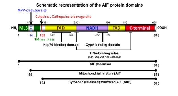
The mammalian mitochondrial AIF is a flavoprotein that belongs to a larger family of proteins with common structural and functional features, containing bacterial, plant and fungal oxidoreductases. It is a protein with 613 aminoacids and is structurally divided into three typical domains: a FAD and a NADH binding domain and a C-terminal domain, where its pro-apoptotic activity resides. In addition, it also possesses an N-terminal MLS (Mitochondria Localization Signal) (Figure 2) (Gurbuxani et al., 2003). Besides AIF, AIF homologous mitochondrion-associated inducer of death (AMID) and AIF-Like (AIFL) also belong to this family (Hangen et al., 2010).
Figure 2 – Representation of the three different AIF forms: the precursor, the mature and the truncated AIF. AIF contains an N-terminal MLS domain (green), a FAD bipartite domain (yellow), an NADH binding domain (violet) and a C-terminal domain (red). It also possesses an Hsp70p and a Cyclophilin A binding domain. Cleavage at the MPP (Mitochondrial Processing Peptidase) cleavage site (blue dotted line) generates mitochondrial mature AIF and cleavage by calpains (at the red dotted line) generates truncated AIF (tAIF) (Yuste et al., 2007).
In healthy cells, AIF is confined to mitochondria, where it plays an important role in bioenergetic and redox metabolism. However, after an apoptotic stimulus, AIF is released from mitochondria to the cytosol and translocated into the nucleus (Modjtahedi et al., 2006).
1.2.1. The role of AIF in cells
AIF has dual functions, in DNA fragmentation and in redox activity, and thus acts both in cell death and cell survival. However, biochemical and mutational analysis of AIF suggests that its apoptotic and redox functions can be separated (Lipton et al., 2002). Three distinct functions have been associated with AIF, as seen in figure 3.
Figure 3 - Representation of the three different functions of AIF in cells. First, AIF has a role as an NADH oxidase. Second, cytosolic AIF seems to promote mitochondrial membrane permeabilization and third, AIF can promote chromatin condensation and DNA fragmentation (Candé et al., 2002).
The first function attributed to AIF is it NADH oxidase activity. AIF has a redox potential of –308 mV ± 15 mV at pH 7.5. It has monodehydroascorbate reductase activity and catalyses cytochrome c reduction in the presence of NADH. Results have shown that the AIF NADH oxidase activity requires an electron donor (NADH), an electron acceptor and a prosthetic FAD group for catalytic electron transfer (Miramar et
al., 2001).
The second function is permeabilization of the mitochondrial membrane and consequent release of cytocrome c and additional AIF. This function is attributed to cytosolic AIF, since studies revealed that when AIF is introduced into the cytosol it can induce the release of AIF and cytochrome c from mitochondria (Daugas et al., 2000). When cells commit to apoptosis, AIF translocates to the nucleus, where it exerts its third function: to trigger cell death by promoting chromatin condensation and DNA fragmentation (Candé et al., 2002). Several reports suggest that this role of AIF in the
nucleus functions as a backup to caspase-dependent mechanisms. However, it was also described that AIF-induced cell death occurs in the complete absence of caspases and the oxireductase activation. AIF seems to play a role in caspase-independent PCD in several organisms such as Caenorhabditis elegans, Dyctiostelium discoideum and mammals (Arnoult et al., 2001, Wang et al., 2007, Wang et al., 2002 and Lorenzo et
al., 2007).
But how are these effects achieved at the molecular level? The crystal structure of AIF shows that the surface of the protein has positively charged amino acids, allowing it to form electrostatic connections with DNA. AIF binds with more affinity to linearized forms than to intact circular plasmids, suggesting that it is introduced into the DNA strand breaks. The AIF-DNA interaction is accompanied by DNA condensation (shortening of the DNA), hairpin formation (intermolecular packageing) and DNA oligomerization (Modjtahedi et al., 2006). Studies have shown that addition of AIF to DNA does not cause DNA degradation. It was therefore suggested that after entering the nucleus AIF can cooperate with several exo- and endonucleases, building up a so-called “degradeosome” including cyclophilins, proteins belonging to the family of peptidylprolyl cis–trans isomerases. Human AIF is able to interact with cyclophilin A (CypA) and forms an active DNase, and several studies confirm that CypA is essential for the apoptotic activity of AIF (Candé et al., 2004). These characteristics can be included into a hypothetical model: AIF invades DNA strand-breaks, induces DNA condensation and finally participates in the formation of a DNA-degrading polyprotein complex (Figure 4). It is still not known if the deadly effect of the translocation of AIF to the nucleus is due to inhibition of the respiratory chain or to apoptotic DNA degradation in the nucleus.
It has recently been discovered that AIF acts in programmed necrosis, in addition to its involvement in apoptotic PCD. PCD is a dynamic process that depends on the characteristics of the involving scenario, such as cell or tissue type, death stimulus or environmental conditions. Cells can use caspase or non-caspase dependent mechanisms, exhibiting necrotic or apoptotic features. Programmed necrosis is thus an active capase-independent pathway. Moubarak et al. described that AIF is a regulator of this type of PCD by causing extensive DNA damage when cells are exposed to high doses of MNNG, an alkylating agent and necrotic inducer. In this study, AIF knockout protected cells from DNA damage and cell death, highlighting the requirement of this protein for programmed necrosis (Moubarak et al., 2007, Boujrad et
Figure 4 -Hypothesized model for the action of AIF on DNA (Modjtahedi et al, 2006).
1.2.2. The mechanism underlying AIF release from mitochondria
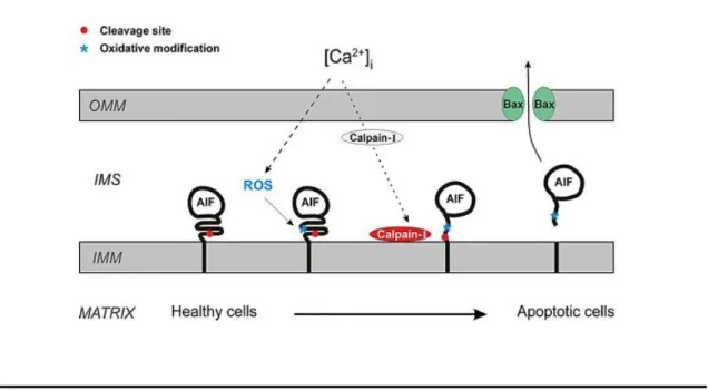
AIF is encoded by a nuclear gene and is synthesized in the cytoplasm. It is imported to mitochondria through the general import pathway as a 67 kDa protein with a mitochondrial localization signal (MLS) in the N-terminus and anchored to the IMM. Upon import to the mitochondria IMS, the MLS is cleaved by a mitochondrial peptidase to generate mature 62 kDa AIF. As referred above, after treatment with certain apoptotic stimuli, AIF can be cleaved from its membrane anchor by proteases (calpains and cathepsins), forming a soluble 57 kDa AIF fragment (Figure 2). Calpains are a family of calcium-dependent cysteine proteases. There are two main isoforms, calpain-I and calpain-II, which co-exist with the calpain-specific inhibitor calpastatin. The interaction between the two prevents both activation and catalytic activity of calpains. Their localization is uncertain, but recently calpain was found to contain an MLS and it was suggested that calpains are present in the mitochondrial IMS. Upon activation, calpains can cleave and destroy calpastatin that, with addition of Ca2+ to the mitochondria, is sufficient to stimulate AIF cleavage, as illustrated in figure 5 (Badugu
Figure 5 - Schematic representation of the mechanism behind the release of AIF from IMM to the cytoplasm (Norberg et al., 2010).
1.2.3. Heat shock protein 70 (Hsp70p) acts as a chaperone to AIF protein
After permeabilization of the OMM, cleaved AIF can be released to the cytosol and translocated to the nucleus. However, this translocation seems to be regulated by cytoprotective proteins. The first proteins thought to play this role were heat-shock proteins (HSPs). This group of proteins is found in most the living organisms, such as, bacteria, yeast, humans, and others. They are involved in the folding and unfolding of other proteins and their expression increases when cells are exposed to several stresses but mostly when cells are exposed to elevated temperatures (Parsell and Lindquist, 1993). This family of chaperones recognizes a broad spectrum of unfolded or misfolded proteins (Chakrabart et al., 2011). Using a series of biochemical tests, it was discovered that, within the family of HSPs proteins, Heat-shock protein 70 (Hsp70p) was the only capable of physically interacting with AIF. Hsp70p is involved in the inhibition of apoptosis by blocking Apaf-1 and by consequence the formation of the apoptosome, (Ravagnan et al., 2001). It also binds to and neutralizes AIF, protecting cells against translocation of AIF to the nucleus and by consequence protecting against its apoptogenic effects, such as chromatin condensation and fragmentation. This is corroborated by several studies, where the authors demonstrate that the interaction between AIF and Hsp70p inhibits its translocation to the nucleus both in cultured cells and in vivo (Ravagnan et al., 2001 and Ruchalski et al., 2001). However, the precise
mechanism of AIF import into the nucleus is not known. Yeast Aif1p shows the same localization and exhibits similar death executing pathways as mammalian AIF. Therefore, this model system is particularly suitable to investigate the mechanism involved in importing AIF to the nucleus.
1.3. Yeast apoptosis
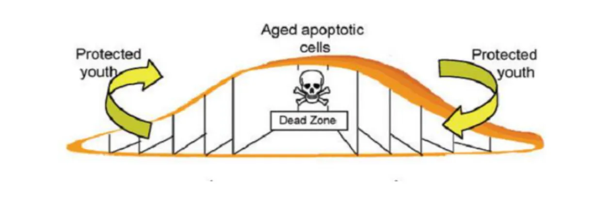
Over the last decades, Saccharomyces cerevisiae has been a preferred research tool in numerous areas of cell biology, due to its easy handling, technical tractability, fast growth, small sequenced genome and the fact it is a eukaryote. However, it was often questioned whether single-cell organisms need to commit suicide or die by apoptosis. In fact, yeast cells tend to form colonies and communities, and it is hypothesized that apoptosis may occur in yeast during chronological and replicative ageing, leading to removal of virus-infected and damaged cells and unsuccessful mating processes from colonies. This altruistic cell death gives younger cells nutrients they can metabolize, contributing to the maintenance of the members of the community (Büttner et al., 2006) (Figure 6). It has now become clear that apoptosis does not occur only in multicellular organisms, but can also be triggered in unicellular organisms like yeast. It is believed that several apoptotic pathways are conserved in yeast, making this organism attractive to study programmed cell death (Carmona-Gutierrez et al., 2010).
The first observations of yeast apoptosis were made in a mutant strain of S.
cerevisiae with a mutation in the CDC48 gene, which encodes a protein necessary for
vesicle trafficking. The authors observed that dying cells of this mutant showed an apoptotic phenotype with several characteristics of mammalian apoptosis, such as phosphatidylserine exposure to the outer leaflet of the plasma membrane, DNA fragmentation and margination, and condensation of chromatin (Madeo et al., 1997).
Figure 6 – Representative image of how cell death and ageing occurs in a yeast colony. The amount of aged cells located in the center of the colony corresponds to the dead zone. These cells die altruistically to give to the younger cells located at the periphery nutrients, allowing the maintenance of the colony (adapted from Gourlay et al., 2006).
Since then, multiple yeast orthologs of mammalian apoptotic proteins have been found and characterized. They include AIF, IAP, caspases, AMID, Endo G and Omi/HtrA2 (Madeo et al., 2004). In addition, cell death has been associated with chronological and replicative ageing and cell cycle arrest (Madeo et al., 2002).
Another similarity of yeast apoptosis with mammalian apoptosis is the important role of mitochondria in both processes, through ROS production and release of pro-apoptotic proteins from the mitochondria IMS. Moreover, mitochondrial morphology is radically reorganized during apoptosis, from filamentous tubules into small punctate spheres. This dynamic nature of mitochondrial morphology is due to two processes, fission and fusion, important for maintaining mitochondrial function and repair damaged mitochondria. The morphological change of mitochondria seems to be related with cell death, as one of the early markers of mammalian apoptosis is fragmentation of tubular mitochondria. In yeast, there are three genes involved in mitochondrial fission which have been associated with regulation of apoptosis: DNM1, MDV1/NET2 and FIS1. Deletion of the first two (DNM1, MDV1/NET2) protects cells from death induced by acetic acid or Hydrogen peroxide (H2O2) treatment, whereas deletion of the latter (FIS1)
leads to an increase in cell death. This observation suggested that Fis1p could have a function similar to that of Bcl-2 proteins (Fanjiang et al., 2004). However, it was recently found that the sensitivity of fis1 mutants to cell death is due to a secondary mutation in the stress-response gene WHI2 (Cheng et al., 2006), which confers a sensitive phenotype in response to apoptosis inducers.
Like mammals, yeast also has caspase-dependent and caspase-independent apoptotic pathways. YCA1, the yeast orthologue of mammalian caspases, is
considered a metacaspase and has a central role in yeast apoptosis. It is known that yeast cells with a disruption of YCA1 are less susceptible to apoptotic cell death under stress conditions like ageing and exposure to oxidative stress, salt, valproic acid, iron and other metals. Recently, another caspase-like protease in yeast was found, Eps1p. When released from the anaphase inhibitor Pds1p, Eps1p works as a caspase-like protease and cleaves Mcd1p. Mcd1p is a yeast homolog of human cohesion Rad21 that is truncated after apoptotic stimuli and translocates from the nucleus to the mitochondria, decreasing the mitochondrial membrane potential and thus promoting the release of cytochrome c (Madeo et al., 2009). However, not all yeast apoptotic cell death is dependent on YCA1 or on another caspase-like protease.
1.3.1. Yeast apoptotic triggers
In yeast, apoptosis can be triggered by three different strategies: by heterologous expression of pro-apoptotic proteins, by environmental stress or drug-induced stress, or by yeast endogenous apoptotic inducers.
Heterologous expression of proteins consists in expressing one or various proteins in an organism that does not possess these proteins in its own genetic background. It is described that yeast cells do not possess obvious orthologous of proteins from the Bcl-2 family. However, heterologous expression of these human pro-apoptotic proteins can trigger an active cell death. While some studies showed that when Bax or Bak proteins are expressed in yeast, they can trigger cell death with apoptotic characteristics, such as release of cytochrome c and generation of ROS, others demonstrated that Bax induces a cell death process associated with activation of autophagy (Kissova et al., 2007). These facts suggest that these proteins have their function conserved in this organism (Eisenberg et al., 2007 and Madeo et al., 2004).
It is also known that several exogenous agents such as hyperosmotic stress, heavy metals, amiodarone, ethanol, elevated temperatures, oxidative stress, UV radiation, various pharmacological agents (such as aspirin), osmotin, viral "killer" toxins, HOCl, pheromones and sometimes sugar can efficiently induce apoptosis in yeast (Liang et al., 2008; Silva et al., 2005; and Carmona Gutierrez et al., 2010). However, the best characterized and the most used exogenous inducers of apoptosis in yeast are H2O2 and acetic acid (Madeo et al., 1999 and Ludovico et al., 2001).
Finally, several endogenous factors that trigger yeast apoptosis have been described, such as defects in cellular processes like chromatid cohesion, N-glycosylation, mRNA stability (Carmona-Gutierrez et al., 2010). Other examples of
endogenous apoptosis triggers are DNA damage resultant from oxygen metabolism and ROS generation and failed replication. Chronological and replicative ageing are additional examples, and apoptosis also occurs during the development of colonies on solid media (Herker et al., 2004 and Vachova and Palkova, 2005). Other endogenous triggers are some pro-apoptotic proteins that are released from the mitochondria to the cytosol, such as AIF, AIF-homologous mitochondrion-associated inducer of death (AMID) and EndoG orthologues, which once in the cytosol are translocated to the nucleus.
1.3.2. Yeast orthologue of AIF, Aif1p
Aif1p, the yeast orthologue of AIF, was first described in 2004 by Wissing et al. It was shown that Aif1p, as mammalian AIF, is mitochondrial in normal cells and relocalizes to the nucleus in the presence of apoptotic stimuli. It was also found that the role of yeast Aif1p is dependent on cyclophilin A and partially on caspase activity. Therefore in yeast, in contrast with mammalian cells, Aif1p-mediated apoptosis is not completely independent on the yeast metacaspase Yca1p. Moreover, in contrast to mammalian AIF, purified Aif1p degrades yeast nuclei and plasmid DNA (Wissing et al., 2004).
AIF1 deletion led to increased survival to treatment with H2O2 and acetate, as
well as to decreased chronological ageing. Like in mammalian cells, Aif1p also has a vital role in respiration via its NADH oxidase domain, and AIF1-deficient S. cerevisiae have decreased growth on non-fermentable carbon sources. This redox function is important for an effective anti-oxidant defense and oxidative phosphorylation (Madeo et
al., 2009).
1.3.2.1. Stimuli that regulate Aif1p release
Ludovico (2002) was the first to observe translocation of Aif1p to the nucleus in cells undergoing chronological ageing or treated with camptothecin, an S phase-specific anticancer drug that inhibits the action of the enzyme DNA topoisomerase-I. Then Wissing et al. (2004) also observed translocation of Aif1p to the nucleus, in cells treated with H2O2. Since then, few additional studies have addressed this issue and the
role of Aif1p in yeast apoptosis. Morton et al. described that antimicrobial peptides, the dermaseptins obtained from amphibians, are capable of inducing yeast cell death. The yeast cell death described by this group is independent of the metacaspase Yca1p but
depends on Aif1p for nuclear fragmentation (Morton et al., 2007). Bostrycine is an anthracenedione with antimicrobial and phytotoxic activity that belongs to the quinone family, which inhibits cell proliferation and induces a decrease in the mitochondrial membrane potential, leading to mitochondrial disruption. When yeast cells were treated with this compound, cell death with hallmarks of apoptosis was observed. However, bostrycin-induced cell death was promoted in yca1Δ. In contrast, this death phenotype was partially rescued in aif1Δ cells (Chunlingku et al., 2009). Another substance that can induce apoptosis is allicin, an antimicrobial extracted from garlic that is capable of inducing apoptosis through its oxidative properties; both Aif1p and Yca1p seem to be involved in this process, suggesting that allicin induces apoptosis through an alternative mechanism (Gruhlke et al., 2010).
So far only H2O2 and chronological ageing have been shown to trigger Aif1p
release to the cytosol and translocation to the nucleus (Wissing et al., 2004). However, there have been no studies addressing the mechanism underlying the import of yeast Aif1p into the nucleus, which, like for mammalian AIF, is most likely an active and highly regulated process.
1.4. Nucleocytoplasmic trafficking
The nucleus is a defining characteristic of eukaryotic cells and is physically separated from the cytosol by the nuclear envelope (NE), a double membrane structure. Most genetic information is confined to the nucleus, where most functions of the cell are thus governed. Non-cytosolic proteins synthesized in the cytoplasm have to be directed to their specific locations, where they exert their functions. For that they contain a signal sequence that will direct them to a specific location. For example, proteins containing a nuclear localization sequence (NLS) are imported into the nucleus (Macara, 2001).
1.4.1. Nuclear Pore Complex (NPC)
The NE is crossed by multiple supramolecular structures named nuclear pore complexes (NPCs), structures specialized in transport of small molecules, ions and macromolecules between the cytoplasm and the nucleus. The number of NPCs in each cell is variable and depends on cell size and transcriptional activity, and differs between species. Human cells may contain about 5x10³ – 5x10⁷ NPCs per nucleus and these
that of higher eukaryotes, with a molecular weight of approximately 44 MDa, and there are about 200 NPCs per nucleus (Freitas et al., 2009).
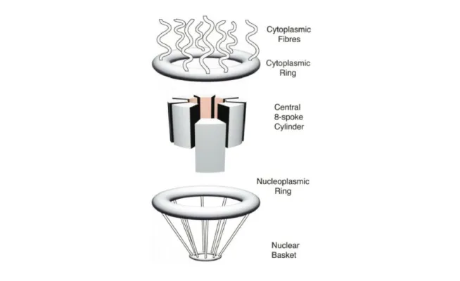
NPCs consist of a cylindrical central body sandwiched between the outer and inner layers of the NE. Nucleoplasmic and cytoplasmic rings are constituted by eight subunits that constitute the central structure. Eight protein filaments rise up from the cytoplasmic ring towards the cytoplasm, and the nuclear ring forms eight filaments that converge in a structure named nuclear basket, as shown in figure 7.
Figure 7 - Representation of a human nuclear pore complex (NPC) showing its morphology (Bayliss et al., 2000).
The central structure of the human NPC has a central channel about 30 nm in diameter that allows the transport of ions and proteins with a molecular weight of 20-40 kDa (Bayliss et al., 2000). Some proteins can be transported into the nucleus by an intrinsic ability to directly interact with the NPC, but most are transported through the NPC by association with several types of transport receptors. The overall structure of the NPC is similar between human and yeast, but yeast NPCs lack the cytoplasmic and nuclear rings (Rout et al., 2000). Amazingly, despite their large size, purified yeast NPCs contain only about 30 different proteins, named nucleoporins (Nups). Nups can be separated into three groups: nucleoporins that span the nuclear membrane; non-membrane proteins that have multiple repeats of a phenylalamine-glycine (phe-gly)
motif, and non-membrane proteins that don’t have the phe-gly motif (Macara et al., 2001).
1.4.2. Protein transport receptors and transport cycle
For the majority of macromolecules, transport into the nucleus and through the NPCs is an energy-dependent process. This process can be mediated by the major class of transport receptors, which includes different soluble proteins (karyopherins or Kaps, also named importins, exportins or transportins). They mediate import into or export from the nucleus of proteins that cannot be transported by simple diffusion through the NPCs, as well as mediate the transport of non-coding RNAs. Karyopherins are acidic proteins with a molecular weight of 90 to 145 kDa. About 10 of the 20 karyopherins identified were demonstrated to participate in the nuclear import of proteins in eukaryotes (Tran et al., 2006). Sequence analysis identified 13 predicted importin-β proteins in the yeast S. cerevisiae.
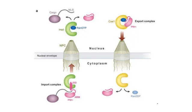
Most transport receptors of the importin-β family are capable of binding directly to cargo and transport it across the NE while others, like importin-β, need an adaptor. Importin-β can interact with importin-α and mediate a process of transport into and out of the nucleus. Importin-α is a transport adaptor that recognizes and binds to the NLS of proteins. The NLS-binding domain is formed by 10 arginine-rich motifs (ARM) in tandem, localized on the central region of importin-α. The NLS of the cargoes is recognized by importin-α through ARM, and the interaction between the importins is mediated by an importin-β binding domain (IBB) that is localized in the N-terminal region of importin-α. The cargo/importin-α/importin-β complex is then transported through the NPCs. In the nucleus, this complex is dissociated by binding of Ran-GTP to importin-β, which displaces the cargo. Importin-α is then transported back to the cytosol by the exportin Cse1/CAS (Figure 8) (Cook et al., 2007). However, most karyopherins are able to directly interact with their cargo and recognize a variety of nuclear localization signals, though most are basic in nature.
Another important characteristic of karyopherins is that they can interact with Ran GTPase and the phe-gly domains of nucleoporins. Ran GTPase, a monomeric protein belonging to the Ras superfamily, can be found in two distinct forms; GTP-bound and GDP-GTP-bound, and these two different forms are distributed asymmetrically in the nucleus (more Ran-GTP) and in the cytoplasm (more Ran-GDP). Specific regulatory proteins that are localized in the cytoplasm and the nucleus maintain the asymmetric distribution of these proteins. Normally, the reaction of hydrolysis of GTP
into GDP is slow; however, this reaction can be accelerated by two proteins, Ran-GAP and Ran-BP1. On the other hand, the nuclear protein RCC1 promotes the reverse reaction. It is the combined action of Ran regulatory proteins that creates and maintains the Ran-GTP gradient across the NE, and it is this gradient that establishes the directionality of nucleocytoplasmic transport (Freitas et al., 2009).
.
Figure 8 - Overview of the transport cycle of the classical nuclear localization signal (NLS) transport with the ligation of importin-β and importin-α (Cook et al., 2007).
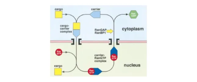
Ran-GTP binds to the transport receptors (importin-β family) and also to other proteins in solution. Ran-GTP, in the nucleoplasmic side of the NPC, dissociates import receptors from their cargo, promoting release of the cargo. In contrast, it promotes the formation of export complexes. Ran-GAP and Ran-BP1, located in the cytoplasm, promote hydrolysis of Ran-GTP bound to export complexes, releasing the cargo and freeing the carrier for another round of transport (Figure 9).
Figure 9 - Schematic representation of processes underlying nuclear import and export mediated by β-karyopherins (Importin-β) (Bayliss et al., 2000).
Several cycles of export complexes with Ran-GTP would lead to depletion of Ran from the nucleus. Ran is small and can therefore diffuse through the NPC, but this mechanism is not fast enough for efficient nuclear trafficking (Bayliss et al., 2000). It is known that the nuclear import of Ran-GDP is promoted by the nuclear transport factor NTF2/p10. After import of Ran-GDP to the nucleus, the protein RCC1 will convert the Ran-GPD into Ran-GTP (Figure 10) (Cook et al., 2007, Freitas et al., 2009). The levels of Ran-GTP are thus replenished in the nuclear side, allowing for continuous cycles of import and export.
1.4.3. Regulation of nucleocytoplasmic trafficking
As mentioned above, the majority of proteins are synthesized in the cytosol and then transported to their final localization in order to exert their function. This process is often an important regulatory step in cellular pathways. Indeed, there are several examples of proteins whose function is regulated by nuclear localization. One example is NF-kB (nuclear factor kappa-light-chain-enhancer of activated B cells), a transcription factor found in almost all types of mammalian cells. It is involved in the cellular response to several stimuli, such as several types of stress, cytokines, ultraviolet radiation, free radicals and LDL oxidation. This transcription factor is bound to an inhibitory protein (IkB) in unstimulated cells and therefore is latent. However, upon activation by extracellular agents, IkB is phosphorylated by a protein kinase and then ubiquitinated and degraded by the proteasome, allowing NF-kB translocation to the nucleus. There, it binds to the kB consensus sequence generally leading to an increase in the expression of the target gene (O’Neill et al., 1997).
Another example is the yeast transcription factor Yap1p, which regulates expression of target genes containing a binding site named Yap1p-response element on their promoter. Normally Yap1p is located in the cytosol and is translocated into the nucleus under stress conditions. In the nucleus, nuclear export of Yap1p is inhibited, which leads to its accumulation in the nucleus and consequently inducing the expression of several genes encoding antioxidant proteins. Exposure to H2O2 induces
the formation of disulfide bonds between the C-terminal cysteine-rich domain (C-CRD) and the N-terminal cysteine-rich domain (N-CRD) of Yap1p. The C-CRD possesses a NES, which ordinarily leads to export of Yap1p from the nucleus, but it is masked by this dually disulfide-bonded structure. Therefore, Yap1p export is inhibited, promoting Yap1p nuclear accumulation (Gulshan et al., 2005).
Another example of regulated transport is the regulation of protein localization by phosphorylation and desphosphorylation of the yeast transcription factor Pho4p. Pho4p localization is regulated in response to modifications in the concentration of inorganic phosphate in the media. Pho4p is imported into the nucleus through the importin-β family member Pse1p/Kap121p. It is known that inhibition of the Pho4p/Pse1p interaction is mediated by phosphorylation and that Pho4p translocation requires Pse1p. This suggests that this import is regulated by phosphorylation in vivo. In yeast cells grown in phosphate-rich medium, Pho4p is phosphorylated by the Pho80-Pho85 cyclin-CDK complex and import into the nucleus is therefore inhibited. Consequently, there is no transcription of phosphate starvation-specific genes. However, in yeast grown with limited phosphate, the CDK inhibitor Pho81p inhibits
Pho80-Pho85, leading to accumulation of unphosphorylated Pho4p. Unphosphorylated Pho4p can then interact with Pse1p/Kap121p, translocate into the nucleus and induce transcription of phosphate-responsive genes (Kaffman et al., 1998).
1.4.4. Nuclear import of AIF
AIF function is regulated at different levels including through its subcellular localization. As mentioned above, AIF is transported to the mitochondria due to the N-terminal MLS. After mitochondrial membrane permeabilization, truncated AIF is released into the cytosol and then translocated to the nucleus.
The crystal structure of AIF indicates that this protein has to be re-localized to the nucleus to exert its apoptotic activity (Ye et al., 2002). To discover which region of this gene is responsible for the apoptotic activity of this protein, Gurbuxani and colleagues mapped functional regions of AIF by deleting several regions and observing the resultant phenotype. The authors determined that the C-terminal domain (beyond residue 567) is responsible for AIF-induced chromatin condensation (once AIF is in the nucleus). Deletion of the Hsp70p-binding region (residues 150 to 228) leads to a gain of function phenotype, i.e., facilitates nuclear translocation in response to an apoptotic stimulus. One region of AIF contains a consensus NLS (residues 367 to 459), normally involved in protein translocation to the nucleus. However, two NLS domains are described for AIF, one more N-terminal (residues 277 to 301) and another closer to the C-terminus (residues 445–451) (Susin et al., 1999). In this study, the authors showed that the C-terminal NLS is functionally more important for AIF translocation to the nucleus than the N-terminal NLS domain. However, deletion of the region containing the C-terminal NLS only partially inhibited AIF translocation, which suggests that there are other domains involved in AIF import (Gurbuxani et al., 2003). However, the mechanism mediating this nuclear translocation of AIF is still unclear.
2. AIMS AND
Apoptosis-inducing factor is a flavoprotein with oxidoreductase activity localized in the mitochondrial intermembrane space. Upon apoptosis induction, AIF translocates to the nucleus, where it leads to chromatin condensation and DNA degradation. AIF has been suggested to control a caspase-independent pathway of apoptosis, important for neurodegeneration and normal development. However, it remains unknown how AIF translocates to the nucleus. Yeast Aif1p shows the same localization and exhibits similar death executing pathways as mammalian AIF, though it mediates a partially caspase- dependent pathway (Wissing et al., 2004). Therefore, our aim was to use the yeast model system to investigate the mechanism involved in importing Aif1p to the nucleus.
Aim 1. Determine which soluble import factor is necessary to import Aif1p
Transform mutants in each of the yeast soluble transport receptors (Kaps) with Aif1p-GFP and assess the localization of Aif1p after apoptosis induction by fluorescence microscopy. Mutation of the Kap mediating Aif1p import will prevent its nuclear accumulation.
Aim 2. Map the NLS of Aif1p
Clone different fragments of Aif1p in frame with GFP and assess their localization by fluorescence microscopy. Identify the smallest sequence that is both necessary and sufficient for transport into the nucleus.
3. MATERIALS AND
METHODS
3.1. Plasmids
All the plasmids and oligonucleotides used in this work are listed in Table I and ll, respectively. Plasmids were amplified in the Escherichia coli XL1Blue strain (as described below) and purified using a Miniprep kit (GenElute Plasmid Miniprep kit, Sigma-Aldrich) according to manufacturer’s instructions. The identity of the plasmids was confirmed by digestion with specific restriction enzymes and by PCR.
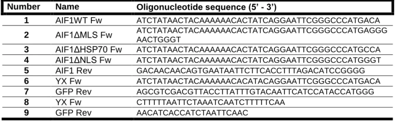
pAIF1WT-GFP, pAIF1ΔMLS-GFP, pAIF1ΔHSP70-GFP, pAIF1ΔNLS-GFP were constructed by Gap Repair (Figure 11). Briefly, four fragments of the AIF1 gene (Wild-type AIF1, AIF1 ΔMLS, AIF1 ΔHSP70, AIF1 ΔNLS) were amplified by Polymerase Chain Reaction (PCR) using the plasmid pAIF1 from Euroscarf as a template and oligonucleotides forward 1-4, respectively, and the reverse oligonucleotide 5 (Table II).
Saccharomyces cerevisiae strain DF5a was transformed with pYX242GFP vector
digested with EcoRI and HindIII and each of the four fragments (Figure 11). Correct integration of the AIF1 fragments in pYX242GFP was confirmed by colony PCR using oligonucleotides that anneal upstream and downstream of the insertion (6 and 7, respectively). After confirmation, the plasmids were purified from yeast cells (described below in 3.6), amplified in E. coli and sequenced.
Table I – List of the plasmids used in this work.
Plasmid Description References/Sources
pAIF1 AIF1 ORF Euroscarf (Germany)
pYX242GFP LEU2, AmpR Rosenblum et al., 1998
pAIF1WT-GFP AIF1 1-1137 cloned in pYX242GFP This study
pAIF1ΔMLS-GFP AIF1 Δ1-78 cloned in pYX242GFP This study
pAIF1ΔHSP70-GFP AIF1 Δ1-336 cloned in pYX242GFP This study
pAIF1ΔNLS-GFP AIF1 Δ1-723 cloned in pYX242GFP This study
pBAX c-myc Bax c-myc cloned in PCM189, URA3 Priault et al., 1999
Table II – List of the oligonucleotides used in this work.
Number Name Oligonucleotide sequence (5’ - 3’)
1 AIF1WT Fw ATCTATAACTACAAAAAACACTATCAGGAATTCGGGCCCATGACA
2 AIF1ΔMLS Fw ATCTATAACTACAAAAAACACTATCAGGAATTCGGGCCCATGAGGG
AACTGGGT
3 AIF1ΔHSP70 Fw ATCTATAACTACAAAAAACACTATCAGGAATTCGGGCCCATGCCA
4 AIF1ΔNLS Fw ATCTATAACTACAAAAAACACTATCAGGAATTCGGGCCCATGGGT
5 AIF1 Rev GACAACAACAGTGAATAATTCTTCACCTTTAGACATCCGGGG
6 YX Fw ATCTATAACTACAAAAAACACATACAGGAATTCGGGCCCATGACA
7 GFP Rev AGCGTCGACGTTACCTTATTTGTACAATTCATCCATACCATGGG
8 YX Fw CTTTTTAATTCTAAATCAATCTTTTTCAA
Figure 11 - Schematic representation of the methodology used in the amplification of AIF1 fragments (AIF1 ΔMLS, AIF1 ΔHSP70, AIF1 ΔNLS) and construction of plasmids.
3.2. Yeast strains and growth conditions
All S. cerevisiae strains used in this work and respective genotypes are shown in Table III. Strains were transformed with the indicated plasmids using the Lithium acetate/Single Stranded carrier DNA/Polyethylene Glycol (PEG) method previously described in (Gietz and Woods, 2006). Transformants were selected on Synthetic Complete (SC) medium [SC containing 0.17% (w/v) Yeast nitrogen base without aminoacids and ammonium sulfate, 0.5% (w/v) ammonium sulfate, 0.14% (w/v), drop-out mixture lacking histidine, leucine, tryptophan and uracil, 0.008% (w/v) Histidine, 0.04% (w/v) Leucine, 0.008% (w/v) Tryptophan and 0.008% (w/v) Uracil] lacking the appropriate aminoacids plus 2% (w/v) of carbon source and 2% agar. Yeast strains were maintained on solid YPD or SC medium (lacking the appropriate amminoacids), grown at 30°C for 48 h, stored at 4°C, and refreshed every 2 weeks. Yeast cultures were grown aerobically in SC medium with 2% Glucose or Galactose as a carbon source. Strains transformed with plasmids were grown in the same medium lacking the appropriate amino acids. Cells were incubated at 30°C with orbital shaking (200 rpm) and a liquid/air ratio of 1:5.
3.3. Hydrogen peroxide and acetic acid treatment
Cells were grown overnight until exponential phase (OD600 = 0.5-0.6) on SC
Glu, collected by centrifugation and resuspended in new medium (medium with pH 3 in the case of acetic acid treatment) to a final concentration of 107 cells/mL (approximately OD600 = 0.2) and incubated with H2O2 (1 mM, 2 mM and 3 mM), or
acetic acid (140 mM, 160 mM, 180 mM) for up to 360 min at 30°C. At specific time points, serial dilutions (1:10) were spotted onto YPD plates and colony growth was scored after 2 days of incubation at 30°C. Viability was determined in relation to time 0 (100%). In parallel, 500 µL of cells were harvested and processed for fluorescence microscopy.
3.4. Heterologous expression of Bax c-myc
Strains harboring the plasmid pBAX c-myc were grown overnight in SC Glucose lacking uracil and supplemented with doxycycline (10 µg/ml) to repress Bax c-myc expression. Cells were centrifuged, washed three times with sterilized water, ressuspended in the same medium without doxycycline (to induce Bax c-myc expression) or with doxycycline (as a negative control). At specific time points, serial