FACULDADE DE FARMÁCIA
Evaluation of antiviral drugs against
BK polyomavirus
Carolina Martins da Conceição
Mestrado Integrado em Ciências Farmacêuticas
FACULDADE DE FARMÁCIA
Evaluation of antiviral drugs against
BK polyomavirus
Carolina Martins da Conceição
Trabalho de Campo de Mestrado Integrado em Ciências Farmacêuticas apresentado
à Universidade de Lisboa através da Faculdade de Farmácia
Orientador: Doutor Gilles Duverlie
Co-orientador: Doutor José Miguel Azevedo Pereira, Professor Auxiliar com Agregação
This project was developed under the Erasmus+ Programme, at the AGIR (AGents Infectieux, Résistance et chimiothérapie) unit, CURS (Centre Universitaire de Recherche en Santé), belonging to Université de Picardie Jules Verne in Amiens, France, under the supervision of Professor Gilles Duverlie.
Abstract
BK polyomavirus (BKPyV) is a DNA virus belonging to the Polyomaviridae family. BKPyV is considered ubiquitous among the population, and primary infection usually occurs during early childhood. This primary infection is usually asymptomatic, following which the virus establishes a lifelong persistent infection mainly on epithelial cells of the urinary tract and the kidney. In immunocompetent individuals the virus remains clinically silent throughout the life of the host. Immunosuppression, however, can lead to reactivation of the latent infection. In such conditions, one of the major clinical complications of BKPyV reactivation is polyomavirus-associated nephropathy (PyVAN) in kidney transplant recipients. The development of more potent immunosuppressant drugs has increased the incidence rate of PyVAN in these patients and to date, because a specific and safe antiviral drug against BKPyV is still lacking, the mainstay approach to avoid PyVAN development and the possibility of graft loss is to reduce the immunosuppressive treatment. In this context, the development and search for antiviral drugs that can efficiently stop BKPyV replication has become of great clinical interest.
This project focused on studying the efficacy of two drugs - NA-17 and TN-18 - against BKPyV replication in hTERT immortalized human renal proximal tubular epithelial cells (RPTEC/TERT1), an immortalized cell line of the major target cells of the virus in vivo. Results showed an inhibition of BKPyV infection with both NA-17 and TN-18, having been estimated effective concentration 50% (EC50) values of 190 μM and 682 μM, respectively. Treatment of BKPyV-infected RPTEC/TERT1 with the highest concentrations tested for both drugs was responsible for a decrease of the extracellular BKPyV DNA load, that although discrete, corresponded also with an inhibition of the release of infectious progeny. Overall our results show a moderate antiviral effect against BKPyV for both drugs, making us believe that they would have only a limited effect on treating this infection in vivo. Further research in this area is needed to develop an efficient treatment against BKPyV infection and to improve disease outcomes in kidney transplant recipients that develop PyVAN as a result of BKPyV infection reactivation.
Resumo
O poliomavírus BK é um vírus de DNA pertencente à família Polyomaviridae. A infeção primária é normalmente assintomática, após a qual o vírus estabelece uma infeção persistente maioritariamente nas células epiteliais do trato urinário e do rim. Em indivíduos imunocompetentes o vírus permanece clinicamente silencioso durante toda a vida do hospedeiro. No entanto, a imunossupressão pode conduzir à reativação do vírus latente, sendo uma das principais complicações desta reativação a nefropatia associada a poliomavírus em transplantados renais. O desenvolvimento de imunossupressores cada vez mais potentes tem aumentado a incidência da nefropatia nestes doentes e atualmente, não existindo nenhum antiviral que possa ser utilizado de forma eficaz e segura contra este vírus, a abordagem clínica consiste na redução do tratamento imunossupressor de modo a prevenir o desenvolvimento da nefropatia. Neste contexto, o desenvolvimento e a procura de antivirais que consigam eficazmente impedir a replicação do poliomavírus BK têm assumido um grande interesse clínico.
O principal objetivo deste projeto consistiu no estudo da eficácia de dois compostos - NA-17 e TN-18 - contra a replicação do poliomavírus BK em células epiteliais tubulares proximais renais imortalizadas com hTERT (RPTEC/TERT1), uma linha celular imortalizada de um dos maiores alvos celulares do vírus in vivo. Os resultados mostraram uma inibição da infeção pelo poliomavírus BK, tendo sido estimadas as concentrações responsáveis por uma inibição de 50% da infeção de 190 μM e 682 μM para o NA-17 e o TN-18, respetivamente. O tratamento com as concentrações mais elevadas testadas para ambos os compostos foi responsável por uma redução da carga extracelular de DNA viral, assim como na redução de libertação de novas partículas virais infeciosas. De um modo geral, os resultados mostram um efeito antiviral moderado de ambos os compostos contra o poliomavírus BK, mostrando que teriam um efeito limitado no tratamento desta infeção in vivo. A continuação da investigação nesta área é necessária para o desenvolvimento de um tratamento eficaz da infeção pelo poliomavírus BK e para a melhoria do prognóstico nos transplantados renais que desenvolvem nefropatia como resultado da reativação desta infeção.
Acknowledgments
First, I would like to thank Professor Gilles Duverlie, supervisor of this work. I am sincerely grateful to you for accepting me in your lab to develop this project. Thank you for sharing with me your knowledge and for guiding me through all this work.
To Professor José Miguel Azevedo Pereira for the co-supervision of this work. Thank you for all the support and words of encouragement even at a distance, and the availability on discussing the results and revising this thesis. Your contribution was essential to this work, and for that I am very thankful.
A very special thanks to all the team from the Virology lab at Amiens. To Veronique Descamps and Lynda Handala for sharing with me your experience, and above all for all the time and patience you had to teach me and answer all my questions. Also, to Fatima Dakroub, Tony Fiore, François Helle, Etienne Brochot, François Peltier, Virginie Morel, Catherine François, Baptiste Demey, Laurence Marquis and Catherine Moriset, thank you all for the way you welcomed me in Amiens and in your team, it was a pleasure having spent these three months with you. Merci à vous!
Also, I would like to thank Marta Calado. It was two years ago that I took my first steps on a research lab with you. All you taught me these years was fundamental to get me here. To my friends that shared with me these last five years, Catarina, Inês, Cátia, Marta, Xana, Cláudia and Sofia. Thank you so much for your friendship, for your patience, and above all, for never letting me give up. Also, to Ana, Henrique and Inês, thank you so much for always being there for me.
To my mother and my sister, thank you for always having my back. Thank you for your unconditional support through all my worries and insecurities. I couldn’t have done this without you.
List of abbreviations
BKPyV BK polyomavirus c/mL copies/mL CC50 cytotoxic concentration 50% CC90 cytotoxic concentration 90% CI confidence intervals CSK cytoskeletal bufferDMEM Dulbecco's modified Eagle medium
DMSO dimethyl sulfoxide
dpi days post-infection
hpi hours post-infection
dsDNA double-stranded DNA
EC50 effective concentration 50%
EC90 effective concentration 90%
ER endoplasmic reticulum
ERAD ER-associated degradation
FBS fetal bovine serum
FAM 6-carboxyfluorescein
GA-100 gentamicin and amphotericin B
HEK293 human embryonic kidney 293
HLA human leukocyte antigen
hTERT human telomerase reverse transcriptase
IARC International Agency for Research on Cancer
kbp kilobase pairs
MCPyV Merkell cell polyomavirus
miRNA microRNA
MOI multiplicity of infection
MPyV mouse polyomavirus
mRNA messenger RNA
mTOR mammalian target of rapamycin
NAb neutralizing antibodies
NCCR non-coding control region
NLS nuclear localization sequences
ORI origin of replication
PBS phosphate buffered saline
PCR polymerase chain reaction
PFA paraformaldehyde
pre-miRNA precursor-miRNA
PyVAN polyomavirus-associated nephropathy
qPCR quantitative real-time PCR
REGM renal epithelial growth medium
RPTEC human renal proximal tubular epithelial cells
RPTEC/TERT1 hTERT immortalized RPTEC
RT-qPCR reverse transcription qPCR
SI selectivity index
tAg small tumor antigen
TCID50 50% tissue culture infectious dose
truncTAg truncated TAg
TSPyV Trichodysplasia spinulosa-associated polyomavirus
Table of Contents
Abstract ... 3 Resumo ... 4 Acknowledgments ... 5 List of abbreviations ... 6 Table of Contents ... 9 List of Figures ...10 List of Tables ...10 1. Introduction ...11 1.1. BK polyomavirus ...11 1.1.1. BKPyV virion ...12 1.1.2. BKPyV genome ...131.1.2.1. Genotypes and variants ...14
1.1.3. BKPyV replication cycle ...15
1.2. Primary infection, latency and reactivation ...17
1.3. Polyomavirus-associated nephropathy ...18
1.3.1. Early screening and diagnosis...19
1.3.2. Therapeutic approaches...20
1.4. Model systems for the study of BKPyV infection ...24
2. Aims of this study ...26
3. Materials and Methods ...27
3.1. Cell culture ...27
3.2. Virus ...27
3.2.1. Virus stock production ...27
3.2.1. Virus stock titration ...27
3.3. Infection and drug treatment ...28
3.4. Immunofluorescence staining, microscopy, and digital image processing ...28
3.7. Calculation of the effective concentrations 50% ...30
4. Results ...31
4.1. Effect of NA-17 and TN-18 on BKPyV infection on Vero cells ...31
4.1.1. Inhibition of BKPyV infection ...31
4.2. Effect of NA-17 and TN-18 on BKPyV infection on RPTEC/TERT1 ...32
4.2.1. Inhibition of BKPyV infection ...32
4.2.2. Effect on BKPyV genome replication ...34
4.2.3. Effect on the release of infectious progeny ...35
5. Discussion ...37
6. Conclusions and Future Outlooks ...41
7. References ...44
List of Figures
Figure 1. Cryo-electron microscopy structure of BKPyV virions. ... 12Figure 2. Structure of BKPyV genome. ... 13
Figure 3. Schematic representation of BKPyV replication cycle. ... 15
Figure 4. Description of the QuantIF macro. ... 29
Figure 5. Effect of increasing concentrations of NA-17 and TN-18 on the inhibition of BKPyV infection on Vero cells. ... 32
Figure 6. Effect of increasing concentrations of NA-17 and TN-18 on the inhibition of BKPyV infection on RPTEC/TERT1. ... 33
Figure 7. Effect of increasing concentrations of NA-17 and TN-18 on BKPyV DNA load. ... 34
Figure 8. Effect of increasing concentrations of NA-17 and TN-18 on the release of infectious progeny. ... 35
List of Tables
Table 1. Calculated EC50 values for NA-17 and TN-18 tested on RPTEC/TERT1 ... 341. Introduction
1.1. BK polyomavirus
BK polyomavirus (BKPyV) is a member of the Polyomaviridae family of non-enveloped, small viruses, with 40 to 45 nm in diameter, and circular double-stranded DNA (dsDNA) genomes comprising approximately 5 kilobase pairs (kbp) (1). The polyomavirus family was first named after its founding member, the mouse polyomavirus (MPyV), which was found to cause tumors in mice (polyoma meaning “many tumors”) (2). This family has now more than eighty different viruses divided in four genera, each of them having a specific host range, including mammals, birds and fish (1).
Polyomaviruses with the ability to infect humans were first described in 1971. BKPyV was first isolated by inoculating Vero cells with urine samples of a patient who exhibited chronic pyelonephritis and advanced renal failure, and that for those reasons had undergone kidney transplant (3). In the same issue of The Lancet, independently, it was reported the isolation of JC polyomavirus (JCPyV) from brain tissue of a patient with Hodgkin’s disease who had developed progressive multifocal leukoencephalopathy (4). These viruses, both named after the patients’ initials, were the first two human polyomaviruses to be isolated. After these, numerous human polyomaviruses have been described, and to date, there are 13 polyomaviruses that are known to infect humans (1).
Human polyomaviruses are considered ubiquitous among the population, but primary infections with these viruses are generally not associated with significant clinical symptoms or disease. Immunosuppression, however, can lead to reactivation of the latent infection, and in such conditions there are four different human polyomaviruses that have been associated with human diseases (1). BKPyV is one of these viruses, and major complications, when reactivated, include polyomavirus-associated nephropathy (PyVAN) and hemorrhagic cystitis in kidney and hematopoietic stem cell transplant recipients, respectively (5). Additionally, JCPyV is associated with progressive multifocal leukoencephalopathy (6), Trichodysplasia spinulosa-associated polyomavirus (TSPyV) with trichodysplasia spinulosa (7), and Merkell cell polyomavirus (MCPyV) with Merkel cell carcinoma (8). Concerning their oncogenic potential, current evidence only supports the role of MCPyV as carcinogenic to humans, being classified by the International Agency for Research on Cancer (IARC) as “probably carcinogenic to humans” (group 2A). Other human polyomaviruses have already been found in tumors and efforts have been made to clarify the link between infection with these viruses and tumor development. However, their role in human cancer is not completely understood and BKPyV and JCPyV are only classified as “possibly carcinogenic to humans” (group 2B)
(9,10). As for the other human polyomaviruses, a relation between infection and disease development as not yet been established (10).
BK virus shares approximately 75% genome homology with JCPyV and also 70% homology with simian virus 40 (SV40) (11), a virus first isolated from rhesus monkey kidney cells that had been used for poliovirus vaccine production (12).
1.1.1. BKPyV virion
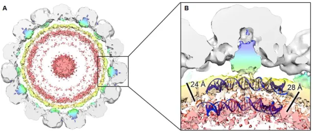
BKPyV virions are non-enveloped, with approximately 45 nm in diameter. The viral capsid, with an icosahedral symmetry, is composed of three structural proteins, the major capsid protein VP1, and the two minor capsid proteins VP2 and VP3. On the outer surface of the capsid, 360 copies of VP1 form 72 pentamers arranged in a T=7d icosahedral structure (13) (Figure 1). The pentamers are linked between them through the C-terminal arm of VP1, and are further stabilized by disulfide bonds and calcium cations (14). Each of these pentamers is associated with a single molecule of either VP2 or VP3 on the inner surface of the capsid (13).
Figure 1. Cryo-electron microscopy structure of BKPyV virions. (A) View of a 40-Å thick slab through the
unsharpened/unmasked virion map shown at a contour level of 0.0034. Pyramidal density below each VP1 penton and two shells of electron density adjacent to the inner capsid layer can be seen. The density within 6 Å of the fitted coordinates for SV40 VP1 is colored gray. The remaining density is colored in a radial color scheme. Densities for VP2 and VP3 are colored blue and green, and for packaged dsDNA yellow and pink. (B) Enlarged view of the pyramidal density beneath a single VP1 penton of the virion shown at a contour level of 0.0032. Strands of dsDNA wrapped around a human histone octamer (PDB: 1AOI) are shown, indicating that the two shells of density have a comparable spacing. Discrete connective density between the pyramidal density and internal shells is also apparent. (Adapted from Hurdiss, et al., 2016)
The capsid encloses the viral genome, a single circular dsDNA molecule, packed with the cellular histones H2A, H2B, H3 and H4 under the form of a minichromossome containing around 20 nucleosomes (13).
1.1.2. BKPyV genome
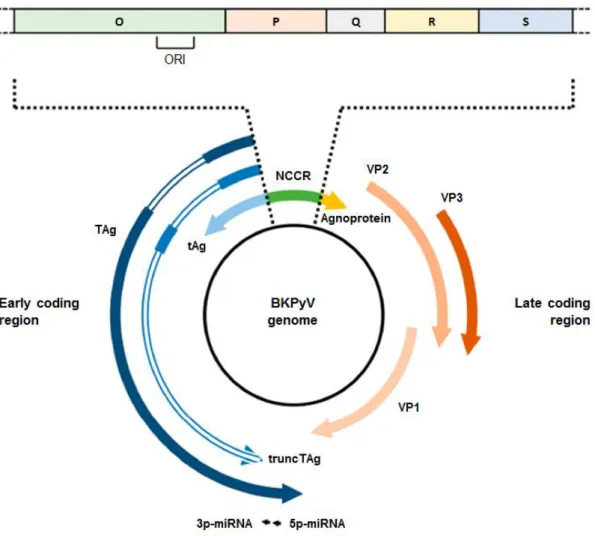
The BKPyV genome comprises about 5 kbp and it is divided into two transcriptional regions, the early and the late coding regions (referring to the phase of the infection in which each of them is transcribed), separated by the non-coding control region (NCCR), a highly variable region (1) (Figure 2).
Figure 2. Structure of BKPyV genome. The BKPyV genome is a closed circular, double-stranded DNA (dsDNA)
molecule comprising approximately 5 kbp. It is divided into two transcriptional regions, the early and the late coding regions, separated by the non-coding control region (NCCR). Transcription of both early and late coding regions proceeds in a bidirectional way from the origin of replication (ORI) within the NCCR. The early coding region encodes the large tumor antigen (TAg), the small tumor antigen (tAg) and the truncated TAg (truncTAg). The late coding region encodes the structural proteins (VP1, VP2, and VP3) as well as the agnoprotein. The late coding region also encodes for two microRNA (miRNA), 5p-miRNA and 3p-miRNA, produced after processing of a common precursor-miRNA (pre-miRNA). (Adapted from Helle, et al., 2017)
Within the NCCR is located the origin of replication (ORI), from where the replication proceeds in a bidirectional way, and also promoters and regulatory regions of early and late gene transcription. The early region encodes for three proteins, the large tumor antigen (TAg), the small tumor antigen (tAg) and the truncated TAg (truncTAg), that are expressed prior to DNA replication. The late region is only transcribed after the onset of DNA replication, and encodes for the structural proteins VP1, VP2 and VP3, as well as the agnoprotein (15,16). The late coding region also encodes for two microRNA (miRNA), referred to as 5p-miRNA and 3p-5p-miRNA, produced after processing of a common precursor-5p-miRNA (pre-miRNA) perfectly complementary to the 3′ end of the TAg messenger RNA (mRNA) (17).
1.1.2.1. Genotypes and variants
BKPyV can be divided into four genotypes (I to IV) due to variations on the genomic region coding for VP1 (18). Genotype I is further divided into the subgroups Ia, Ib1, Ib2, and Ic (19), and genotype IV into subgroups IVa1, IVa2, IVb1, IVb2, IVc1, and IVc2 (20). The most prevalent is genotype I, distributed worldwide, followed by genotype IV which is found in East Asia and Europe, while genotypes II and III are less commonly detected (21). It has been demonstrated that genotypes II, III, and IV and subgroups Ib1 and Ib2 behave as five fully distinct serotypes. The implications of infection with the different genotypes are yet to be understood, but successive infections with different genotypes are possible as they don’t cross-neutralize (18).
Besides from the variations on the VP1 region, variations on the NCCR lead to two different forms of BKPyV, referred to as archetype and rearranged variants. Archetype-NCCR is divided into five sequence blocks (Figure 2) named O, which contains the ORI, and P, Q, R and S, which contain regulatory regions for gene transcription (22). Rearrangements in the NCCR during viral replication usually involve duplications and deletions leading to the appearance of different rearranged-NCCR. These variants are more commonly found in the plasma and tissues of individuals with BKPyV-associated diseases (22,23). Archetypal variants of BKPyV are predominant in the urine, and are thought to be the transmissible forms of the virus, as they can be found in both healthy and diseased individuals (24). While the role of these variants in the development of BKPyV-associated diseases is not well established, it has already been shown that unlike rearranged strains, archetypal forms of BKPyV replicate poorly in cell culture (25). More recent studies have demonstrated that rearrangements on the NCCR lead to the decrease of viral miRNA production. This could result in an increase on the viral replication through the increase of TAg expression, since miRNAs are known to target TAg mRNAs to degradation (17). In this context, it has been
proposed that rearrangements in the NCCR might constitute an adaptation of the virus to enhance viral replication.
1.1.3. BKPyV replication cycle
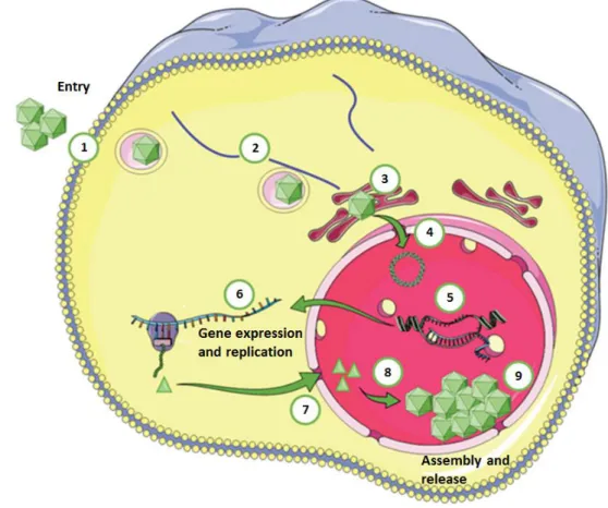
The infection with BKPyV starts with the attachment of the virion to the host cell, followed by entry, genome expression and replication, assembly and release of the viral progeny (Figure 3).
Figure 3. Schematic representation of BKPyV replication cycle. BKPyV infection starts with the attachment of
the virions to the host cell by binding to the ganglioside receptors (mainly GT1b and GD1b). Then, the virions are internalized through a caveolae-mediated endocytic pathway (1). Inside the cell, the virus is transported along the microtubules towards the endoplasmic reticulum (ER; 2) where the uncoating process begins (3). The partially uncoated viruses subsequently gain access to the cytosol and the viral genome is transported into the nucleus through the nuclear pore complex due to VP2 and VP3 nuclear localization sequences (NLS) and the importin α/β1-mediated import pathway (4). Once the viral genome enters the nucleus, the early gene expression begins. Early proteins are then translocated to the nucleus promoting genome replication and late gene expression (5). After translation of viral mRNAs in the cytosol (6), VP1, VP2 and VP3 gain access to the nucleus where they self-assemble around newly synthetized viral DNA (8). Progeny virions start to accumulate in the nucleus (9) and their release from infected cells is thought to occur mainly upon cell lysis. However, a non-lytic egress as also been described. (Adapted from Solis, et al., 2019)
Entry of the virus
In order to start the infection, the viral particle must attach to the host cell through the binding of viral VP1 with specific cell receptors. These receptors have been identified as gangliosides, one type of glycosphingolipids, composed of a ceramide chain and an oligosaccharide to which one or more sialic acid residues are linked. α(2,8)-disialic acid motif on b-series gangliosides has shown to be required for this interaction, thus BKPyV can interact with the gangliosides GT1b, GD1b, GD2 and GD3 (26,27). Despite this, it has already been demonstrated that BKPyV genotype IV is capable of establishing an infection through a ganglioside-independent pathway, showing that different BKPyV genotypes might present different cellular tropisms (18).
Following attachment to the cell surface, BKPyV is internalized through a caveolae-mediated endocytic pathway. Inside the cell, the virus is transported along the microtubules towards the endoplasmic reticulum (ER) where the uncoating process begins. In this process there seems to be the involvement of the ER protein disulfide isomerase which promotes conformational changes in VP1 molecules, providing the initial step for the capsid disassembly (28). Furthermore, proteins of the ER-associated degradation (ERAD) pathway, that are normally involved in the removal of misfolded proteins from the ER for proteasomal degradation, have been implicated as a mechanism used by the virus to escape from the ER to the cytosol (29).
Once in the cytosol and after partial uncoating, nuclear localization sequences (NLS) of VP2 and VP3 are exposed and thought to be used by the importin α/β1-mediated import pathway to transport the viral genome into the nucleus through the nuclear pore complex. However, an alternative route for genome entry into the nucleus has also been suggested to be present, as the BKPyV NLS mutation only attenuates the infection (30).
Gene expression and genome replication
When in the nucleus, the viral genome remains episomal in host cells (11). Soon after the genome enters the nucleus, cellular transcription factors bind to the early promotor leading to the expression of early proteins. After translation in the cytosol, TAg is transported into the nucleus where it binds to the ORI, leading to the recruitment of the cellular replication complex and to the unwinding of the double stranded DNA through its helicase activity, mediating the initiation of BKPyV genome replication. Additionally, TAg stimulates cell cycle progression and prevents apoptosis, by binding to the tumor suppressor proteins pRb, p107 and p130, and p53, respectively (31). tAg has also been associated with cell cycle
progression through inactivation of the protein phosphatase 2A (32). After the onset of DNA replication, in a process promoted by TAg, late genes are transcribed leading to the expression of the structural proteins and also agnoprotein (16). Agnoprotein is a small regulatory protein which seems to play important roles during the viral replication cycle, including regulation of gene expression, genome replication, and also virion assembly and release (33).
Assembly and release of viral particles
After translation in the cytosol, the structural proteins VP1, VP2 and VP3 are imported into the nucleus where the assembly of new viral particles takes place. At 2 days post-infection (dpi), progeny virus start to accumulate in the nucleus, forming nuclear inclusions that can be seen by electron microscopy in tubular epithelial cells from kidney biopsies or excreted in the urine as decoy cells, which are virally infected epithelial cells characterized by large nuclear viral inclusions (34,35).
As for the release of the newly formed virus from infected cells, the knowledge is still very limited. Generally, it is suggested that the virus egress from the cell occurs by passive means such as host cell lysis, which has already been observed in RPTEC (human renal proximal tubular epithelial cells) (34). However BKPyV-infected cells rarely exhibit strong cytopathic effects (36). Furthermore, non-lytic egress for BKPyV progeny, although in smaller scale, has already been demonstrated (37). Even though further studies are needed in this subject, evidence points out that both lytical and non-lytical release of BKPyV progeny might occur.
1.2. Primary infection, latency and reactivation
BKPyV infection is ubiquitous, with a seroprevalence of over 80% among the adult population. Primary infection typically occurs during early childhood, with studies having shown that by the age of 10 approximately 70% of children present detectable serum antibodies against BKPyV (38). This primary infection is usually asymptomatic or associated with mild upper respiratory symptoms. The route of transmission is not yet fully established, but evidences of a primary upper respiratory infection, particularly in the tonsils, suggests that the virus is most likely transmitted through a respiratory route (39). However, other routes of transmission are also being proposed including fecal-oral, urino-oral, blood transfusion or organ transplantation (16,40). From the respiratory tract, presumably following a primary viremia, the virus is thought to disseminate to other sites of infection, including
mainly the urinary tract and the kidney. Following primary infection, BKPyV establishes a lifelong persistent infection mainly on epithelial cells of the urinary tract and the kidney, however, it is still unclear if the virus persists with a minimal replication, or if it enters a true viral latency. Occasional reactivations of the persistent virus occur that manifest as asymptomatic viruria with BKPyV loads of <5 log10 copies (c)/mL (41,42).
In immunocompetent individuals, the virus remains clinically silent throughout the life of the host. However, under immunosuppressing conditions, typically immunosuppressive disease or treatment, viral reactivation might occur. The major clinical complications of BKPyV reactivation are PyVAN and hemorrhagic cystitis in kidney and hematopoietic stem cell transplant recipients, respectively (5). Less commonly BKPyV-associated clinical manifestations include interstitial pneumonitis, retinitis, native kidney nephritis and meningoencephalitis, described particularly in severely immunocompromised individuals such as patients with acquired immune deficiency syndrome (43).
1.3. Polyomavirus-associated nephropathy
PyVAN most commonly occurs in kidney transplant recipients due to BKPyV reactivation, with a prevalence rate that ranges from 1 to 10% in these patients (44). After reactivation, BKPyV replication in tubular epithelial cells results in cytopathic changes in those cells. The progress of the infection leads to tubular cell injury and interstitial inflammation that characterize PyVAN. Persistent PyVAN eventually leads to tubular atrophy, interstitial fibrosis and loss of function(45), which can potentially result in graft loss in 10 to 80% of the cases (46).
Several factors have been described to modify the risk of BKPyV reactivation. However, immunosuppressive therapy is thought to be the one that has the major role in this reactivation. Rather than a specific therapy, it has been described that the overall degree of immunosuppression is responsible for increasing the risk. In fact, the development of more potent immunosuppressant drugs, such as tacrolimus and mycophenolate mofetil, have led to an increase on the PyVAN incidence rate in kidney transplant recipients (47).
Other than immunosuppressive therapy, a variety of weekly associated risk factors have also been described to increase BKPyV reactivation. The preferential occurrence of PyVAN after kidney transplants over other solid organ transplants, even with comparable levels of immunosuppression (48), points out that the process of kidney transplantation itself and the resultant renal injury, might have a role on BKPyV reactivation. This includes prolonged cold ischemia time, delayed graft function and ureteral stent placement. Other transplant-related
risk factors that are usually referred to include degree of human leukocyte antigen (HLA) mismatch, donor BKPyV seropositivity, recipient BKPyV seronegativity, donor and recipient older age, recipient male gender and previous graft loss due to PyVAN. Prolonged pre-transplant dialysis has also been pointed out as a potential risk factor for PyVAN development (5,16,49).
Interestingly, the donor’s BKPyV strain reactivation has been suggested to be the one involved on PyVAN development. Consequently, genotype mismatch between the donor and the recipient neutralization profiles and their subsequently replicating strain before transplant might significantly increase the risk of BKPyV reactivation (50).
1.3.1. Early screening and diagnosis
Clinically BKPyV reactivation is asymptomatic, and in case of PyVAN development it only manifests as a deterioration in allograft function. The first evidence of BKPyV reactivation is a high-level BKPyV viruria with decoy cells shedding, followed by viremia (51). In kidney transplant recipients it is estimated that approximately 50% of the patients who present high-level viruria evolve to viremia 2 to 6 weeks later, and that 50% of those who develop viremia are diagnosed with PyVAN 2 to 6 weeks later (52).
Because no efficient and safe antiviral therapies against BKPyV are yet available, screening for BKPyV replication in urine and blood has become the key recommendation for kidney transplant recipients. This allows an early intervention, which to date consists mainly in modulating the immunosuppressive treatment, in order to prevent the development of PyVAN and ultimately graft loss (52).
According to the Guidelines from the Infectious Diseases Community of Practice of the American Society of Transplantation, current recommendations point that kidney transplant recipients should be screened for BKPyV DNA loads in plasma by quantitative polymerase chain reaction (PCR)-based techniques, first monthly until month 9, then every 3 months until 2 years post-transplant. This screening allows the identification of patients that should be considered for a preemptive treatment for PyVAN, however, prior to reducing immunosuppression and risking the precipitation of acute rejection, BKPyV viremia should be confirmed within 3 weeks. BKPyV DNA plasma loads sustained above 3 log10 c/mL, but below 4 log10 c/mL define probable PyVAN, and above 4 log10 c/mL define presumptive PyVAN. Screening for BKPyV reactivation can also be performed in the urine, although these methods are usually less preferred because of their low positive predictive values. In this case, either urine cytology for decoy cells or BKPyV DNA loads in urine by quantitative
PCR-based techniques can be performed, 2 times a week for the first 3 months, then monthly until month 6, then every 3 months until 2 years post-transplant. If decoy cells are detected or if urine BKPyV DNA loads increase above 7 log10 c/mL, patients should be tested for BKPyV viremia (52).
Although a BKPyV DNA plasma load above 4 log10 c/mL can diagnose a presumptive PyVAN, kidney biopsy remains the gold standard for definitive diagnosis of PyVAN. Because of the focal nature of the infection, it is recommended that a minimum of two biopsy cores are taken, preferentially containing medullary tissue, which is a significant reservoir for BKPyV infection, in order to avoid false negatives. The diagnosis of PyVAN is made upon identification of cytopathic changes in the tubular epithelial cells of the biopsy tissue. Since PyVAN and acute rejection might resemble morphologically, the diagnosis can be confirmed by immunohistochemistry or in situ hybridization (52).
1.3.2. Therapeutic approaches
Because BKPyV lacks a specific target, the search for a specific and safe antiviral against this virus becomes more difficult, and to date, none is still available. As such, the mainstay approach for persistent BKPyV replication and PyVAN in kidney transplant recipients is to reduce the immunosuppressive treatment. This strategy allows the reconstitution of the patients’ immune system, which becomes capable of controlling the infection. Although not all patients exhibit the same response, an efficient clearance of BKPyV viremia and the preservation of allograft function has been described in a high percentage of patients in several studies (52,53). Despite this, by reducing immunosuppression there is a risk of precipitating acute rejection. To prevent this from occurring, the immunosuppression reduction must be done in a stepwise manner with close monitoring of serum creatinine levels and BKPyV viremia, at least every two weeks until viremia clearance (52).
The immunosuppressive regimen for kidney transplant recipients may vary between centers but usually consists of a calcineurin inhibitor, such as cyclosporine A and tacrolimus, an antimetabolite, such as azathioprine and mycophenolate mofetil, and a corticosteroid (54). Regarding the adjustment of the immunosuppressive treatment, there is no single approach and different strategies have been suggested. One of these approaches consists to first reduce the calcineurin inhibitor or the antiproliferative drug, followed by a dose reduction of the other drug and then discontinuation of the antiproliferative drug (52). Alternatively, other approaches include switching to less potent drugs, like for example, from tacrolimus to cyclosporine A (a less potent calcineurin inhibitor) (55), from the calcineurin inhibitor to
sirolimus, or from mycophenolate mofetil to sirolimus (56,57) or leflunomide (58–60). Successful outcomes have been reported with all these different strategies, however, that is still no randomized controlled trial that can recommend one over the others.
In addition to the adjustments in the immunosuppressive treatment, and despite no specific antiviral against BKPyV is yet available, the off-label use of agents with potential anti-BKPyV activity in patients with PyVAN has been reported. These includes cidofovir, fluoroquinolones and intravenous immunoglobulins (IVIGs). However, as they are often used in combination with immunosuppression reduction and randomized controlled trials are still lacking, it is difficult to make strong conclusions about their efficacy (61).
Novel therapeutic approaches against BKPyV are starting to appear. These include adoptive T cell therapy, that is starting to be studied to treat BKPyV-associated diseases (62,63). Furthermore, a vaccine for prevention of cytomegalovirus and BKPyV reactivation is currently under evaluation on a phase I clinical trial (64).
Leflunomide
Leflunomide presents both immunosuppressive and antiviral properties, being currently approved for the treatment of rheumatoid arthritis. Its antiviral effects are attributed to its active metabolite A771726 which inhibits protein kinase activity and the synthesis of pyrimidines. In vitro, leflunomide has already shown to interfere with BKPyV replication in RPTEC (65). Several studies have reported the use of leflunomide, often combined with reduction of the immunosuppressive treatment, and clearance or reduction of BKPyV viremia has been described (58–60). However, determining its efficacy remains difficult since therapeutic outcomes change between studies (53) and no randomized controlled trials have been performed. Additionally, significant toxic effects have been described when using this drug including hepatitis, hemolysis, thrombotic microangiopathy, bone marrow suppression, and fungal pneumonia (52).
Mammalian target of rapamycin inhibitors
Mammalian target of rapamycin (mTOR) inhibitors, such as sirolimus and everolimus, are immunosuppressive agents that also exhibit antitumoral activity by inhibiting cell-cycle progression and proliferation. In vitro, they have shown to decrease BKPyV replication by interfering with mTOR–SP6‐kinase activation (66). Different studies have reported that
kidney transplant recipients receiving mTOR inhibitors compared to those receiving standard immunosuppressive regimens exhibit a more efficient clearance of BKPyV viruria and viremia or even lower risks of developing BKPyV viremia (56,57,67). Although its use seems promising in preventing PyVAN, further studies are needed, particularly regarding their toxicity, as severe adverse effects have been reported, including pneumonitis and proteinuria (68,69).
Cidofovir
Cidofovir is a nucleoside analog and viral DNA polymerase inhibitor only approved for the treatment of cytomegalovirus retinitis. The antiviral effect against BKPyV has already been demonstrated in vitro in RPTEC, however, the mechanism behind this effect remains unclear (70). Treating kidney transplant recipients with cidofovir alongside with reducing immunosuppression has shown favorable outcomes in some studies (71,72), whereas others claim no advantage with this treatment (53). Despite this potential benefit in PyVAN, major adverse effects associated with the use of this drug include nephrotoxicity and uveitis, with the first one limiting its use in kidney transplant recipients (52,73).
Brincidofovir is an experimental pro-drug of cidofovir, orally administrated and substantially less nephrotoxic. In vitro, it has shown to decrease BKPyV replication in RPTEC more efficiently when compared to cidofovir, and also to exhibit fewer toxicity (74). Successful outcomes have been obtained with this drug in two different studies (75,76), however, randomized controlled trials are currently on hold due to adverse events already described (52).
Fluoroquinolones
Fluoroquinolones are a family of broad-spectrum antibiotics that have shown to present an antiviral effect against BKPyV in vitro, possibly by inhibiting the helicase activity of TAg (77). Initial studies have reported a prophylactic effect with the use of fluoroquinolones in both kidney and hematopoietic stem cell transplant recipients (78,79). However, a double-blind, placebo-controlled randomized trial failed to demonstrate the efficacy of a 3-month course of levofloxacin on lowering BKPyV viruria, as well as it shown an increased risk of bacterial resistance (80). These results do not support the use of fluoroquinolones on the prevention of BKPyV reactivation.
Intravenous immunoglobulins
Because of the high seroprevalence of BKPyV in the population, commercially available IVIG preparations have been demonstrated to contain high titers of potent BKPyV neutralizing antibodies (NAb). Its antiviral effect is attributed to direct neutralizing activity but also to indirect immunomodulatory effects (81). Some studies have described the use of IVIG in combination with reduced immunosuppression with beneficial outcomes (82,83). Additionally, a study suggesting that higher NAb titers against the replicating strain of BKPyV were associated with a lower risk of BKPyV reactivation, supports the potential benefit of administering NAb either as a preventive or a therapeutic strategy (50). However, no controlled studies have yet been reported.
Cellular immunotherapy
Immunosuppression is thought to be the main cause of BKPyV reactivation. In this context, the adoptive transfer of BKPyV-reactive T cells may help to restore the immune response of these patients, thus allowing it to control viral replication and reducing the complications associated with this reactivation. This approach has already shown to be effective to treat other virus-associated diseases, particularly associated with Epstein-Barr virus and cytomegalovirus (84,85), in immunocompromised patients. More recently, the use of this immunotherapy started to be studied to treat diseases caused by BKPyV infection. The immunodominant epitopes of BKPyV to be used on T cell expansion are not yet defined, nevertheless, it is already reported the in vitro expansion of BKPyV-reactive T cells using overlapping peptide mixes of the five BKPyV proteins (VP1, VP2, VP4, TAg and tAg) (62). One study described the use of adoptive T cell therapy with full resolution of the symptoms and viremia on a patient that who, following hematopoietic stem cell transplantation, developed hemorrhagic cystitis due to BKPyV reactivation (63). Another promising study in this area described the use of adoptive transfer of virus-reactive T cells targeting BKPyV and four other viruses (Epstein-Barr virus, adenovirus, cytomegalovirus, and Human Herpesvirus 6) known to cause frequent infections in immunocompromised patients, with a great clinical response (86). With further research in this area, adoptive T cell therapy can become a potential approach to treat BKPyV-associated diseases.
1.4. Model systems for the study of BKPyV infection
To better understand the virus’ structure, the replication cycle, the pathogenicity, and also to identify compounds with antiviral activity, both in vitro and in vivo models are crucial. Much of the knowledge regarding BKPyV in the past has been inferred from the much more studied polyomavirus SV40 (11,13). However, in the last years, a big effort to study BKPyV has been made as it is increasingly recognized as responsible for an important infection affecting mostly kidney and hematopoietic stem cell transplant recipients using the new potent immunosuppressive protocols.
To date, BKPyV has been known to replicate only in human and monkey cells. Vero cells were the first cells used to isolate BKPyV and currently, this African green monkey kidney cell line is still widely used in research to replicate the virus (3). RPTEC, however, constitute a more appropriate model for the study of BKPyV, as they are one of the major target cells of the virus in vivo (34). The problem regarding these cells is that primary RPTEC divide slowly and quickly enter into replicative senescence. Because of that, human telomerase reverse transcriptase (hTERT) immortalized RPTEC (RPTEC/TERT1), which have shown to maintain their functional characteristics, also started to being used (87). Other kidney cell lines of both human and monkey origin, human embryonic kidney 293 (HEK293) cells and CV-1 cells, respectively, are also used (88,89). Interestingly, other cell types allow the replication of BKPyV, including human salivary gland cells (90) and human embryonic lung fibroblast cells, such as MRC-5 (91). The cells mentioned above successfully allow the propagation of rearranged forms of BKPyV. Archetype forms, however, due to low TAg expression replicate poorly in cell culture. The human endothelial cell line HUV-EC-C was the first one to allow the propagation of archetypal BKPyV (25). More recently, archetype forms of BKPyV have also shown to be able to replicate in cell lines transformed with the SV40 TAg including 293TT and COS-7 cells, cell lines derived from HEK293 and CV-1 cells, respectively (24). Regarding the virus, recombinant viruses with both rearranged and archetype NCCR are commonly used. Rearranged strains include Gardner, Dunlop and TU strains, and as for the archetypal strains, WW and WWT strains are among the most common (92).
Alternatively, virus-like particles (VLPs) can be used. VLPs consist on capsid-like particles that spontaneously self-assemble upon overexpression of the major capsid protein VP1, but that devoid of the virus’ genetic material. These models can be produced in a variety of different systems including Escherichia coli, yeast, insect cells and mammalian cells (93). Additionally, these systems can be co-transfected with vectors encoding a reporter gene, enabling the quantification of the particles that enter target cells. Although these models cannot be used to study BKPyV replication, as they do not contain the viral genome, they
have provided useful information on the viral structure and on the early steps of the viral cycle, including interactions with cell surface receptors and entry (13,94). Finally, VLPs seem to have a promising application in the development of vaccines that could be used prior to transplantation (93).
Although in vitro models are sufficient to provide information on the virus replication cycle, animal models are needed in order to better understand the mechanisms underlying the virus-associated diseases but also for conducting preclinical tests on potential new antiviral drugs and vaccines. Because of the narrow host range of BKPyV the development of small animal models to study BKPyV infection was hampered. In fact, BKPyV DNA replication is inhibited by a restriction factor found in murine cells (95). Previous attempts to develop an animal model for BKPyV infection have already been carried out (91). However, an animal model that closely mimics BKPyV infection in humans is still lacking.
2. Aims of this study
Over the last years, the development of more potent immunosuppressive agents has made BKPyV emerge as an important pathogen responsible for serious complications such as PyVAN in kidney transplant recipients. The lack of specific and safe antiviral agents that can efficiently stop BKPyV replication still poses significant challenges, as PyVAN can lead to alterations in the kidney’s structure, loss of function and potentially graft loss.
In this context, this project focused on studying the efficacy of two drugs - NA-17 and TN-18 - against BKPyV. For that purpose, this study was divided in different tasks in order to provide a characterization of the drugs’ effect against BKPyV.
First, the effect of both drugs was assessed on Vero cells, a monkey kidney cell line, to determine whether they had potential to proceed for further testing. Then, the drugs that exhibited an antiviral effect on the first step, were submitted to a more detailed study on a better in vitro model of the virus target in vivo, RPTEC/TERT1. On this second stage a selected range of concentrations of each drug was used to characterize their effect against the virus. The inhibition of BKPyV infection for each concentration of both drugs was determined and used to calculate the respective effective concentrations 50% (EC50). Then, as both drugs were known to exhibit an antimetabolite effect, the alterations on the extracellular BKPyV DNA load were measured for each concentration tested, and a correspondence was tried with the variations on the release of infectious progeny.
3. Materials and Methods
3.1. Cell culture
Vero cells (ATCC, CCL-81) were maintained in Dulbecco's modified Eagle medium (DMEM; Gibco, Thermo Fisher Scientific) supplemented with 10% fetal bovine serum (FBS). RPTEC/TERT1 (Evercyte, CHT-003-0002) were maintained in renal epithelial growth medium (REGM) supplemented with 0.1% recombinant human epidermal growth factor, 0.1% hydrocortisone, 0.1% epinephrine, 0.1% insulin, 0.1% triiodothyronine, 0.1% transferrin, 0.1% GA-100 (gentamicin and amphotericin B) and 0.5% FBS (REGM Bulletkit; Lonza). All cells were kept in a humidified incubator at 37°C with 5% CO2 and propagated as described by the manufacturer.
3.2. Virus
3.2.1. Virus stock production
BKPyV stock was produced by transfecting Vero cells with BKPyV Dunlop genome. Briefly, the pUC19pBKv(34-2) plasmid, provided by Professor Walter Atwood (Brown University, USA), was amplified after transformation of chemically competent Escherichia coli (Invitrogen, C404010). The plasmids produced were digested with BamHI to separate BKPyV genome from the vector plasmid and were used to transfect Vero cells. Cells were kept in culture until cytopathic effects were observed. After that time, cell culture supernatants were harvested and submitted to 3 freeze/thaw cycles that allowed cell lysis and release of the virions. BKPyV was further amplified by successive infection of Vero cells, and the cell culture supernatant of infected cells was kept. The viral stock produced was stored at -80°C.
3.2.1. Virus stock titration
BKPyV stock was titrated in Vero cells and RPTEC/TERT1. For the titration, cells were plated and infected 24 h later with the viruses in 10-fold serial dilutions ranging from 10-1 to 10-8. After 2 h of incubation for viral adsorption, the supernatants were removed and replaced with the adequate cell growth medium. At 72 h post-infection (hpi), cells were washed, fixed and immunostained as described below. The stocks’ titer was expressed in 50% tissue culture infectious dose (TCID50)/mL and determined using Reed and Muench calculator (96) (Brett D. Lindenbach, 2008).
3.3. Infection and drug treatment
Cells were plated in 96-well plates for confluence at day 5 and infected 24 h later with BKPyV Dunlop at a multiplicity of infection (MOI) of 1 in DMEM. After 2 h of incubation in a humidified incubator at 37°C with 5% CO2, the supernatants were removed and replaced with the adequate cell growth medium (for Vero cells, medium supplementation with FBS was of 5%) with or without the drugs for 3 days. Controls containing only cells or only cells with the drugs were also included. All experiments were performed in triplicates.
Prior to each experiment, NA-17 and TN-18 drugs were freshly dissolved in dimethyl sulfoxide (DMSO) and H2O, respectively. NA-17 was further diluted in cell growth medium with DMSO at 1:1000 in 10-fold serial dilutions ranging from 10 nM to 100 µM. TN-18 was further diluted in cell growth medium in 10-fold serial dilutions ranging from 10 nM to 1 mM. DMSO controls matching the DMSO concentration in 100 µM NA-17 (1:1000) were included in all experiments. The drugs’ structures are protected under laboratory confidentiality and therefore, they are only represented by a code.
3.4. Immunofluorescence staining, microscopy, and digital image
processing
Cells were washed with phosphate-buffered saline (PBS) at 72 hpi, fixed with 3.7% paraformaldehyde (PFA) in PBS for 10 min and permeabilized with 0.5% Triton X-100 in filtered CSK (cytoskeletal buffer; 10 mM PIPES pH 6.8, 100 mM NaCl, 300 mM sucrose, 3 mM MgCl2, 1 mM EDTA) and H2O (1:1) for 15 min, both at room temperature. Fixed cells were then incubated with the primary antibody for 1 h at room temperature, and the secondary antibody for 30 min at room temperature in the dark. The primary antibody used was mouse monoclonal anti-SV40 TAg (Pab416; 1:500; Abcam), and the secondary antibody used was goat anti-mouse IgG conjugated with Alexa Fluor Plus 488 (1:1000; Thermo Fisher Scientific), both diluted in PBS/Tween 0.1%. Cell nuclei were stained with DAPI (1:1000; Santa Cruz Biotechnology).
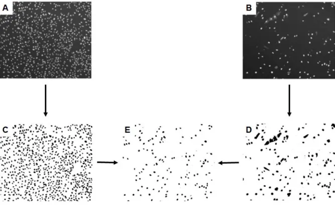
Images were collected using an inverted microscope (Axio Vert.A1 and Colibri 7 light source, Zeiss). For cell counting, pictures from two random fields per well were taken and DAPI and TAg stained nuclei were automatically counted using QuantIF (Figure 4), an ImageJ (National Institute of Health, Bethesda, MD, USA) macro (97).
Figure 4. Description of the QuantIF macro. Two pictures of each field must be taken, one corresponding to the
specific staining (infected cells) and the other corresponding to the DAPI staining (total cells), for analysis by the QuantIF macro. Both the DAPI staining image (A) and the specific staining image (B) are then converted to a staining mask, (C) and (D), respectively. A third mask corresponding to the nuclei of the immunostained cells is created (E). Finally, DAPI stained and immunostained cells’ nuclei are counted using the DAPI and the immunostained cells’ nuclei staining masks. (Adapted from Handala, et al., 2019)
3.5. Extracellular BKPyV DNA load
To quantify the extracellular BKPyV DNA loads, cell culture supernatants of infected cells both untreated and treated with the drugs were harvested at 72 hpi and stored at 4°C until quantification by quantitative real-time PCR (qPCR) using primers and a probe targeting a common region of the VP1 and VP2 genes in the BKPyV genome. Reactions were performed in a total volume of 25 μL containing 2X TaqMan Universal PCR Master Mix (Applied Biosystems), 10 µM of each primer and the 6-carboxyfluorescein (FAM)-labeled probe, and 5 μL of supernatant. Amplification was performed using an ABI Prism 7900HT Sequence Detection System (Applied Biosystems) with the following PCR program: initial step of 2 min at 50°C followed by 1 cycle at 95°C for 10 min, and 45 two-step cycles at 95°C for 15 s and 60°C for 1 min. BKPyV DNA loads were determined using a standard curve generated by amplification of four quantification standards containing different known concentrations of BKPyV DNA (9 000 000 to 9000 c/mL of the pUC19pBKv(34-2) plasmid in
10-fold serial dilutions). For every experimental condition triplicates were analyzed. A BKPyV-negative control was added to each run.
3.6. Release of infectious progeny
To detect the release of infectious progeny into the supernatant, cell culture supernatants of infected cells both treated and untreated with each drug were harvested at 72 hpi and stored at 4°C. Supernatants were used to infect new cells for 2 h and were then removed and replaced with fresh cell growth medium. At 72 hpi, cells were washed, fixed and immunostained as described above.
3.7. Calculation of the effective concentrations 50%
GraphPad Prism 8 software (GraphPad, La Jolla, CA) was used for curve fitting to determine the EC50. EC50 corresponds to the drug concentration responsible for 50% inhibition of BKPyV infection.
4. Results
4.1. Effect of NA-17 and TN-18 on BKPyV infection on Vero cells
4.1.1. Inhibition of BKPyV infection
First, the effect of both drugs, NA-17 and TN-18, on inhibiting BKPyV infection was evaluated on Vero cells. For such, Vero cells were infected with BKPyV and different concentrations of each drug were added 2 hpi. Cells were fixed at 72 hpi and immunostained for TAg. Cell nuclei were stained with DAPI. Then, pictures were taken for cell counting, and DAPI and TAg stained nuclei were automatically counted using QuantIF, an ImageJ macro. Results show an inhibition of BKPyV infection only with the highest concentration tested for each drug (Figure 5A and B). Of note, NA-17 at concentrations from 10 μM and TN-18 at a concentration of 1000 μM reduced the total cell number per field (Figure 5C and D).
Figure 5. Effect of increasing concentrations of NA-17 and TN-18 on the inhibition of BKPyV infection on Vero cells. (A, B) Vero cells infected with BKPyV were treated 2 hpi with the indicated concentrations of NA-17
(A) and TN-18 (B). Cells were fixed at 72 hpi and immunostained for TAg. Cell nuclei were stained with DAPI. Pictures were taken for cell counting, and DAPI and TAg stained nuclei were automatically counted using QuantIF, an ImageJ macro. The percentage of inhibition of BKPyV infection for each concentration of the drugs was calculated in relation to a control of infected Vero cells without any treatment. Determinations were performed in triplicate, each corresponding to the analysis of two pictures. The mean values are shown, and the error bars represent the standard deviations of means between triplicates. Curve fitting was obtained using GraphPad Prism 8 software. (C, D) Total cell number of infected Vero cells both untreated and treated with different concentrations of NA-17 (C) and TN-18 (D). The number of cells in each picture was counted using QuantIF. The mean values are shown, and the error bars represent the standard deviations of means between triplicates.
Since an inhibition of BKPyV infection on Vero cells was observed for NA-17 and TN-18, the effect of both drugs was then investigated in RPTEC/TERT1, an immortalized cell line of the major target cells of BKPyV in vivo.
4.2. Effect of NA-17 and TN-18 on BKPyV infection on RPTEC/TERT1
4.2.1. Inhibition of BKPyV infection
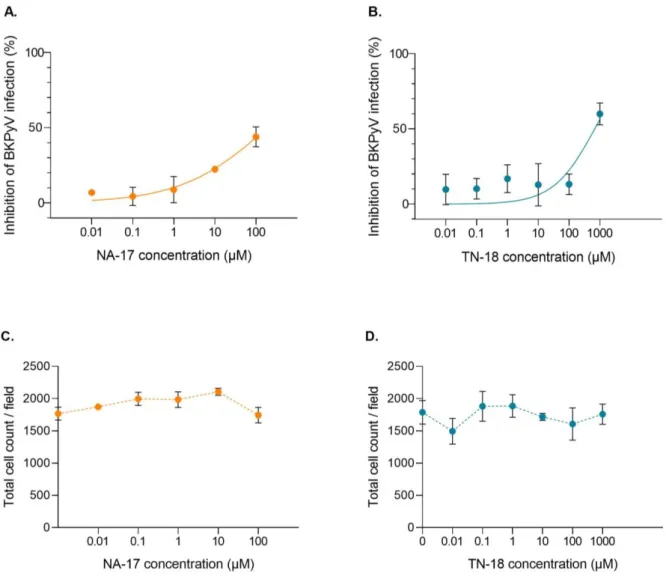
To investigate the effect of both drugs on inhibiting BKPyV infection on RPTEC/TERT1, we proceeded like previously for Vero cells. Similarly to what we observed for Vero cells, the lowest concentrations tested for both drugs provided none to a very discrete effect. For NA-17 an inhibitory effect on BKPyV infection starts to be observed with a concentration of 10 μM and seems to increase in a concentration dependent manner, as it increases with a concentration of 100 μM. For TN-18, like previously observed on Vero cells, an antiviral effect is only evident for the highest concentration tested (Figure 6A and B). Of note, unlike observed on Vero cells, none of the concentrations tested for both drugs seems to have a significant impact on the total cell count (Figure 6C and D).
Figure 6. Effect of increasing concentrations of NA-17 and TN-18 on the inhibition of BKPyV infection on RPTEC/TERT1. (A, B) RPTEC/TERT1 infected with BKPyV were treated 2 hpi with the indicated concentrations
of NA-17 (A) and TN-18 (B). Cells were fixed at 72 hpi and immunostained for TAg. Cell nuclei were stained with DAPI. Pictures were taken for cell counting, and DAPI and TAg stained nuclei were automatically counted using QuantIF, an ImageJ macro. The percentage of inhibition of BKPyV infection for each concentration of the drugs was calculated in relation to a control of infected RPTEC/TERT1 without any treatment. Determinations were performed in triplicate, each corresponding to the analysis of two pictures. The mean values are shown, and the error bars represent the standard deviations of means between triplicates. Curve fitting was obtained using GraphPad Prism 8 software. (C, D) Total cell number for infected RPTEC/TERT1 both untreated and treated with different concentrations of NA-17 (C) and TN-18 (D). The number of cells in each picture was counted using QuantIF. The mean values are shown, and the error bars represent the standard deviations of means between triplicates.
In order to determine the EC50 for both drugs, curve fitting based on the data obtained with the RPTEC/TERT1 was performed using GraphPad Prism 8 software (Figure 6). An EC50 of 190 μM and an EC50 of 682 μM were calculated for NA-17 and TN-18, respectively (Table 1).
Table 1. Calculated EC50 values for NA-17 and TN-18 tested on RPTEC/TERT1a
Drug EC50 (μM) (95% CI)
NA-17 190 (97.9 - 566)
TN-18 682 (311 - 23 310)
a EC
50 values and the correspondent 95% confidence intervals (CI) were calculated based on the curve fitting
performed using GraphPad Prism 8 software
4.2.2. Effect on BKPyV genome replication
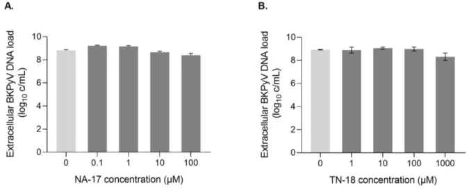
Although the mechanism by which these drugs may present an anti-BKPyV effect is not established, they are known to exhibit an antimetabolite effect. In this context, next, we investigated their effect on the virus genome replication by measuring the extracellular BKPyV DNA load. Cell culture supernatants of infected RPTEC/TERT1 both untreated and treated with NA-17 and TN-18 were harvested at 72 hpi and BKPyV DNA load was measured by qPCR. Supporting the results obtained with immunofluorescence, the lowest concentrations tested for both drugs don’t seem to have any effect on inhibiting BKPyV replication (Figure 7). For NA-17 a decrease on BKPyV DNA load can be observed with the two highest concentrations. For TN-18 only the highest concentration seems to cause a decrease on BKPyV DNA load. Although an effect on viral DNA replication can be observed using the highest concentrations tested of these drugs, the decrease of extracellular BKPyV DNA load is very discrete, being for both cases <1 log10 c/mL, reaching a maximum inhibition of approximately 5% and 7% for NA-17 and TN-18, respectively.
Figure 7. Effect of increasing concentrations of NA-17 and TN-18 on BKPyV DNA load. RPTEC/TERT1
Supernatants were harvested at 72 hpi and BKPyV DNA load was measured by qPCR. Determinations were performed in triplicate. The mean values are shown, and the error bars represent the standard deviations.
4.2.3. Effect on the release of infectious progeny
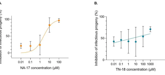
To determine whether the decrease in extracellular BKPyV DNA load corresponded to the release of infectious progeny, supernatants harvested at 72 hpi from BKPyV-infected RPTEC/TERT1 untreated and treated with different concentrations of each drug were used to infect freshly seeded RPTEC/TERT1. Immunofluorescence staining was performed at 72 hpi like previously described and pictures were taken for cell counting. Results show an inhibition of infectious progeny release corresponding to the results found on extracellular BKPyV DNA load (Figure 8). This means that for NA-17 the release of infectious progeny decreased with the two highest concentrations tested, and for TN-18 only with the highest concentration. Of note, with the lowest concentrations tested of NA-17 and TN-18 an inhibition of approximately 20% and 40% in relation to the controls is already found, which does not correspond to the results obtained for the BKPyV DNA load, where differences from the controls were only observed with the highest concentrations. However, caution must be taken when analyzing these results since the infection of RPTEC/TERT1 using supernatants from both untreated and treated cells produced infection rates <1% for every case.
Figure 8. Effect of increasing concentrations of NA-17 and TN-18 on the release of infectious progeny.
Supernatants harvested at 72 hpi from BKPyV-infected RPTEC/TERT1 untreated and treated with different concentrations of NA-17 (A) and TN-18 (B) were used to infect freshly seeded RPTEC/TERT1. Immunofluorescence staining for TAg was performed at 72 hpi. Cell nuclei were stained with DAPI. Pictures were taken for cell counting, and DAPI and TAg stained nuclei were automatically counted using QuantIF, an ImageJ macro. The percentage of inhibition of infectious progeny release for each concentration of the drugs was
calculated in relation to a control of fresh RPETC/TERT1 infected with the supernatants from untreated BKPyV-infected RPTEC/TERT1. Determinations were performed in triplicate, each corresponding to the analysis of two pictures. The mean values are shown, and the error bars represent the standard deviations of means between triplicates. Curve fitting was obtained using GraphPad Prism 8 software.
5. Discussion
A specific and safe antiviral that can efficiently stop BKPyV infection and prevent PyVAN progression is still in need. In this project, the effect of two drugs - NA-17 and TN-18 - against BKPyV replication was characterized in RPTEC/TERT1. We found out that the treatment with each drug 2 hpi resulted in a moderate inhibition of BKPyV infection, having been estimated EC50 values of 190 μM and 682 μM for NA-17 and TN-18, respectively. A more detailed study of the effect of these drugs against BKPyV showed that with the highest concentrations tested, both NA-17 and TN-18 were responsible for a decrease on the extracellular BKPyV DNA load, that although discrete, corresponded also with an inhibition on the release of infectious progeny.
Before proceeding to the study of the drugs on BKPyV-infected RPTEC/TERT1, a first assay was performed on Vero cells in order to determine if the drugs exhibited a potential antiviral effect against the virus. Vero cells were the first cells used to isolate BKPyV, and besides having shown to be permissive and to well replicate this virus, this cell line is easily cultivated, achieves confluence readily and does not require the use of specialized media (3,98). Because of this, this cell line is still widely used on basic research and constitutes an appropriate model for a first approach on evaluating the effect of a potential anti-BKPyV drug. First results obtained using Vero cells showed that 72 hpi an inhibition of BKPyV infection was obtained with 100 μM of NA-17 and with 1000 μM of TN-18. Although only the highest concentrations tested for each drug allowed an inhibition of the infection, a peak of BKPyV infection on Vero cells is expected between the 7 and 10 dpi (98), meaning that assays carried only until 3 dpi may not entirely reflect the effect of the drugs tested against the virus. As such, the anti-BKPyV effect observed in these preliminary results was considered sufficient to proceed with both drugs for further testing.
On a second stage, we wanted to characterize the anti-BKPyV effect of both drugs on a better in vitro model. As mentioned before, the ideal cells for this study would be RPTEC, one of the major targets of the virus in vivo, however, working with primary cells comes with its challenges as they are harder to cultivate and exhibit a limited lifespan. To overcome these problems, we decided to use RPTEC/TERT1, an immortalized cell line of RPTEC. To start the characterization of the drugs’ effect on RPTEC/TERT1, the same assay previously performed on Vero cells was repeated on this cell line. Results obtained using RPTEC/TERT1 showed a better anti-BKPyV effect of both drugs on these cells. For NA-17 an inhibition of the infection was observed from a concentration of 10 μM, that increased with the increase of the concentration. For TN-18, similarly to what we had observed on Vero cells, only the concentration of 1000 μM seemed to have an impact on inhibiting the infection,