ISSN 0101-2061 Food Science and Technology
DI:
D http://dx.doi.org/10.1590/1678-457X.35716
1 Introduction
Arteriosclerosis obliterans (ASD) is a disease in which the atherosclerosis lesions involves the peripheral arteries and causes the chronic occlusion. It is a part of systemic arteriosclerosis, and is the lesion arising from the vascular degenerative change (Juergens et al., 1960). Lower limb ASD is the local performance of systemic atherosclerosis at the lower limb. The incidence of lower limb ASD is increasing year by year. If this disease doses not receive proper treatment, it will lead to the occurrence of amputation and even endangers the life of patients, which brings great economic burden to the family and society (Davì et al., 1985; Safar, 1991; Huang et al., 2007). Lower limb ASD will cause extensive multi-segment vascular lesions. It is the main reason of severe ischemia in the lower extremity, and is also a difficult and hot spot in clinical treatment (Phornphibulaya et al., 1981).
Puerarin (PUE) is a kind of flavonoid compounds, which is extracted from the root and stalk of leguminous plant Pueraria lobata or Pueraria edulis Pamp.. Studies have indicated that, PUE has obvious protective effects on cardiac and cerebral vessels (Zhou et al., 2014a), nervous system (Li et al., 2014) and cognitive function (Zhou et al., 2014b). In addition, it can play an anti-atherosclerosis role by anti-inflammation (Singh et al., 2013), anti-hyperlipidemia (Liu et al., 2011) and protecting the blood vessel endothelium (Huang et al., 2012). In this study, a rat model of lower limb ASD was established, and the effects of PUE on blood lipids and inflammatory factors in rats with lower
limb ASD were observed. The objective was to provide a basis for further application of PUE to treatment of lower limb ASD.
2 Materials and methods 2.1 Animals and reagents
Sixty male Sprague-Dawley rats (8 weeks age; 280-300 g) were provided by Beijing Vital River Laboratory Animal Technology Co., Ltd., (Beijing, China). The rats were single-cage raised in the condition avoiding strong light and noise (12/12-h day-night cycle; free to feed and water). PUE was provided by Shaanxi Tianyi Biotech Co., Lid. (Xianyang, China). Simvastatin was purchased from Hangzhou Moshadong Pharmaceutical Co., Ltd. (Hangzhou, China). The enzyme-linked immunosorbent assay (ELISA) kits were provided by Shanghai Sangon Biological Engineering Technology And Service Co., Ltd. (Shanghai, China). Dther reagents were provided by Sigma-Aldrich Corp. (MD, USA).
2.2 Establishment of rat model of lower limb ASO
Rat model of lower limb ASD was established according to the reported method (Che et al., 2008). The rats were anesthetized with hydrate hydrate. The left lower limb of rat was disinfected using iodophor. The skin from the inguinal point to the lower knee point was longitudinally incised, and the bifurcation of
Effects of puerarin on blood lipids and inflammatory factors in rats with lower
limb arteriosclerosis obliterans
Bo LI1, Shiqi WEN2*, Quan CHEN2, Qibing NIU2, Fang DDNG2, Wanli SUN2, Junjun DU2
Received 17 Dec., 2016 Accepted 02 Mar., 2017
1Department of Orthopaedics, Gansu Provincial Hospital, Lanzhou, Gansu Province, China 2Department of Vascular Surgery, Gansu Provincial Hospital, Lanzhou, Gansu Province, China *Corresponding author: [email protected]
Abstract
This study investigated the effects of puerarin (PUE) on blood lipid and inflammatory factor levels in rats with lower limb arteriosclerosis obliterans (ASD). Sixty rats were randomly divided into control, model, simvastatin, low-PUE, middle-PUE and high-dose PUE group. The animals in later 5 groups were with lower limb ASD, and the later 4 groups were given 1 mg/kg simvastatin and 5, 10 and 20 mg/kg PUE, respectively. The blood lipid and inflammatory factor levels were determined. Results showed that, the serum total cholesterol (TC), triglyceride (TG), low-density lipoprotein cholesterol (LDL-C), interleukin-6 (IL-6), tumor necrosis factor-α (TNF-α) and high-sensitivity C-reactive protein (hs-CRP) level in model group were significantly increased (P <0.01), while the high-density lipoprotein cholesterol (HDL-C) was significantly decreased (P <0.01). Compared with model group, TC, TG, LDL-C, IL-6, TNF-α and hs-CRP in high-dose PUE and simvastatin group were significantly decreased (P <0.01 or P <0.05), and HDL level was significantly increased (P <0.01 or P <0.05). There was no significant difference of each index between simvastatin and high-dose PUE group (P >0.05). PUE can obviously decrease the blood lipid and inflammatory factor levels in rats with lower limb ASD.
Keywords: puerarin; blood lipid; inflammatory factor; lower limb; arteriosclerosis obliterans.
femoral artery and popliteal artery was separated and exposed. The proximal and distal ends of femoral artery were blocked using an artery clamp. 0.2 mL sterile distilled water was slowly injected in the vessel at blocking site until the vessel was filled. After 5 min, the artery clamp was released, followed by hemostasis and suture.
2.3 Animal grouping and drug administration
Sixty rats were randomly divided into control, model, simvastatin, low-dose PUE, middle-dose PUE and high-dose PUE group, 10 rats in each group. The animals in later 5 groups were the rats with lower limb ASD. The rats in control group were given normal diet. The rats in other 5 groups were fed with high-fat feed (basic feed + 5% lard + 2% cholesterol + 0.5% sodium cholate hydrate + 0.2% propylthiouracil + 130000 U/kg vitamin D3 powder). In addition, the simvastatin, low-dose PUE, middle-dose PUE and high-dose PUE group were intragastrically administrated with 1 mg/kg simvastatin and 5, 10 and 20 mg/kg PUE, respectively. The feeding and administration were continued for 40 days.
2.4 Determination of serum lipid levels in rats
At the end of experiment, 3 mL of blood was drawn from the carotid artery of rats. After centrifugation at 2000 rpm for 10 min, the serum was obtained. The serum levels of total cholesterol (TC), triglyceride (TG), low-density lipoprotein cholesterol (LDL-C) and high-density lipoprotein cholesterol (HDL-C) were determined used in Hitachi 7600-020 automatic biochemical analyzer (Hitachi High-Technologies Corp., Tokyo, Japan).
2.5 Determination of serum inflammatory factor levels in rats
Serum levels of inflammatory factor interleukin-6 (IL-6), tumor necrosis factor-α (TNF-α) and high-sensitivity C-reactive protein (hs-CRP) were determined using ELISA. The operation was in accordance with the manufacturer’s instructions of kits.
2.6 Statistical analysis
All statistical analysis was carried out using SPSS17.0 software (SPSS Inc., Chicago, IL, USA). The data were presented as mean±SD. Comparisons between two groups were performed using t test. P <0.05 and P <0.01 were considered as statistically significant and highly statistically significant, respectively.
3 Results
3.1 Effects of PUE on serum lipid levels in rats with lower limb ASO
After 40 days of feeding, compared with the control group, the serum levels of TC, TG and LDL-C in the model group were significantly increased (P <0.01), while the serum HDL-C level was significantly decreased (P <0.01). Compared with the model group, the serum levels of TC, TG and LDL-C in the high-dose PUE group and simvastatin group were significantly decreased (P <0.01), and the serum HDL-C level was significantly increased (P <0.01). In addition, the serum LDL-C level in middle-dose PUE group was significantly lower than that in model group (P <0.05). Compared with the simvastatin group, the serum levels of TC, TG and LDL-C in the PUE groups were increased, and the serum HDL-C level was decreased. There were significant differences of TC, TG and HDL-C level between low-dose PUE group and simvastatin group and between middle-dose PUE group and simvastatin group (P <0.05), with significant difference of LDL-C level between low-dose PUE group and simvastatin group (P <0.05), but the difference of each index between high-dose PUE and simvastatin group was not significant (P >0.05) (Table 1).
3.2 Effects of PUE on serum IL-6 level in rats with lower limb ASO
Compared with the control group, the serum IL-6 level in model group was significantly increased (P <0.01). Compared with the model group, the serum IL-6 level in simvastatin group was significantly reduced (P <0.05), and that in middle-dose PUE group and high-dose PUE group was also significantly reduced (P <0.05). There was no significant difference between low-dose PUE group and model group (P >0.05), with no significant difference among simvastatin group, middle-dose PUE group and high-dose PUE group (P >0.05). The IL-6 level in low-dose PUE group was significantly higher than that in simvastatin group (P <0.05) (Figure 1).
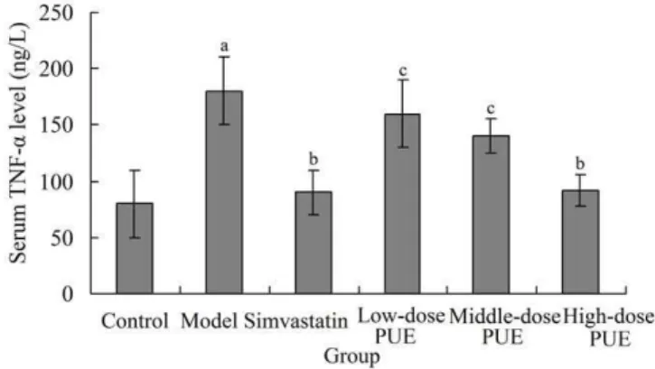
3.3 Effects of PUE on serum TNF-α level in rats with lower limb ASO
Serum TNF-α level in model group was significantly higher than that in the control group (P <0.01). Compared with the model group, the serum TNF-α level in simvastatin group was significantly decreased (P <0.05), and that in high-dose PUE
Table 1. Effects of PUE on serum TC, TG, LDL-C and HDL-C level in rats with lower limb ASD.
Group TC (mmol/L) TG (mmol/L) LDL-C (mmol/L) HDL-C (mmol/L)
Control 1.2 ± 0.3 0.6 ± 0.1 0.8 ± 0.1 2.0 ± 0.2
Model 10.1 ± 0.8a 2.3 ± 0.2a 6.2 ± 0.7a 0.3 ± 0.1a
Simvastatin 3.0 ± 0.5b 1.0 ± 0.2c 2.3 ± 0.4b 0.9 ± 0.1c
Low-dose PUE 7.2 ± 0.2d 1.9 ± 0.0d 5.1 ± 0.1d 0.4 ± 0.1d
Middle-dose PUE 6.5 ± 0.6d 1.7 ± 0.0d 2.8 ± 0.0c 0.5 ± 0.1d
High-dose PUE 4.1 ± 0.4c 1.1 ± 0.0c 2.3 ± 0.0b 0.8 ± 0.1c
aP <0.01 compared with control group. bP <0.01 compared with model group. cP <0.05 compared with model group. dP <0.05 compared with simvastatin group. PUE, puerarin; TC,
group was also significantly decreased (P <0.05). There was no significant difference between low-dose PUE group and model group (P >0.05) or between middle-dose PUE group and model group (P >0.05), with no significant difference
between simvastatin group and high-dose PUE group (P >0.05). The TNF-α levels in low-dose PUE group and middle-dose PUE group were significantly higher than that in simvastatin group (P <0.05) (Figure 2).
3.4 Effects of PUE on serum hs-CRP level in rats with lower limb ASO
Compared with the control group, the serum hs-CRP level in model group was significantly increased (P <0.01). Compared with the model group, the serum hs-CRP level in simvastatin group was significantly reduced (P <0.05), and that in high-dose PUE group was also significantly reduced (P <0.05). The hs-CRP levels in low-dose PUE group and middle-dose PUE group were significantly higher than that in simvastatin group (P <0.05). There was no significant difference between low-dose PUE group and model group (P >0.05) or between middle-dose PUE group and model group (P >0.05), with no significant difference between simvastatin group and high-dose PUE group (P >0.05) (Figure 3).
4 Discussion
Arteriosclerosis includes three kinds such as atherosclerosis, artery medial calcification and thin artery sclerosis. AS is a systemic vascular lesion. If AS occurs in the peripheral blood vessels and causes the distal limb ischemia, leading to a series of symptoms, this is known as ASD. Therefore, the pathology of ASD is closely linked to arteriosclerosis (Uchida et al., 1995). The pathogenesis of arteriosclerosis is not completely clear. At present, the damage response theory it is generally accepted, which proposes that arteriosclerosis is a chronic inflammatory reaction (Berliner et al., 1995). The lower limb ASD is the performance of systemic atherosclerosis in the local parts of body. It is the degenerative and proliferative change of the arterial intima and media (Juergens et al., 1960). The establishment of animal model of lower limb ASD is of great significance to the study of the pathological process of this disease. This study used rats as the experiment animals, and had successfully constructed the lower limb ASD model.
PUE is one of the main active components of Pueraria lobata
or Pueraria edulis Pamp.. Its chemical name is 7,4’-dihydroxy-8-C-glucosylisoflavone, with molecular weight of 416. Puerarin has multiple effects such as lowering blood glucose, dilating micro-arteries, decreasing blood viscosity, improving microcirculation, clearing free radicals after glycosylation and relieving oxidative stress response (Zhang et al., 2013). It is mainly used in clinical treatment of cardiovascular diseases such as coronary heart disease (Shi et al., 2002), angina pectoris (Wang et al., 2006) and myocardial infarction (Zhang et al., 2006). In this study, PUE was used to intervene in the lower limb ASD of rat model. The results showed that, PUE had the effect to lower limb ASD which was similar to that of simvastatin.
TC refers to the sum of cholesterols in the blood, including free cholesterols and cholesterol esters. The level of TC will be significantly elevated in atherosclerosis (Futema et al., 2015). TG is the fat molecule formed by three molecules of long-chain fatty acids and glycerol. As a kind of fatty substance in the blood,
Figure 1. Effects of PUE on serum IL-6 level in rats with lower limb
ASD. aP <0.01 compared with control group; bP <0.05 compared with
model group; cP <0.05 compared with simvastatin group. PUE, puerarin;
IL-6, interleukin-6; ASD, arteriosclerosis obliterans.
Figure 2. Effects of PUE on serum TNF-α level in rats with lower limb
ASD. aP <0.05 compared with control group; bP <0.05 compared with
model group; cP <0.05 compared with simvastatin group. PUE, puerarin;
TNF-α, tumor necrosis factor-α; ASD, arteriosclerosis obliterans.
Figure 3. Effects of PUE on serum hs-CRP level in rats with lower limb
ASD. aP <0.05 compared with control group; bP <0.05 compared with
model group; cP <0.05 compared with simvastatin group. PUE, puerarin;
most of TG can be obtained from the diet. The level of TG also increases significantly in atherosclerosis (Ma et al., 2015). LDL-C is a kind of lipoprotein rich in cholesterol which originates from HDL-C. The increase of LDL-C is also one important index which reflects the atherosclerosis (Kosmas & Frishman, 2015). HDL-C is one of the serum proteins, which is rich in phospholipids. HDL-C plays a role in delivering cholesterol in extrahepatic tissue to the liver, which can prevent the deposition of cholesterol in extrahepatic tissue cells. The level of HDL-C is reduced in coronary heart disease and other cardiovascular diseases (Ishigaki et al., 2014). Results of this study showed that, after 40 days of feeding, compared with the control group, the serum levels of TC, TG and LDL-C in the model group were significantly increased (P <0.01), while the serum HDL-C level was significantly decreased (P <0.01). This indicates that, the rat model of lower limb ASD is basically successfully established. Compared with the model group, the serum levels of TC, TG and LDL-C in the high-dose PUE group were significantly decreased (P <0.01), and the serum HDL level was significantly increased (P <0.01). This indicates that, PUE can obviously decrease the serum levels of TC, TG and LDL-C in rat model of lower limb ASD, and enhance the serum HDL-C level, namely, PUE has the anti-ASD effects.
Inflammatory reaction plays an important role in the occurrence, development and evolution of atherosclerosis. The inflammatory mechanism has become one important pathogenesis of atherosclerosis. IL-6 is an important inflammatory factor, and it is also a key factor in the induction of hs-CRP in the liver cells (Liu et al., 2016). TNF-α is produced by macrophages, which can regulate the immune function of body. It promoted the function of T cells and other killer cells, and its level is also significantly increased in stress state (Shiraki et al., 2012). The serum hs-CRP is a sensitive indicator reflecting the inflammatory reaction in body (Pearle et al., 2007). The level of hs-CRP is consistent to the severity of atherosclerosis. It exists widely in the site of atherosclerotic lesion, and it may also a direct factor which promotes the arteriosclerosis (Zhong et al., 2013). Therefore, seeking the method of curing atherosclerosis and other cardiovascular diseases from aspect of relieving the inflammatory reaction and finding the effective drugs to reduce the hs-CRP level in the body can provide new ideas and ways for the prevention and treatment of disease. Results of this study showed that, compared with the control group, the serum levels of IL-6, TNF-α and hs-CRP in model group were significantly increased (P <0.01). This indicates that, there was obvious inflammatory reaction in rats with lower limb ASD. Compared with the model group, the serum levels of IL-6, TNF-α and hs-CRP in high-dose PUE group were also significantly reduced (P <0.05). This indicates that, PUE can obviously reduce the inflammatory reaction, thus exerting the anti-ASD effects.
5 Conclusions
This study has successfully established the rat model of lower limb ASD. PUE has obvious function of decreasing the blood lipids and inflammatory factors in rats with lower limb ASD. This study has provided a basis for further application of PUE to treatment of lower limb ASD. This study still has some
limitations. Firstly, the sample size of this study is relatively small. Larger sample size will make the results more convincing. Secondly, there may be other mechanisms for effects of PUE on lower limb ASD. In next studies, the sample size should be further increased for obtaining more satisfactory outcomes, and the further action mechanisms of PUE on lower limb ASD should be investigated.
References
Berliner, J. A., Navab, M., Fogelman, A. M., Frank, J. S., Demer, L. L., Edwards, P. A., Watson, A. D., & Lusis, A. J. (1995). Atherosclerosis:
basic mechanisms. Dxidation, inflammation, and genetics. Circulation,
91(9), 2488-2496. PMid:7729036. http://dx.doi.org/10.1161/01. CIR.91.9.2488.
Che, Z. Y., Song, Q. B., Zhang, J. W., Xin, S. J., & Zhang, J. (2008). Comparison of constructing arteriosclerosis obliterans models
with two methods in rats. Journal of Clinical Rehabilitative Tissue
Engineering Research, 12(50), 9841-9844.
Davì, G., Pinto, A., Francavilla, G., Paterna, S., Campisi, D., & Strano, A. (1985). Inhibition of platelet function by ticlopidine in arteriosclerosis
obliterans of the lower limbs. Thrombosis Research, 40(2), 275-281.
PMid:2933846. http://dx.doi.org/10.1016/0049-3848(85)90339-1. Futema, M., Kumari, M., Boustred, C., Kivimaki, M., & Humphries,
S. E. (2015). Would raising the total cholesterol diagnostic cut-off from 7.5 mmol/L to 9.3 mmol/L improve detection rate of patients
with monogenic familial hypercholesterolaemia? Atherosclerosis,
239(2), 295-298. PMid:25682026. http://dx.doi.org/10.1016/j. atherosclerosis.2015.01.028.
Huang, F., Liu, K., Du, H., Kou, J., & Liu, B. (2012). Puerarin attenuates endothelial insulin resistance through inhibition of inflammatory
response in an IKKβ/IRS-1-dependent manner. Biochimie, 94(5),
1143-1150. PMid:22314193. http://dx.doi.org/10.1016/j.biochi.2012.01.018. Huang, P. P., Yang, X. F., Li, S. Z., Wen, J. C., Zhang, Y., & Han, Z. C. (2007). Randomised comparison of G-CSF-mobilized peripheral blood mononuclear cells versus bone marrow-mononuclear cells for the treatment of patients with lower limb arteriosclerosis
obliterans. Journal of Thrombosis and Haemostasis, 98(6),
1335-1342. PMid:18064333.
Ishigaki, Y., Kono, S., Katagiri, H., Dka, Y., & Dikawa, S. (2014). Elevation of HDL-C in response to statin treatment is involved in
the regression of carotid atherosclerosis. Journal of Atherosclerosis
and Thrombosis, 21(10), 1055-1065. PMid:24930383. http://dx.doi.
org/10.5551/jat.22095.
Juergens, J. L., Barker, N. W., & Hines, E. A. Jr (1960). Arteriosclerosis obliterans: review of 520 cases with special reference to pathogenic
and prognostic factors. Circulation, 21(2), 188-195. PMid:14408094.
http://dx.doi.org/10.1161/01.CIR.21.2.188.
Kosmas, C. E., & Frishman, W. H. (2015). New and emerging LDL
cholesterol-lowering drugs. American Journal of Therapeutics,
22(3), 234-241. PMid:25486520. http://dx.doi.org/10.1097/ MJT.0000000000000063.
Li, C., Pan, Z., Xu, T., Zhang, C., Wu, Q., & Niu, Y. (2014). Puerarin induces the upregulation of glutathione levels and nuclear translocation of Nrf2 through PI3K/Akt/GSK-3β signaling events
in PC12 cells exposed to lead. Neurotoxicology and Teratology, 46,
1-9. PMid:25195717. http://dx.doi.org/10.1016/j.ntt.2014.08.007. Liu, C. M., Ma, J. Q., & Sun, Y. Z. (2011). Protective role of puerarin
on lead-induced alterations of the hepatic glutathione antioxidant
Singh, A. K., Jiang, Y., Gupta, S., Younus, M., & Ramzan, M. (2013). Anti-inflammatory potency of nano-formulated puerarin and curcumin in rats subjected to the lipopolysaccharide-induced inflammation.
Journal of Medicinal Food, 16(10), 899-911. PMid:24138167. http://
dx.doi.org/10.1089/jmf.2012.0049.
Uchida, Y., Nakamura, F., & Morita, T. (1995). Dbservation of atherosclerotic lesions by an intravascular microscope in patients with
arteriosclerosis obliterans. American Heart Journal, 130(5), 1114-1119.
PMid:7484744. http://dx.doi.org/10.1016/0002-8703(95)90216-3. Wang, Q., Wu, T., Chen, X., Ni, J., Duan, X., Zheng, J., Qiao, J.,
Zhou, L., & Wei, J. (2006). Puerarin injection for unstable angina
pectoris. Cochrane Database of Systematic Reviews, 3(3), CD004196.
PMid:16856037.
Zhang, C. T., Shi, D., Zheng, Y., Zheng, C. Y., & Li, Q. H. (2013).
Chronopharmacokinetics of puerarin in diabetic rats. Indian Journal
of Pharmaceutical Sciences, 75(3), 357-361. PMid:24082353. http://
dx.doi.org/10.4103/0250-474X.117407.
Zhang, S., Chen, S., Shen, Y., Yang, D., Liu, X., Sun-Chi, A. C., & Xu, H. (2006). Puerarin induces angiogenesis in myocardium of rat with
myocardial infarction. Biological & Pharmaceutical Bulletin, 29(5),
945-950. PMid:16651724. http://dx.doi.org/10.1248/bpb.29.945. Zhong, J., Wang, Y., Wang, X., Li, F., Hou, Y., Luo, H., & Chen, H. (2013).
Significance of CAVI, hs-CRP and homocysteine in subclinical
arteriosclerosis among a healthy population in China. Clinical and
Investigative Medicine. Medecine Clinique et Experimentale, 36(2),
E81-E86. PMid:23544609.
Zhou, F., Wang, L., Liu, P., Hu, W., Zhu, X., Shen, H., & Yao, Y. (2014a). Puerarin protects brain tissue against cerebral ischemia/
reperfusion injury by inhibiting the inflammatory response. Neural
Regeneration Research, 9(23), 2074-2080. PMid:25657724. http://
dx.doi.org/10.4103/1673-5374.147934.
Zhou, Y., Xie, N., Li, L., Zou, Y., Zhang, X., & Dong, M. (2014b). Puerarin alleviates cognitive impairment and oxidative stress in APP/PS1
transgenic mice. The International Journal of Neuropsychopharmacology,
17(4), 635-644. PMid:24345484. http://dx.doi.org/10.1017/ S146114571300148X.
49(12), 3119-3127. PMid:22001170. http://dx.doi.org/10.1016/j. fct.2011.09.007.
Liu, H., Zhang, Y., Gao, Y., & Zhang, Z. (2016). Elevated levels of Hs-CRP and IL-6 after delivery are associated with depression
during the 6 months post partum. Psychiatry Research, 243, 43-48.
PMid:27359302. http://dx.doi.org/10.1016/j.psychres.2016.02.022. Ma, W., Ding, H., Gong, X., Liu, Z., Lin, Y., Zhang, Z., & Lin, G. (2015). Methyl protodioscin increases ABCA1 expression and cholesterol efflux while inhibiting gene expressions for synthesis of cholesterol and triglycerides by suppressing SREBP transcription and microRNA
33a/b levels. Atherosclerosis, 239(2), 566-570. PMid:25733328. http://
dx.doi.org/10.1016/j.atherosclerosis.2015.02.034.
Pearle, A. D., Scanzello, C. R., George, S., Mandl, L. A., DiCarlo, E. F., Peterson, M., Sculco, T. P., & Crow, M. K. (2007). Elevated high-sensitivity C-reactive protein levels are associated with local
inflammatory findings in patients with osteoarthritis. Osteoarthritis
and Cartilage, 15(5), 516-523. PMid:17157039. http://dx.doi.
org/10.1016/j.joca.2006.10.010.
Phornphibulaya, P., Tongprasroeth, S., Phimolsanti, R., & Limwongse, K. (1981). Severe ischemia of the lower extremity due to arteriosclerosis
obliterans. Journal of the Medical Association of Thailand, 64(12),
591-599. PMid:6153025.
Safar, M. E. (1991). Arteriosclerosis obliterans of the lower limbs as a
model of peripheral vascular disease with hypertension. Journal of
Cardiovascular Pharmacology, 18(Suppl 4), S73-S77. PMid:1721984.
http://dx.doi.org/10.1097/00005344-199118041-00015.
Shi, W. G., Qu, L., & Wang, J. W. (2002). Study on interventing effect of puerarin on insulin resistance in patients with coronary heart
disease. Chinese Journal of Integrated Traditional and Western
Medicine, 22(1), 21-24. PMid:12585165.
Shiraki, A., Dyama, J., Komoda, H., Asaka, M., Komatsu, A., Sakuma, M., Kodama, K., Sakamoto, Y., Kotooka, N., Hirase, T., & Node, K. (2012). The glucagon-like peptide 1 analog liraglutide reduces TNF-α-induced oxidative stress and inflammation in endothelial
cells. Atherosclerosis, 221(2), 375-382. PMid:22284365. http://dx.doi.