U
T
L
F
ACULDADE DEM
OTRICIDADEH
UMANASHOULDER MORPHOFUNCTIONAL ADAPTATIONS ON
OVERHEAD-THROWING ATHLETES
Implications for physiotherapy throwing-shoulder examination
Dissertação elaborada com vista à obtenção do Grau de Doutor em Motricidade Humana, Especialidade de Ciências da Fisioterapia
Orientador: Doutor Augusto Gil Brites de Andrade Pascoal
Júri
Presidente: Reitor da Universidade Técnica de Lisboa.
Vogais: Doutor João Paulo Vilas Boas Soares Campos, Professor Catedrático da Faculdade de
Desporto da Universidade do Porto
Doutor António Prieto Veloso, Professor Catedrático da Faculdade de Motricidade Humana da Universidade Técnica de Lisboa
Doutora Paula M. Ludewig, Professora Associada da University of Minnesota (EUA), Program in Physical Therapy, Department of Physical Medicine & Rehabilitation
Doutor Augusto Gil Brites de Andrade Pascoal, Professor Auxiliar da Faculdade de Motricidade Humana da Universidade Técnica de Lisboa
Doutor Raul Alexandre Nunes da Silva Oliveira, Professor Auxiliar da Faculdade de Motricidade Humana da Universidade Técnica de Lisboa
Aos meus anjos da guarda, os meus filhos; José Miguel e Afonso Miguel.
Ao meu marido Afonso.
O trabalho desenvolvido no decorrer desta tese teve o financiamento da Fundação para
a Ciência e Tecnologia através da Bolsa Individual de Doutoramento
TABLE OF CONTENTS
FIGURES ... iv
TABLES ... vi
ABBREVIATIONS ... vii
AGRADECIMENTOS/ ACKNOWLEDGEMENTS ... viii
ABSTRACT ... xi
RESUMO ... xii
CHAPTER 1 - GENERAL INTRODUCTION ... 13
Overview ... 15
Research goals ... 20
Dissertation Structure ... 20
Publications ... 23
Papers in publications with impact factor ... 23
Communications in proceedings with refereeing: ... 24
CHAPTER 2 - REVIEW OF LITERATURE ... 27
The Overhead Throwing Shoulder ... 28
Overhead Throwing Shoulder Adaptive Pattern ... 34
Structural adaptations ... 38
Functional adaptations ... 47
CHAPTER 3 - METHODOLOGY ... 77
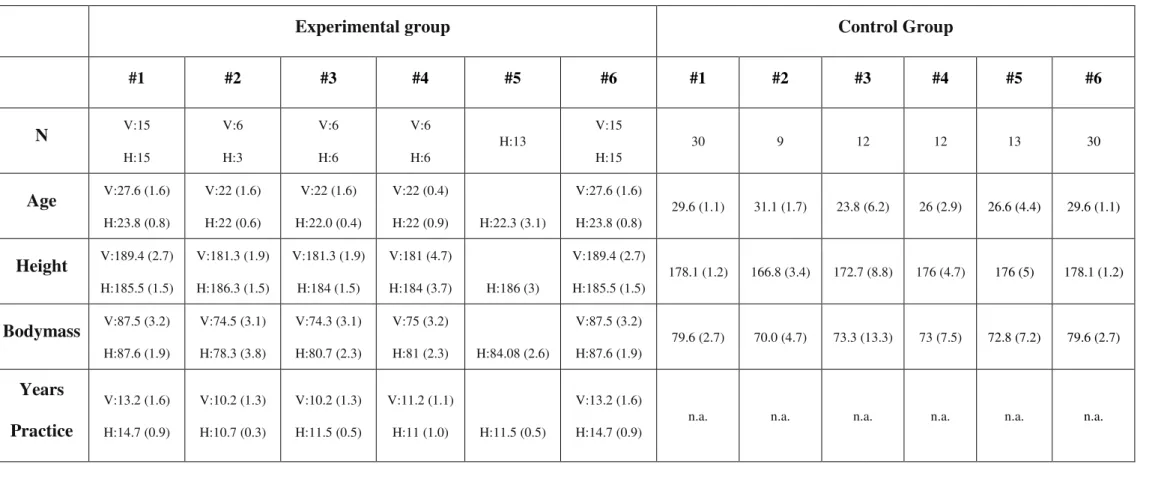
Study Design and Participants ... 79
Measurements ... 82
X-Ray measurements ... 82
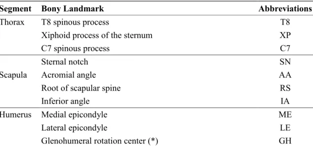
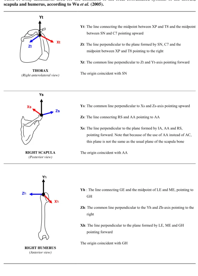
Shoulder Kinematic measurements ... 85
Statistical Procedures ... 89
CHAPTER 4 - OSSEOUS ADAPTATIONS ... 93
Humeral Retroversion Angle and Its Relationship with Active Shoulder External Rotation Range-Of-Motion in Volleyball and Team-Handball Players ... 94
Abstract ... 94
Introduction ... 95
Materials And Methods ... 98
Discussion ... 109
Conclusion ... 114
CHAPTER 5 - SHOULDER ROTATIONAL PATTERN ... 115
The Effects of Testing Subject Position (Seated vs. Supine) in Shoulder External Rotation ... 117
Abstract ... 117
Introduction ... 118
Materials and Methods ... 119
Results ... 122
Discussion ... 122
Conclusion ... 124
Shoulder Rotation Range-of-Motion in Throwing Athletes. The Effect of Active or Passive End-Range Determination ... 125
Abstract ... 125
Introduction ... 126
Materials And Methods ... 129
Results ... 133
Discussion ... 135
Conclusions ... 137
CHAPTER 6 - SCAPULAR ADAPTATIONS ... 139
The Scapular Contribution to the Amplitude of Shoulder External Rotation on Throwing Athletes ... 141
Abstract ... 141
Introduction ... 142
Materials And Methods ... 150
Results ... 153
Discussion ... 155
Conclusions ... 158
Scapular Contribution for the End-Range of Shoulder Axial Rotation. Scapular Behavior in Over-Head Athletes... 159
Abstract ... 159
Introduction ... 160
Materials and Methods ... 162
Results ... 165
Conclusions ... 171
Resting Scapular Posture in Healthy Overhead Throwing Athletes ... 173
Abstract ... 173
Introduction ... 174
Materials and Methods ... 175
Results ... 178
Discussion ... 180
CHAPTER 7 - GENERAL DISCUSSION ... 183
Overview ... 184
Main research findings ... 191
Implications and future directions ... 192
Limitations ... 193
CHAPTER 8 - REFERENCES ... 197
CHAPTER 9 - APPENDICES ... 213
FIGURES
TABLES
ABBREVIATIONS
3D ... Three-Dimensional DOF ... Degree-Of-Freedom ER ... External Rotation
ERG ... External Rotation Gain ES ... Effect Size GH ... Glenohumeral Joint
GH ... Glenohumeral Angles GIRD ... Glenohumeral Internal Rotation Deficit HRA ... Humeral Retroversion Angle HRs ...Scapulohumeral Rotation
HRt ... Humeral Axial Rotation IR ... Internal Rotation IR-ER-ROM ... Internal-External Rotation Range-Of-Motion
ISB ... International Society of Biomechanics
ISBS ... International Society of Biomechanics in Sport LCS ... Local Coordinated System
OTSAP ... Overhead Throwing Shoulder Adaptation Pattern
ROM ... Range-Of-Motion SH ... Scapulohumeral Angles SPSS ... Statistical Package for Social Sciences
SSC ... Stretch-Shorten System Sxt ...Scapular Upward-Downward Rotation Syt ...Scapular Protraction-Retraction
Szt ... Scapular Spinal Tilt
TH ... Thoracohumeral Angles Tukey HSD ... Tukey Honestly Significant Difference
AGRADECIMENTOS/ ACKNOWLEDGEMENTS
Chegado o momento da entrega deste documento, olho para trás e vejo sorrisos, lágrimas; marcas que jamais deixarão de fazer parte de mim e de todos os que me rodeiam. Foi um percurso longo, por vezes tortuoso e difícil. Uma subida a uma enorme montanha, uma prova de resistência intensa, nunca só, sem alguma vez sair do trilho até à alvorada. Trilhar este caminho só foi possível com a mobilização de uma grande energia, vontade e perseverança que trazem consigo força, vigor e paz, indispensáveis em cada momento da caminhada, para encontrar o melhor rumo, fazer as melhores opções e tomar as melhores decisões.
Este caminho não foi percorrido na solidão. Foi um trabalho coletivo, embora a sua redação e responsabilidade sejam um ato individual.
Assim este trabalho não teria sido possível sem a ajuda e apoio de muitas pessoas, a quem estarei eternamente grata:
Em primeiro lugar ao Professor Doutor Augusto Gil Pascoal que acreditou em mim aceitando-me como sua aluna de doutoramento. À sua total disponibilidade, desde o primeiro momento. A sua presença foi constante, não obstante a distância, nunca me senti sozinha. Não tenho palavras para agradecer a hora que me recebeu, lendo e acompanhando todo o trabalho passo a passo, Muito obrigada.
Gostaria também de agradecer a todos os membros do júri pelo tempo dispensado na leitura e apreciação da minha tese.
A minha eterna gratidão para o Dr. Melo Monteiro, para o João, Lídia, Avó Sílvia, Cláudia e Isidro, sem vós nada disto teria sido possível.
Aos amigos que sempre disseram presente, Nuno Morais, Isabel Loureiro, Ermelinda, Rui Castelar, Ricardo Matias, José Miguel Leite, José Teixeira, D. Fernanda, Rosinha, Eurico Peixoto, Rita Oliveira, Alan Coccato, todos grandes amigos e companheiros, sempre disponíveis para ajudar e acima de tudo para me ouvirem.
Ao Nuno, Marina, Márcio e Lago, eu sei que não fui fácil de aturar....
Ao Vitória Sport Clube, minha eterna paixão, pelo apoio das suas direções, compreensão e disponibilidade. Aos jogadores e treinadores, a todos, muito obrigada.
Ao Fermentões, pela disponibilidade imediata, assim como aos seus atletas.
Ao Xico Andebol por todo o apoio.
À Faculdade de Motricidade Humana por ter aprovado esta investigação e me ter permitido crescer pessoal e profissionalmente.
Aos meus pais, sim a vós que sempre me encorajaste e apoiaste de forma incondicional, que me substituíram vezes sem conta no papel de mãe... não sei como vos agradecer. Apenas posso dizer que vos adoro!
A ti Carlos, meu irmão, que tantas vezes me ouviste e me impediste de desistir, obrigada pelas palavras de incentivo quando já estava “sem pilhas” e por estares sempre do meu lado. És e sempre serás o meu menino!
Tio Guilherme e Tia Sameiro, Tio Nandinho e Mila e aos primos,António Carlos, Sofia, Rita, Maria João, Pedro, João Pedro a todos obrigada.
Ao Chico, à Zé, Mariazinha e Fernando, ainda deu para nos divertirmos! Obrigada por me verem e por me tratarem como uma filha.
Finalmente, um grande e carinhoso agradecimento ao Afonso, pelo seu amor, paciência e apoio,,e a vós meus lindos filhos: José Miguel e Afonso Miguel; desculpem ter sido tão ausente, desculpem não ter brincado quando me pediram, desculpem por estar sempre a trabalhar, obrigada por serem a luz na minha vida e com o vosso sorriso conseguirem mostrar-me que o caminho é sempre para a frente
SHOULDER MORPHOFUNCTIONAL ADAPTATIONS ON
OVERHEAD-THROWING ATHLETES. IMPLICATIONS FOR PHYSIOTHERAPY THROWING-SHOULDER EXAMINATION
Andrea Ribeiro & Augusto Gil Pascoal
ABSTRACT
The overhead throwing motion is a highly skilled movement, particularly demanding to the shoulder due to high strength levels and/or acceleration applied to the hand and by the elevated degree of control and precision required to position the arm in space. The shoulders of those involved in repeated forceful overhead throwing, the overhead-throwing athletes, undergo a range of neural, soft tissues and skeletal adaptations that
could be described as, the “overhead-throwing shoulder adaptive pattern” (OTSAP).
The main goal of overall studies in this thesis was to characterize the dominant overhead-throwing shoulder adaptive pattern of non-symptomatic overhead throwing athletes, comparing with a non-athletic population. Additionally, while comparing volleyball and team-handball players, we looked for specific sport-related components of the OTSAP. Knowledge on OTSAP is important for those involved on training, but also for sport physiotherapist during shoulder functional assessment. Some components of the OTSAP could be mistaken by injury signs or risk factors. Structural (osseous) and functional changes were identified on the dominant shoulder of volleyball and team-handball players. Some were similar of those described in baseball players, and others were sport-related. Thus, the OTSAP should be considered by the physiotherapist during overhead-throwing shoulder assessment.
Keywords: shoulder, athlete, physiotherapy, overhead, throwing, volleyball,
SHOULDER MORPHOFUNCTIONAL ADAPTATIONS ON
OVERHEAD-THROWING ATHLETES. IMPLICATIONS FOR PHYSIOTHERAPY THROWING-SHOULDER EXAMINATION
Andrea Ribeiro & Augusto Gil Pascoal
RESUMO
O movimento de lançamento é altamente especializado e particularmente exigente para o ombro devido aos excessivos níveis de carga/aceleração aplicados. Atletas cujos ombros estão envolvidos em movimentos repetidos de lançamento, consideram-se, na literatura anglo-saxónica, atletas “overhead”. Estes são sujeitos a um conjunto de adaptações neurais, tecidulares e ósseas que podem ser descritas como, o “padrão de
adaptação do ombro do atleta overhead” (PAOAO). O principal objetivo da tese foi
caracterizar o padrão de adaptação do ombro dominante, não sintomático, dos atletas
“overhead”, comparando-os com não atletas. Adicionalmente, comparando voleibolistas e andebolistas, procurou-se encontrar componentes específicos da modalidade praticada inerentes ao PAOAO. O conhecimento detalhado deste PAOAO é crucial para os intervenientes em processos de treino, e para o fisioterapeuta responsável por uma avaliação detalhada do ombro, sob pena de alguns dos componentes do PAOAO serem erroneamente considerados como sinais de lesão ou fatores de risco. Foram identificadas alterações estruturais e funcionais no ombro dominante de voleibolistas e andebolistas. Algumas são similares às encontradas em jogadores de beisebol, enquanto outras estão diretamente relacionadas com a prática desportiva específica. Assim, este PAOAO deverá ser tido em consideração pelo fisioterapeuta aquando da avaliação do
ombro do atleta “overhead”.
Overview
Overhead-throwing athletes include, among others, baseball pitchers, swimmers, team-handball players, volleyball or tennis players. These athletes perform specific sports gesture, known as overhead activity; such as throwing, passing, hitting, spiking or even swimming stroke, where the hand describes overhead trajectories
The overhead throwing motion is a highly skilled movement performed at high velocity, which requires synchronicity, neuromuscular control, flexibility, muscular strength and coordination (Wilk et al., 2009). For the shoulder, these overhead activities are particularly demanding due to the huge strength levels and/or acceleration applied to the hand and by the elevated degree of control and precision required positioning arm in space. Competitive overhead throwing athletes perform at the extremes of glenohumeral range-of-motion and place tremendous repetitive stresses on their shoulders (McConnell, Donnelly, Hamner, Dunne, & Besier, 2012). From a functional standpoint these overhead-activities require repetitive overhead motions where the arm is forcefully propelled forward from near maximal external rotation to internal rotation (Borsa, Laudner, & Sauers, 2008). It is estimated that the magnitude of strength in shoulder external to internal rotation in baseball throwing is about 111 Nm (Levine et al., 2006), which clearly shows the stress imposed on the athlete´s shoulder.
pre-select an athlete to a certain overhead sport, or acquired through adaptive change in shoulder joint. Discussion continues as to whether these adaptations arise from soft-tissue (e.g. capsule and ligaments) or from osseous adaptations within and around the shoulder. However, in literature a selection of adaptive changes were identified and described on the morphology and function of the dominant shoulder of overhead-throwing athletes. These changes resulting from the extreme physiological demands of overhead-throwing activity, which are sport-related adaptations, configure an unique adaptive pattern on the dominant overhead-throwing shoulder: the overhead-throwing shoulder adaptive pattern (OTSAP). In essence this adaptive pattern represents a shoulder attempt to maintain balance between the needed flexibility (to allow more external rotation) and the necessary stability for the throwing motion (Osbahr, Cannon, & Speer, 2002).
Brindle, 2004; Reagan et al., 2002; Tokish et al., 2008), team-handball (Pieper, 1998) and volleyball players (Schwab & Blanch, 2009). It is thought that change in retroversion angle occur in the proximal physis of the humerus over time in young pre-adolescent athletes when the proximal epiphysis is not yet completely fused (Yamamoto et al., 2006). In fact, it is known that most of the humeral growth takes place in the proximal physis, particularly after 11 years of age (Pritchett, 1991). Recently Wyland et al. (2012) demonstrated that beyond the humeral retroversion also glenoid retroversion angle was significantly greater on the throwing side than on the non-throwing side, suggesting that further studies must be addressed to the scapular osseous adaptation on the dominant shoulder of overhead-throwing athletes.
Functional adaptations refer to adaptive changes in the rotational motion pattern at the dominant glenohumeral joint of overhead-throwing athletes, but also to changes on scapulothoracic stability and mobility, particularly on scapular resting position and scapular kinematics during arm motion.
and non-throwing shoulder (Borsa et al., 2008). The total arc of rotation in the overhead-throwing shoulder seems to adapt by shifting backwards favoring external rotation at the expense of internal rotation. Some authors refer this adaptation in the
rotational arc shift phenomenon as the “total motion concept” (Borsa et al., 2008; Wilk et al., 2011) or posterior shift (Borich, Bright, Lorello, Cieminski, & Buisman, 2006; Cieminski, 2007; McCully, Kumar, Lazarus, & Karduna, 2005; Tokish et al., 2008; Wilk et al., 2009).
Most of the studies about the overhead-throwing shoulder adaptive pattern were made on the dominant shoulder of baseball players, particularly on baseball pitchers. This thesis explores the assumption that the overhead-throwing activities involved in volleyball and team-handball, could induce shoulder sport-related adaptations, similar to those described in baseball players (Braun, Kokmeyer, & Millett, 2009; Warden, Bogenschutz, Smith, & Gutierrez, 2009; Werner, Gill, Murray, Cook, & Hawkins, 2001; Wilk et al., 2011). In fact, in volleyball the spike could be considered as an overhead activity in which the efficacy depends on the magnitude of the contact force between hand and ball. Volleyball spike is used to the strike the ball in the way that it lands on the opponent´s court and cannot be defended. A player makes a series of steps
towards the goal of clarifying what are the limits of shoulder function optimization by training and the injury risk factors.
Research goals
The shoulders of those involved in repeated forceful overhead throwing undergo a range of neural, soft tissues (muscular and capsular and ligaments), and skeletal adaptations that could be described as the overhead-throwing shoulder adaptive pattern (OTSAP). Thus, the major goal of this dissertation was to characterize the dominant overhead-throwing shoulder adaptive pattern of non-symptomatic overhead overhead-throwing athletes, comparing with a non-athletic population. Additionally, while comparing the dominant shoulder of volleyball and team-handball players, we look for specific sport-related components of the OTSAP.
Dissertation Structure
This thesis is a compilation of six papers, Chapter 4 to 6. To them, was added a General Introduction chapter (Chapter 1), Review of literature (Chapter 2), Methodology (Chapter 3) and General Discussion (Chapter 7). Appendices were also added to give further information about clinical tests mentioned on Chapters 3 to 6.
In Chapter 2 (Literature Review) the most relevant studies about throwing shoulder and throwing shoulder adaptations are reviewed. The main concern was to give the reader the necessary framework to the understanding of the following chapters, where results from the experimental work are presented.
In this thesis we started to look for structural, osseous adaptations, Chapter 4. From a skeletal perspective, it is shown that throwing shoulders have more humeral retroversion when compared with the non-throwing shoulder. Alterations in humeral retroversion are thought to develop over time when the proximal humeral epiphysis is
not yet completely fused. In “Humeral retroversion angle and its relationship with active
shoulder external rotation range-of-motion in volleyball and team-handball players”, we
compared the humeral retroversion angle of the dominant shoulder of volleyball and team-handball players with a control group. We also looked for the relationship between humeral retroversion angle and functional adaptations, such as, active shoulder external rotation range-of-motion.
Throwing athletes have been shown to display altered rotational range-of-motion patterns in the dominant shoulder that favors increased external rotation and limited internal rotation range-of-motion. Concerning external rotation range-of-motion, studies often use goniometry as a part of shoulder assessment (Ellenbecker & Roetert, 2002; Ellenbecker, Roetert, Piorkowski, & Schulz, 1996; Tokish et al., 2008; Torres & Gomes, 2009; Wilk et al., 2011). This end-range is determined by capsular end-feel (Awan, Smith, & Boon, 2002; Barlow, Benjamin, Birt, & Hughes, 2002; Reagan et al., 2002), by capsular liftoff (Warner, Micheli, Arslanian, Kennedy, & Kennedy, 1990) or by pain (Andrews AW & RW, 1989), as opposed to an objective assessment of torque. Also these studies use passive motion in supine, where the scapula is stabilized on the table.
In Chapter 5 two studies are presented which intend to clarify some methodological aspects concerning the overhead throwing shoulder rotational pattern changes, and how to evaluate these specifically in the overhead throwing athlete. The first study, “The
analyzed the effects of subject testing position on shoulder external rotation range-of-motion, particularly on shoulder external rotation end-range determination. The other study presented in Chapter 5, “Shoulder rotation range-of-motion in throwing athletes.
The effect of active or passive end-range determination”, explore the effect of passive vs. active end-range determination on shoulder external rotation ROM.
In Chapter 6, two studies are included about the contribution of scapular motion on shoulder rotational pattern. A third study was included about the postural changes identified on scapular resting position in overhead-throwing athletes. The assessment of the glenohumeral internal and external rotation range-of-motion is a standard part of a shoulder clinical examination. However, the contribution of shoulder girdle in the rotational motion pattern often is not considered by clinicians. In fact, during physiotherapy examination, arm passive motion is often used to test glenohumeral range-of-motion while at the same time scapula is stabilized. However, on sport overhead activities, scapular stability and mobility are crucial on the kinetic chain that involves the lower limb, the trunk and the upper limb. The first study presented on Chapter 6, “The scapular contribution to the amplitude of shoulder external rotation on
throwing athletes” explores the contribution of scapular stability and/or mobility to the shoulder external rotation ROM, on thrower athletes. The second study in Chapter 6, entitle “Scapular contribution for the end-range of shoulder axial rotation. Scapular
behavior in overhead athletes” also looked for scapular contribution on shoulder external rotation ROM, but also for scapular motion in internal rotation in overhead throwing athletes, adding information about internal rotation which was not acquired in the first mentioned study.
throwing athletes by comparison with a non-athletic population. It was assumed that this information could be helpful on shoulder physiotherapy examination, particularly on shoulder static examination.
Publications
Papers in publications with impact factor
Parts of this thesis have been published, accepted for publication, or submitted for publication:
Ribeiro A, Pascoal AG. The relationship between the humeral retroversion angle and the active shoulder rotation in volleyball players. Journal Biomechanics. 2012: 45(S1), S626.
Ribeiro A, Pascoal AG. Scapular contribution for the end-range of shoulder axial rotation. Scapula behavior in overhead athletes. International Journal Sports Science and Medicine. 2012: 11, 676-681.
Ribeiro A, Pascoal AG. Shoulder rotation range-of-motion assessment in throwing athletes. The effect of active or passive end-range determination. Physiotherapy Theory and Practice. (Under review).
Ribeiro, A., Pascoal, AG and Ludewig, PM. Humeral Retroversion Angle And Its Relationship With Active Shoulder External Rotation Range-Of-Motion In Volleyball And team-handball Players. American Journal Sports Medicine (Under review)
Ribeiro, A. and Pascoal AG. Resting Scapular Posture in Healthy Overhead Throwing Athletes. Manual Therapy (Under review).
Ribeiro, A. and Pascoal, AG. The Effects Of Testing Subject Position (Seated Vs. Supine) In Shoulder External Rotation. Physical Therapy (Under review).
Book chapters
Communications in proceedings with refereeing:
Parts of this thesis and additional exploration of data were presented in congresses and published in abstract books and in special issues of scientific journals:
Ribeiro A, Pascoal AG, Ludewig PM. Humeral retroversion angle and its relationship with active shoulder external rotation range-of-motion in volleyball and
European team-handball players. IXth Conference of the International Shoulder Group. Aberystwyth University, Wales, UK, 2012:69-70.
Ribeiro A, Pascoal AG, Ribeiro P. Humeral retroversion in overhead throwing athletes. In: ISBS, ed. 29th Conference of the International Society of Biomechanics in Sports. Oporto, Portugal, 2011:125.
Ribeiro A, Pascoal AG, Ribeiro P. The throwing shoulder. Scapular contribution for the end-range of shoulder axial rotation. Coloquium 511 on Biomechanics of Human Motion. Azores, Portugal, 2011:22.
Ribeiro A, Pascoal AG. Shoulder external rotation range-of-motion assessment on thrower athletes, the effects of testing end-range determination (active vs.
passive). XXIIIrd International Society of Biomechanics. Belgium, 2011. (http://isbweb.org/images/conf/2011/ScientificProgram/ISB2011_ScientificProgram.htm)
Ribeiro A, Pascoal AG. The contribution of the scapular patterns to the amplitude of shoulder external rotation on thrower athletes. In: Natal J, Tavares J, Pinotti M, Slade A, eds. 6th International Conference of Technology and Medical Sciences. Oporto, Portugal: Taylor & Francis, 2010:275-278.
Ribeiro A, Pascoal AG. Assessment of the external rotation range-of-motion on thrower athletes. The effects of testing end-range determination (active vs. passive). 1st International Conference of Applied Bionics and Biomechanics. Venice, Italy, 2010.
Ribeiro A, Pascoal AG. Humeral and scapular 3D position in thrower athletes at end-range internal and external rotation during active fast and slow arm movements.
Pascoal AG, Madureira E, Cascais C, Ribeiro A, Neves S. Differences on range of motion of active humeral rotation in throwing athletes. In: ISG, ed. Shoulder biomechanics, ergonomics and sport. Bologna, Italy, 2008.
Ribeiro A, Pascoal AG. The external shoulder rotation range-of- motion on thrower athletes. The effects of testing subject position (seated vs. supine) and en-range
determination (active vs. passive). Shoulder Biomechanics International Shoulder Group. Minneapolis, 2010:31.
Ribeiro A, Pascoal AG. Effect of active v. passive end-range determination on shoulder axial rotation in thrower athletes. International Conference on Biomechanics in Sport. Michigan, USA, 2010: July.
The Overhead Throwing Shoulder
The term “overhead throwing athlete” involves all participants in overhead throwing
activities, where the sports gesture encompasses repetitive overhead throwing actions of the dominant shoulder. Examples of these sport activities are team-handball, volleyball, baseball or tennis. But in this group we can also find athletes which perform cyclic arm activities such as swimmers.
This overhead throwing motion is an extremely skillful and intricate movement which is very stressful on the shoulder joint complex. The overhead throwing athlete places extraordinary demands on this complex. Excessively high stresses are applied to the shoulder joint because of the tremendous forces generated by the athlete.
Kinematics of the throwing arm (with ball) is frequently described as a particular
sequence of phases, the “throwing cycle” (Wagner et al., 2012; Werner et al., 2006), that includes the initial and late cocking phases, where the arm assumes an elevated-external rotated position, followed by an acceleration and a follow-through (deceleration) phases. At the end of the acceleration phase the object (ball) is released or stroked. On throwing athletes, during the deceleration phase, the posterior rotator cuff musculature
acts eccentrically. The goal is to decelerate or “brake” the internal rotation and
that alters kinetic chain components, especially one that affects the so called “core”
(trunk, back and proximal parts of the lower limbs), will alter more distal segments and may result in the development of a dysfunctional shoulder (Braun et al., 2009; McMullen & Uhl, 2000).
Altered mobility patterns have been consistently reported in the dominant shoulder of elite baseball pitchers (Borsa et al., 2006; Borsa, Jacobson, Scibek, & Dover, 2005; Brown, Niehues, Harrah, Yavorsky, & Hirshman, 1988; Downar & Sauers, 2005; Ellenbecker et al., 2002; Joseph B. Myers, Laudner, Pasquale, Bradley, & Lephart, 2006; Osbahr et al., 2002; Reagan et al., 2002).Shoulder mobility in the overhead athlete has been found to be both excessive (hypermobile) and limited (hypomobile) compared with shoulders that are not exposed to overhead sports. This altered shoulder mobility is thought to develop secondary to adaptive structural and functional changes to the shoulder joint resulting from the extreme physiological demands of overhead activity. Researchers have speculated as to whether these structural adaptations compromise shoulder stability, thus exposing the overhead athlete to injury, or if these adaptations predispose the subject to be an elite overhead throwing athlete.
The mentioned changes/adaptations which result from the extreme physiological demands of overhead-throwing activity, and seem to be sports-related adaptations, configure an unique adaptive pattern on the dominant overhead-throwing shoulder: the overhead-throwing shoulder adaptive pattern (OTSAP). This adaptive pattern represents a shoulder attempt to maintain balance between the needed flexibility (to allow more external rotation) and the necessary stability for the throwing motion (Osbahr et al., 2002).
Whiteley, Ginn, Nicholson, & Adams, 2006; Yamamoto et al., 2006), but in Europe, this is not an usual sport. Among throwing sports, volleyball and team-handball are
quite popular in the “old continent”. What kind of shoulder adaptations do these athletes present? Are these similar to the ones shown by baseball players?
Volleyball and team-handball have also been referred as “overhead activities” (Pieper, 1998; Schwab & Blanch, 2009; Seil, Rupp, Tempelhof, & Kohn, 1998; Wang & Cochrane, 2001a). For some authors they are considered to represent typical overarm movements of throwing or hitting a ball and where ball velocity is the main performance variable (Wagner et al., 2012).This labeling suggests that some assumptions regarding the throwing shoulder adaptation on volleyball and team-handball players could be similar as adaptations described about baseball players (Tripp, Yochem, & Uhl, 2007; Warden et al., 2009; Werner et al., 2006; Wilk et al., 2009). However, this holds not to be true. Osseous side-to-side adaptations were described on throwing humerus of volleyball and team-handball players (Pieper, 1998; Schwab & Blanch, 2009), similar as in baseball players. However nature and implications of these are different and must be analyzed in detail by comparison with a non-thrower population.
The volleyball spike has been divided into phases that resemble a slightly simplified version of the general overhead throwing motion seen in baseball (Yamamoto et al., 2006). In this method, five phases were defined, with a general cocking phase encompassing both the early and late cocking phases of the overhand throw. In spike at the moment of contact, the hitter's arm is fully extended above his or her head and slightly forward, making the highest possible contact while maintaining the ability to deliver a powerful hit. The hitter uses arm swing, wrist snap, and a rapid forward contraction of the entire body to drive the ball.
Although volleyball attackers can employ different styles, and therefore different kinematics, the ball gets generally in contact with the hand, above and slightly anterior to the hitting shoulder. As a result, the arm motion is constantly adjusted throughout the spike so that contact can occur in an optimal location. This could lead one to hypothesize that the mechanics of the swing are not necessarily as important in volleyball as in other overhead sports such as baseball or team-handball, or rather, that
the dynamic aspect of the ‘set’ in volleyball requires attackers to be equally dynamic in their upper limb mechanics during an attack sequence.
In elite team-handball, shooting on goal is one of the most important aspects of the game. For a shot to be successful, it requires maximum ball velocity and precision as well as an element of surprise do the defensive players and goalkeeper. But how is this shot performed, how is this throw executed?
Volleyball and team-handball are different with respect to the kinematic and kinetic pattern of the throwing cycle and consequently in the repetitive stress imposed to the shoulder which is beneath osseous and soft tissue adaptations.
In team-handball throw, the throwing arm must accelerate the additional weight of the handball whereas in volleyball spike there is no additional weight that has to be accelerated (Wagner et al., 2012). Wagner found differences in shoulder internal rotation and shoulder flexion angle in the cocking pass of volleyball spike when compared to team-handball throw. In fact volleyball players perform some shoulder flexion at takeoff which leads a delay in maximal shoulder hyperextension angle during spike. They also concluded that overarm movements are similar but not identical due to specific adaptations based on technical and tactical components of different games as well as different body movements (Wagner et al., 2012).
activity of the external rotator muscles on the last phase of the throwing cycle. In fact, during arm deceleration phase on volleyball striking, shoulder internal rotation energy could be totally or partially dissipated into the ball, which could explain the atrophy of some shoulder external rotators muscles. In a prospective cross-sectional study, Lajtai et al. (2009) reported a 30% prevalence of infraspinatus muscle atrophy among the dominant (hitting) shoulder of beach volleyball players. The authors also report a significantly reduction on external rotation strength on all players when compared with the non-dominant shoulder. Players with atrophy had significantly more loss of external rotation strength (2.3 kg) than players without atrophy (0.8 kg; P = .0210).
Overhead Throwing Shoulder Adaptive Pattern
During the overhead throwing motion, due to the repetitive sports gesture, the mechanical stimuli will induce alterations into the micro-structure (composition) and architecture in soft tissues such as; capsule, ligaments, tendon and bone. These will reverberate in shoulder function, inducing changes that could or could not be related to pathology, or are just shoulder adaptations of the throwing motion towards sports gesture optimization.
specific functions. The heterogeneous nature of connective tissue structures causes these structures to exhibit properties (strength and elasticity) that vary according to their orientation in space when a constant force is applied (Levangie & Norkin, 2011).
Although connective tissue appears in many forms throughout the body, all connective tissue exhibits the common property of viscoelasticity. The behavior of viscoelastic materials is a combination of the properties of elasticity and viscosity. Elasticity refers to a material´s ability to return to its original state following deformation after removal of the deforming load. When a material is stretched, it has work done on it and its energy increases. An elastic material stores energy and keeps the energy available so that the stretched elastic material can recoil immediately to its original dimensions following removal of the distractive force. Elasticity implies that length changes or deformations are directly proportional to the applied forces or loads. Viscosity refers to a material´s ability to dampen shearing forces. When forces are applied to viscous materials they exhibit time-and-rate dependent properties (Hamill & Knutzen, 2009).
When load forces are applied to a structure, meaning load by an external force or forces applied to a structure, these are called mechanical load. The type of internal mechanical resistance (stress) and strain (deformation) that develops in human structures is dependent on the nature of the material, type of load, and the rate and duration of loading. When a structure can no longer support load, the structure is said to have failed (Levangie & Norkin, 2011).
are sensitive to the duration of the force application. When a viscoelastic material is subjected to either a constant compressive or tensile load the material deforms and continues to deform over a finite length of time even if the load remains constant. Deformation of the tissue continues until a state of equilibrium is reached. This phenomenon is called creep and is attributed to different mechanisms is according with
the materials.
In bone, creep in compression has been attributed to the slip of lamellae within the osteons and the flow of the interstitial fluid. In articular cartilage subjected to a compressive force, creep is attributed to the gradual loss of fluid from the tissue. Viscoelastic materials respond differently to different rates of loading. When viscoelastic materials are loaded rapidly, they exhibit greater resistance to deformation than occurs if they are loaded more slowly. Generally, the higher the rate and the longer the duration of the applied force, the greater the deformation.
Viscoelastic materials do not store all of the energy that is transferred to them when they are deformed by an applied force, and thus the transferred energy is not available for recovery. When a force is applied and then removed, some of the energy created during the stretching or compression of the material may be dissipated in the form of heat and therefore the material may not return to its original dimensions. The loss of energy is called hysteresis, which is exhibited by viscoelastic materials when they are subjected to the application and removal of forces (Ambrosio, Netti, & Nicolais, 2002).
what occurs when ligaments are overstretched and become lax. The ligaments are no longer capable of returning to their original length after being elongated and remain in a partial state of elongation.
Stress/strain curves for bone demonstrate that cortical bone is stiffer than cancellous bone meaning that cortical bone can withstand greater stress but less strain than cancellous bone. The application of high loads maintained for a short period of time or low loads held for a long period of time will produce high stress and strain. The rate, frequency, duration and type of loading affects bone in that repeated loadings, either high repetition coupled with low load or low repetition with high load, can cause permanent strain and lead to bone failure. Bone loses stiffness and strength with repetitive loading as result of creep strain. Creep strain occurs when a tissue is loaded repetitively during the time the material is undergoing creep (Ambrosio et al., 2002).
All components of shoulder are subjected to continuous changing forces during the throwing activity. The ability of these materials to withstand these forces that provide critical support and protection for shoulder joint, are of extreme importance.
example, to avoid injury due to stressful loads (Dwelly, Tripp, Tripp, Eberman, & Gorin, 2009; Oyama et al., 2008; Schwab & Blanch, 2009; Torres & Gomes, 2009; Warden et al., 2009).
Structural adaptations
Several studies have documented osseous and capsuloligamentous adaptations on the dominant shoulder of the thrower by comparing with the non-dominant side (Dwelly et al., 2009; Oyama et al., 2008; Schwab & Blanch, 2009; Torres & Gomes, 2009; Warden et al., 2009) or with the dominant shoulder of non-athletes (Crockett et al., 2002; Murachovsky et al., 2008).These adaptations are assumed to be beneficial for throwing athletes. These changes occur in the connective tissue composition and/or architecture and are described as structural adaptations. Other authors though do not look at these
adaptations as single benefits but as abnormal stresses at the joints and the surrounding tissues which may cause shoulder pain, decreased performance or some unspecific shoulder disorders (P. McClure, Tate, Kareha, Irwin, & Zlupko, 2009; P. W. McClure, Michener, Sennett, & Karduna, 2001).
Osseous adaptations on the throwing shoulder
Some studies suggested an osseous adaptation as a possible explanation for the increased external rotation observed on the throwing arm, namely an increase on the angle of the humeral head retroversion (Crockett et al., 2002). In a radiographic study, involving 100 shoulders, Kronberg et al. (1990) reported an average retroversion of 33° in the dominant and 29° in the non-dominant shoulder. Murachovsky et al. (2007) in a study involving seventeen team-handball athletes reported an average retroversion of 36º in players who started earlier practicing (10 years age) and 26º in the ones that started later in life practicing team-handball.
Humeral Retroversion Angle
The humeral retroversion or humeral retroversion angle (HRA) refers to the acute angle, in a medial and posterior direction, between the proximal and distal articular surfaces of the humerus (Hernigou, Duparc, & Hernigou, 2002; R. Whiteley et al., 2006; Yamamoto et al., 2006). The HRA, also referred as the “humeral torsion”, describes the amount of ‘twisting’ of the longitudinal axis of the humerus and is a measure of orientation of the humeral head with respect to the elbow joint (Hernigou et al., 2002; R. Whiteley et al., 2006). Normally, the proximal surface is internally rotated with respect to the distal surface. This is often described as anti-version (Yamamoto et al., 2006) with the external rotation of the distal surface with respect to the proximal surface being described as retroversion.
non-dominant shoulder motion within each group, a significant increase in dominant shoulder external rotation in abduction was found in all 3 age groups. Comparison of the differences in external rotation in abduction between the dominant and non-dominant shoulders demonstrated an increase with increasing age. Comparison of differences in internal rotation in abduction between dominant and non-dominant shoulders demonstrated a decrease with increasing age.
The secondary humeral torsion or acquired torsion is due to the muscular forces exerting a pull via their attachments to various anatomic points on the humerus (Cieminski, 2007; Yamamoto et al., 2006). This humeral torsion involves the action of opposite forces exerted by the stronger internal shoulder rotators and weaker external rotators, which set up torsional stresses across the proximal humeral epiphysis. Some authors suggest that this secondary torsion is responsible for the deceleration in rate of de-rotation of the humerus (V. E. Krahl, 1947; Yamamoto et al., 2006). The rate of humeral de-rotation can be slowed down to greater extent, resulting in a larger humeral retroversion angle, when the muscular activity increases around the glenohumeral joint, such as during repetitive overhand athletic activities. The work by Edelson (1999) seems to confirm this progression throughout the human life.
Where is the torsion: proximal epiphysis or humerus diaphysis?
osseous adaptation based on the concept of an envelope of function or load acceptance for joints. According to this concept, during skeletal development if forces or stresses stay within the range of load acceptance but begin to reach the highest level of load, then physiologic and adaptive remodeling occurs.
The work of Pieper et al. (1998) was the first to provide evidence about osseous adaptation of the humerus in the form of increased retroversion angle in the throwing arm of team-handball players. Since then, other studies provided similar evidence for the throwing arm of baseball players, including professional (Chant, Litchfield, Griffin, & Thain, 2007; Cieminski, 2007; Crockett et al., 2002) and college baseball pitchers (Osbahr et al., 2002), or position players (Reagan et al., 2002), and elite volleyball players (Schwab & Blanch, 2009). These studies reported differences on the HRA between dominant (throwing) and non-dominant arm and between throwing athletes and non-throwing athletes (control). Most of the information available about HRA differences between dominant and non-dominant arm refers to baseball players. Chant
et al. (2007) reported an average side-to-side difference of 10.6º on 19 competitive players. On team-handball players, the average side-to-side difference was reported by Pieper et al. (1998) as 14.4º average while Murachovsky et al. (2007) presented a value of 3.06º. Schwab & Blanch (2009) found on twenty-four elite volleyball players a side-to-side difference of 9.6º.
stress effects than the one the throwing motion causes, while the humeral torsion resulting from sports practice develops.
It is believed that increased ROM through osseous changes may provide an adaptive benefit, sparing the joint capsule from excessive strain and disruption, maintaining glenohumeral joint stability (Borsa et al., 2008). More external rotation range in the dominant arm, may also improve performance allowing increased cocking of the throwing arm therefore leading to higher ability to generate power and speed or release (Wang & Cochrane, 2001b). This retroversion seems to increase the available external rotation range-of-motion (ROM) but at the same time reduces the ability of the rotator cuff to control high forces or velocities through the extremes of shoulder ROM which could lead to excessive humeral head translation and culminate in shoulder pain (Crockett et al., 2002; Ellenbecker et al., 2002). Thus, it remains unclear whether there are benefits or disadvantages associated to changes in humeral retroversion.
It has been speculated that retroversion acts as controlling mechanism for overhead activity such as throwing preventing excessive strain on the glenohumeral capsulo-ligamentous structures. Kronberg et al. (1990) found that, in normal shoulders, greater retroversion of the humerus was consistently related with an increased range of external rotation at 90° of shoulder abduction, but no differences were found between subjects’
dominant and non-dominant shoulders for each tested range-of-motion.
theorized that more external rotation range in the dominant arm, could allow increased cocking of the throwing arm and thus increasing the ability to generate power and speed on release (Wang & Cochrane, 2001a). Second, an increased range-of-motion through osseous changes may provide an adaptive benefit for glenohumeral joint stability, sparing the joint capsule from excessive strain and disruption (Borsa et al., 2008). Consequently the joint could be more stable to anterior forces, because the anterior soft tissue structures would have to stretch less for a given amount of external rotation. If the soft tissues are able to stay within their elastic range, they will be better stabilizers of the glenohumeral joint.
the dominant shoulder of an overhead athlete with the dominant shoulder of a non-athlete.
On the other hand, no statistical significant differences were found between the total range-of-motion and glenohumeral laxity, external and internal rotation (recorded at 90º de abduction), flexion or horizontal adduction between dominant and non-dominant arm. Also, no differences were found between the existence of GIRD and the contribution of tissues in the asymptomatic population. Laxity of the posterior capsule did not show differences between the group with GIRD and the group without GIRD, and also no significant correlation was found between laxity of the posterior capsule and changes in internal rotation range-of-motion. Tokish et al. (2008) conclusions are according to Crockett et al. (2002) but different from Pieper (1998) and Myers et al.
(2006) in what concerns to an eventual contribution of the external rotation of the
posterior capsule towards reduction of the internal rotation range-of-motion.
Adaptations on glenohumeral capsuloligamentous structures
Humeral retroversion may not be the only mechanism that explains the external rotation gain in throwing athletes. It seems that the looseness of the connective tissue that surrounds and stabilizes the glenohumeral joint may also play a role.
Ligaments are important structures that provide stabilization. The inferior glenohumeral ligament complex (IGHLC) is considered to be the most restraining structure at the late cocking position (Kuhn, Bey, Huston, Blasier, & Soslowsky, 2000; Turkel, Panio, Marshall, & Girgis, 1981) followed by the coracohumeral ligament (Kuhn et al., 2000). It is likely that with the continuous excessive external rotation in throwing mechanics, the anterior capsule and the anterior band of the IGHLC may become looser than normal subjects (Herrington, 1998; Mihata, Lee, McGarry, Abe, & Lee, 2004). This laxity may not only affect osteokinematics (increased external rotation) but also arthrokinematics with increased translations of the humeral head in the glenoid cavity, predisposing the glenohumeral joint to instability (Mihata et al., 2004). Normally, at late cocking position, the humerus head must spin around its center of rotation and translates posteriorly (J. P. Baeyens, Van Roy, & Clarys, 2000; Harryman et al., 1990; Howell, Galinat, Renzi, & Marone, 1988). Although not fully understood, this translation may be due to increased stiffness of the collagen fibers of anterior band of the IGHLC that forces the head posteriorly when fully stretched, a mechanism known as the hammock effect (Burkhart, Morgan, & Kibler, 2003a). Looseness of the anterior band of the IGHLC may be responsible for shoulder complaints in team-handball players that showed a more anterior translation (no posterior translation) at late cocking position (J. Baeyens, Van Roy, De Schepper, Declercq, & Clarijs, 2001). The link between elongation of the anterior band of the IGHLC, increased anterior and inferior humerus head translations and humeral external rotation was demonstrated in cadaveric models (Mihata et al., 2004).
Functional adaptations
& Uhl, 2000). The ultimate velocity of the distal segment depends on the velocity of the proximal segment and on the interaction of these. This repetitive throwing at high velocities over time leads to chronic shoulder adaptations (Dillman, Fleisig, & Andrews, 1993; Osbahr et al., 2002). Physical examination of the dominant shoulder of overhead throwing athletes consistently shows changes on glenohumeral rotational range-of-motion, namely on external rotation, when compared with non-athletes (Osbahr et al., 2002; Oyama et al., 2008). Most overhead athletes exhibit an obvious motion disparity, whereby external rotation is excessive (external rotation gain, ERG) and shoulder internal rotation is limited when measured at 90º of abduction (Crockett et al., 2002; Meister, 2000b; Pieper, 1998; Reagan et al., 2002). According to Seroyer et
al. (2009) the total arc of motion in the dominant arm is preserved, so any gain of external rotation should be offset by a comparable decrease in IR, resulting in the same total rotational arc.
The loss of internal rotation of the throwing shoulder has been referred to as glenohumeral internal rotation deficit (GIRD). The posterior shift in the total arc of motion is considered to be a physiological adaptation of the shoulder joint to throwing. Burkhart et al (2003c) described glenohumeral internal rotation deficit as an alternative
mechanism for primary progression of “internal impingement-like” changes in the
of the dominant arm of an asymptomatic elite-level overhead athlete typically is shifted posteriorly, with increased external rotation and decreased internal rotation of the abducted shoulder.
Changes on glenohumeral rotational pattern
In general, the shoulder rotational adaptation on the asymptomatic dominant throwing shoulder of an elite-level athlete was described by an increased external rotation range-of-motion and a correspondent decrease in the internal rotation range-range-of-motion, while the total range-of-motion is kept unchanged, in a condition called the “posterior shift” (Borich et al., 2006; McCully et al., 2005; Tokish et al., 2008; Wilk et al., 2009).
Figure 1: The arc of motion of the throwing shoulder is shifted posteriorly, with increased external rotation and decreased internal rotation of the abducted shoulder
The arc of motion of the throwing shoulder is shifted posteriorly, with increased external rotation and decreased internal rotation of the abducted shoulder. According to Wilk et al. (2009) most throwing athletes exhibit an obvious motion disparity, whereby shoulder external rotation (ER) is excessive and internal rotation (IR) is limited when measured at 90º of abduction. This loss of IR on the throwing shoulder, referred as the
“glenohumeral internal rotation deficit” (GIRD) (Crockett et al., 2002; Nakamizo, Nakamura, Nobuhara, & Yamamoto, 2008; Pieper, 1998) is suggested to be caused by the external rotation of the posterior capsule induced by the increased range-of-motion of external rotation in the late cocking phase. This allows hyper-external rotation as the posterior capsule reaches maximum length while the anterior capsule still allows for additional external rotation. Burkhart et al. (2003a) described the GIRD as an
alternative mechanism for primary progression of “internal impingement-like” changes
contracture occurs, the center of rotation of the humerus, or the contact point of the humerus on the glenoid, is shifted postero-superiorly. This shift functionally increases the length of the anterior aspect of the capsule, which provides more clearance for the greater tuberosity, diminishing the glenohumeral contact point of the anterior-inferior aspect of the capsule with proximal part of the humerus. As a result, the biceps anchor is peeled back under tension, causing injury to the postero-superior structures, especially the postero-superior aspect of the labrum The so-called peel-back progression mechanism permits further laxity of the anterior aspect of the capsule (Burkhart et al., 2003a, 2003c). With the glenohumeral internal rotation deficit model, one attempts to identify throwing athletes at risk for shoulder injury by quantifying the internal rotation deficit individuals are considered to have a clinically relevant glenohumeral internal rotation deficit when there is a loss of internal rotation of the throwing shoulder as compared with the non-throwing side. Such deficits are commonly found in overhead throwing athletes, when compared with measurements on the contralateral side, as well as concomitant increases in external rotation.
found in the group with GIRD. Concerning the evaluation of the angular movements no differences were found in any of the components of the throwing motion, neither the late cocking phase or in the deceleration phase (immediately after throwing the ball) when comparing both groups. The explanation advanced by the authors corroborates Pieper et al. (1998) and Crocket et al. (2002) arguments, suggesting the contribution of the external rotation of the posterior capsule in the increase of shoulder external rotation at the end of the late cocking phase.
Concerning variations of the range-of-motion of glenohumeral axial rotation, Dwelly et
al. (2009) showed, using a sample of 29 male baseball players and 19 female softball athletes, the existence of a significant increase in external rotation range-of-motion and no differences in the internal rotation range-of-motion during one season. It is not clear, if the existence of this dislocation of glenohumeral range-of-motion of motion towards external rotation, with the external rotation of the posterior capsule can lead to any kind of impingement. To clarify this issue, Myers et al. (2006) compared 11 baseball throwing athletes with impingement symptoms with a similar number of asymptomatic players (control), verifying that in the athletes group the internal rotation range-of-motion was decreased and also the posterior capsule was retracted. They did not observe any external rotation range-of-motion increase in the athletes group with internal impingement when compared with the control group. These results suggest that external rotation of the posterior shoulder capsule is associated with internal impingement and can eventually be indicated as a possible cause of this clinical condition.
Acquired Glenohumeral Hyperlaxity
stretching out of the anterior capsuloligamentous structures of the glenohumeral joint, producing a lax and mechanically unstable shoulder.
The anterior band of the inferior glenohumeral ligament complex, located on the anterior-inferior side of the joint is one of the primary static stabilizing structures responsible by restriction on the anterior humeral translation (Gardener, 1998). This structure is under maximal strain during arm abduction and external rotation. Stretching of the capsuloligamentous restraints as a result of this chronic strain is thought to result in subtle anterior humeral head translation (micro-instability) and postero-superior labral pathology Some authors suggest that the combination of micro-instability and labral tearing could be responsible for the gain in external rotation ROM in the dominant arm of overhead athletes (Stefko, Tibone, Cawley, ElAttrache, & McMahon, 1997). Walch et al. (1991) suggested that anterior hyperlaxity results in the subsequent development of internal impingement in throwers. It is hypothesized that the repetition of the extreme arm positions inherent in overhead activity, such as the late-cocking stage of throwing, involve extreme glenohumeral external rotation, abduction and horizontal extension. In this position, the humeral head has been shown to contact the undersurface of the supraspinatus tendon in the posterior-superior glenoid region.
In several populations of asymptomatic overhead athletes, a few studies reported a minimal anterior humeral translation in the functional test position of abduction and external rotation (Borsa, Wilk, et al., 2005; Ellenbecker et al., 1996).
between increased shoulder laxity and gain in external rotation ROM in the dominant arm of overhead athletes.
Grossman et al. (2005) in a study with ten cadaveric shoulders tested the humeral rotational range-of-motion using a customized shoulder-testing device. With the humerus positioned in 90º abduction and at the end-of-range of external rotation, the glenohumeral translations in anterior, posterior, superior, and inferior directions were measured. To simulate anterior laxity due to posterior capsular contracture, the capsule was nondestructively stretched 30% beyond maximum external rotation with the shoulder at 90º abduction. This was followed by the creation of a 10-mm posterior capsular contracture. Rotational humeral shift and translational tests were performed for the intact normal shoulder, after anterior capsular stretching, and after simulated posterior capsular contracture. Authors concluded that anterior laxity could be protective to the glenohumeral joint given that allows the humeral rotation and its position more inferiorly and away from the rotator cuff, to the coraco-acromial range-of-motion and the debrum´s postero-inferior portion. They also showed, that the posterior capsular contracture caused the postero-superior migration of the humeral head, and could possibly increase the contact of this one with the debrum and the rotator cuff at the end of the late cocking phase suggesting, that internal impingement can result in the posterior capsule contracture and not in anterior laxity.
follow-through phases due to posterior capsule contracture. Tracking the multiple advantages in a study with corpses one of its limitations is the absence of muscle forces. However, it allows outline the role of the anterior capsular laxity in combination with the posterior capsule contracture.
Some studies analyzed the effect of posterior muscular and capsule, coracoacromial and coracohumeral ligaments stretch programs with the purpose to reduce GIRD. It is in this context that Lintner et al. (2007) can be included, eighty-five male professional pitchers were evaluated in this study. Players were divided into 2 groups based on length of participation in an appropriate internal rotation stretch program. Group 1 consisted of players who had been in a stretching program or its equivalent for 3 or more years, and group 2 were those who were not.
Results revealed that a stretching program is decisive for internal rotation ability or for the total range-of-motion. It is important to highlight without questioning the importance of the results found by Lintner et al. (2007) in fact we do not have enough information about players performance or the quality of the throwing motion after intervention. On the other side, we believe that a prospective study accompanying the evolution of ranges of motion, and also the size of soft tissues and periarticular structures, it would be better to test the hypothesis.
Adaptation in scapulothoracic joint function
posterior and lateral surface of the thorax. Scapula and thorax are separated by the serratus anterior and subscapularis muscles and their surrounding fascias. Functionally the scapulothoracic joint is a gliding mechanism between the concave anterior surface of the scapula on the convex posterior and lateral surface of the thoracic cage (Gardener, 1998).
Scapula plays an important role in normal shoulder function. Proper tridimensional (3D) positioning of the scapula is crucial in allowing full and non-impaired motion of the upper extremity. The resting 3D orientation of the scapula on the thorax has been reported to include slight upward rotation, anterior tilting, and protraction (internal rotation). During planar humeral elevation above shoulder level, the scapula moves into progressive upward rotation, slight external rotation (external rotation) at higher elevation angles, and decreased anterior tilting (scapulohumeral rhythm). These scapular motions are believed necessary during glenohumeral elevation to maximize the distance between the greater tuberosity and acromion process, thus maintaining adequate size of the subacromial space (Borich et al., 2006). In fact, the scapulohumeral rhythm enables an appropriate force-length relationships for the scapulohumeral muscles (e.g. deltoid) and simultaneously optimize the concavity-compression mechanism of the rotator cuff muscles of the humeral head against the cavity (Kibler, 1998; Labriola, Lee, Debski, & McMahon, 2005; Lazarus, Sidles, Harryman Ii, & Matsen Iii, 1996; Terry & Chopp, 2000; Wilk, Arrigo, & Andrews, 1997; Zatsiorsky, 1998).
thorax and the thoracic spine via the scapula bone. The rhomboids and the serratus anterior muscles attach to the medial border of the scapula and the levator scapulae and the inferior portions of trapezius muscles connect to its superior border, specifically at the spine. In conjunction with the pectoralis minor muscle, that anteriorly inserts on the tip of the coracoid process, this postero- lateral muscles couple their actions allowing scapular motion and stability. Unbalancing of these coupling forces may impair glenohumeral structures (e.g. subacromial structures) or be impaired by glenohumeral disorders such as instability and impingement syndrome (Hebert, Moffet, McFadyen, & Dionne, 2002; Kebaetse, McClure, & Pratt, 1999; P.M. Ludewig & Cook, 2000; Lukasiewicz, McClure, Michener, Pratt, & Sennett, 1999; Matias & Pascoal, 2006; P. W. McClure, Michener, & Karduna, 2006).
In sports in which demands placed on the shoulder are extremely high, the quality of movement depends on the interaction between scapular and glenohumeral kinematics. Abnormal scapular kinematics and associated muscle dysfunction are assumed to contribute to shoulder pain and pathology (Forthomme, Crielaard, & Croisier, 2008). Nevertheless the exact cause, or the precise underlying mechanism, changes in scapula alignment in theory it will promote changes in musculoskeletal tissues, followed by permanent (reversible or irreversible) altered alignment at rest and changed dynamics (McConnell et al., 2012).
Scapula has an important role in all throwing actions due to the fact that enlarges the arm movement. Changes in scapular stability and mobility (dyskinesis) can be cause or
consequence in the performance of the athletes’ shoulder or in some other shoulder
in shoulder function is that of being a link in the proximal-to-distal sequencing of velocity, energy, and forces that allows the most appropriate shoulder function. For the most shoulder activities, this sequencing starts at the ground. The individual body segments, or links, are coordinated in their movements by muscle activity and body positions to generate, summate, and transfer force through these segments to the
terminal link. This sequencing is usually termed “kinetic chain” (McMullen & Uhl, 2000). The scapula is pivotal in transferring the large forces and high energy from the major source for force and energy, the legs, back, and trunk to the actual delivery mechanism of the energy and force the arm and the hand (Kibler, 1998). Breaking this sequence , as it seems to happen in the action of the inferior limbs and trunk is interrupted in the glenohumeral joint, with implications in the behavior of the upper limb and force concentration towards instability (Forthomme et al., 2008; Kibler, 1998; J. B. Myers, Laudner, Pasquale, Bradley, & Lephart, 2005).