RADIATION SCIENCES
08-01A (2020) 01-13ISSN: 2319-0612 Accepted: 2020-02-19
BJRS
Evaluation of biokinetics and dosimetry in [
131I]-NaI
therapies: quantification of whole body images versus
thigh region
Lima
a, B.L., Almeida
b, J.S., Potiens
b, M.P.A., Pelissoni
a, R.A., Sapienza
a,c, M.T.,
Buchpiguel
a,c, C.A., Willegaignon
a, J.
a Instituto do Câncer do Estado de São Paulo (ICESP), 01246-000, São Paulo, SP, Brazil b Instituto de Pesquisas Energéticas e Nucleares (IPEN-CNEN), 05508-000, São Paulo, SP, Brazil c Faculdade de Medicina da Universidade de São Paulo (FMUSP),01246-903, São Paulo, SP, Brazil
ABSTRACT
This study evaluate the possibility of restricting the current image quantification (ROI) from the whole body to a small area in thigh for therapy planning. Whole body planar images were obtained from 13 patients after the administration of 131I tracer dose, and OLINDA/EXM software was used for internal dose calculation. The
average value of half-life time was 16.46 ± 3.45 h in whole body quantification, compared to 14.07 ± 2.89 h in thigh. The average absorbed dose in bone marrow from the whole body quantification was 0.0519 ± 0.0250 mGy/MBq, whilst the absorbed dose provided by the thigh quantification represented 87% of this value, with up to 95% in time reduction for dose calculation. Such doses become similar when a correction factor of 1.13 is applied in thigh dose results (p=0.67), which is within the variation coefficient assumed to internal dosimetry (20%). This study shows the possibility of a significant time reduction in internal dose calculation applied to therapy planning.
1. INTRODUCTION
Radioiodine therapy with iodine-131 (131I) is the most commonly performed radionuclide therapy in Nuclear Medicine[1]. It is used in the treatment of differentiated thyroid cancer (DTC). It consists in the administration of a quantity of radioactive iodine to the patient in order to reduce the risk of relapse and mortality of the disease. In the presence of metastases, a higher activity of 131I may be required, thus recommending a previous dosimetric evaluation, to define the maximum activity to be administered to the patient. This activity should be sufficient for a higher exposure of neoplastic tissues and, at the same time, reducing exposure to healthy and critical internal organs[2].
Although, internal dosimetry is a method capable of giving information about the dose of radiation to be absorbed by internal organs, making possible an individualized and safe protocol for the treatment of each patient, this dosimetry is not routinely used in the nuclear medicine clinics and the patients indicated to the radioiodine therapy are treated with fixed or semifixed activities of 131I,
independently of the individual biokinetic characteristics presented by those patients[3]. The
exception to this scenario is usually in cases of patients with metastatic disease, when it is necessary to evaluate the dose to be received by the internal organs. In this context, the bone marrow is one of the most important organs for the dosimetric calculation, since it is highly radiosensitive and should receive a maximum of 2 to 3 Gy[4,5].
As the current dosimetric protocol can be performed by delimiting a region of interest (ROI) in whole body images to evaluate the biokinetics and absorbed dose by the patient, the objective of this study was to analyze the possibility of this measurement be restricted to only one thigh region. That way, it would be possible to optimize the time of adjustment of the ROIs and the processing of information for internal dose calculation.
2. MATERIALS AND METHODS
2.1 Patients
For this retrospective study, 13 patients with metastatic thyroid cancer who had already done therapeutic treatment at the Nuclear Medicine Service of the Cancer Institute of the State of São Paulo (ICESP) were selected. The patients were submmited to scintigraphic imaging procedures at 4, 24, 48, 72 and 96 h after the administration of 131I tracer activity (~74 MBq) in a gamma camera to establish the dose to be administered in the therapy.
2.2 Procedures and technics
The scanning images were obtained on a single photon emission computed tomography equipment (SPECT) (Symbia T16 - Siemens Healthcare, Illinois, USA) using a high energy collimator, with the purpose of estimating the 131I activity present in the whole body of the patient
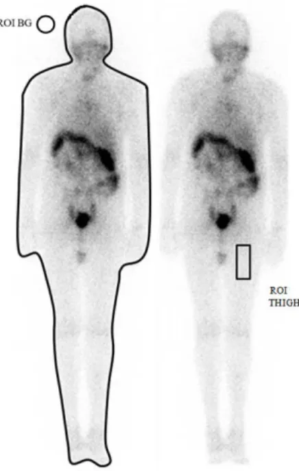
as a function of time. The images obtained at 4, 24, 48, 72 and 96 h after administration of the radioactive material were collected from the database of the Nuclear Medicine Service of ICESP and analyzed using the ImageJ® software, version 1.45s (Wayne Rasband, National Institutes of Health; Bethesda, MD, USA). This software permits creating ROIs in different shapes and sizes, providing the number of counts per pixel in the area of the drawn ROI. As the study objective was to analyze the possibility of restricting ROI only to a region of the thigh, the images were analyzed in two steps: in the first, a ROI was designed around the patient's whole body (WB); and in the second, a ROI was drawn in a patient's thigh region, as exemplified in Figure 1.
In both steps, in addition to acquiring the number of counts per pixel from the patient's body, there was also acquisition of the number of counts per pixel from background radiation (BG), that was subtracted from the number of counts per pixel of the patient's image, according to Equation 1:
WB net counts = WB pixel number x [(WB counts/pixels) - (BG counts/pixel)] (1)
WB net counts represents the net value of the number of counts of the patient's whole body; WB number pixels represents the total number of pixels within the ROI, WB counts/pixels is the number of counts per pixel of the whole body ROI, and BG counts/pixels is the number of counts per pixel present in the BG ROI. For the thigh region, we have the following Equation 2:
Thigh net counts = thigh pixel number x [( thigh counts/pixels) - (BG counts/pixel)] (2)
Thigh net counts represents the net value of the number of counts of the patient's thigh region; thigh pixels number represents the number of pixels within the drawn ROI, thigh counts/pixels the number of thigh region ROI counts per pixel, and BG counts/pixel is the number of counts per pixel present in the BG ROI.
Figure 1: Flat images, exemplifying the design of the ROIs. On the left, whole body ROI (WB); on
the right, thigh ROI.
Source: Prepared by the author.
After the delimitation of ROIs, a simple exponential function was adjusted to describe the variation of activity (𝛢) in the delimited region as a function of time (t) elapsed after administration of the initial activity (𝐴0). For this, it was considered that the net count acquired in the ROI of the
first image would represent 100% of the administered activity, both for the whole body and for the thigh region. This allowed to estimate the biokinetics as well as the effective half-lif
e (
𝑇1/2𝑒𝑓)of131I in the studied region. The Equations 3 and 4 used in this case are below:
𝐴 =𝐴0𝑒−λ𝑡 (3)
Lambda (𝜆) is:
𝜆
=
0.693Another parameter determined was the cumulated activity (Ã) in the region delimited by the ROIs. The cumulated activity represents the amount of atoms that disintegrated in the region, generating radiation dose in the place delimited by the ROIs. The cumulated activity can be determined by the Equation 5:
à = 1.443 𝑥 𝑇1/2𝑒𝑓 𝑥 𝐴0 (5)
Based on these values, it was possible to calculate the absorbed radiation dose in the patient's organism, estimated by OLINDA/EXM software[6]. Dose estimation using this software is
processed by feeding forms with geometric patient data and estimated 131I biokinetics in the body.
One of the parameters entered in the software is the 131I residence time (both for the whole body and for the thigh region) in the patient's body, by the Equation 6:
𝜏= Ã/𝐴0 (6)
In order to obtain a better representation of the dose in the patient's organism, adjustments such as body weight, internal organ mass, such as thyroid and bone marrow were necessary, since the software is based on a standard body model human. In the present study, adjutments to whole body, thyroid, and bone marrow weight were made. In this case, it was considered that 1% of thyroid 131I
uptake is equal to 1 gram of thyroid tissue. The software provided the estimated dose to be received by the bone marrow and whole body per unit of administered 131I activity (mGy/MBq), as shown in
the results of this study.
3. RESULTS
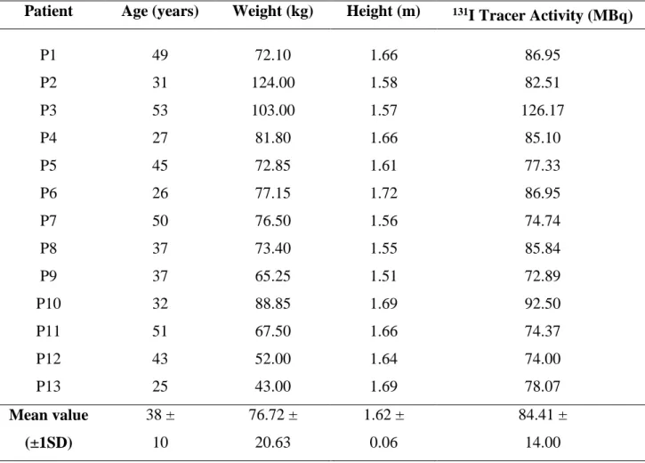
The 13 patients evaluated were female. The age of the patients ranged from 25 to 63 years. The mean ± standard deviation (SD) of the patients' weight was 76.72 ± 20.63 kg and the average height was 1.62 ± 0.06 m. The tracing activity administered ranged from 74 to 126 MBq, with an average
of 84.41 ± 14.00 MBq. The characteristics of the patients and the tracer activity administered are presented in Table 1.
The Table 2 presents the half-life and the residence time of the radioactive material in each patient's organism determined by using the whole body and thigh region quantification. The mean value of half-life and residence time was, respectively, 16.46 ± 3.45 h and 23.76 ± 4.98 h for whole body, compared to, respectively, 14.07 ± 2.89 h and 20.31 ± 4.18 h for thigh region. Both the effective half-life and residence time from whole body quantification were 17% higher than the values obtained when using the thigh area quantification. When evaluating the percentage difference of each patient individually, a greater variation was noted between 2.85% and 54.07% increase.
Table 1: Patient characteristics and 131I activity administered for therapeutic planning.
Patient Age (years) Weight (kg) Height (m) 131I Tracer Activity (MBq)
P1 49 72.10 1.66 86.95 P2 31 124.00 1.58 82.51 P3 53 103.00 1.57 126.17 P4 27 81.80 1.66 85.10 P5 45 72.85 1.61 77.33 P6 26 77.15 1.72 86.95 P7 50 76.50 1.56 74.74 P8 37 73.40 1.55 85.84 P9 37 65.25 1.51 72.89 P10 32 88.85 1.69 92.50 P11 51 67.50 1.66 74.37 P12 43 52.00 1.64 74.00 P13 25 43.00 1.69 78.07 Mean value 38 ± 76.72 ± 1.62 ± 84.41 ± (±1SD) 10 20.63 0.06 14.00
Table 2: Effective half-life (h) and residence time (h).
Patient
Whole body Thigh region Effective half-life (h) Residence time (h) Effective half-life (h) Residence time (h) P1 18.72 27.01 12.15 17.53 P2 15.75 22.72 14.74 21.26 P3 21.65 31.24 14.74 21.26 P4 12.15 17.53 11.74 16.94 P5 18.23 26.30 14.74 21.26 P6 11.00 15.87 11.55 16.66 P7 20.38 29.40 13.32 19.22 P8 13.86 20.00 14.77 21.31 P9 16.50 23.80 17.32 24.99 P10 15.40 22.22 12.60 18.18 P11 11.94 17.22 8.66 12.49 P12 19.25 27.77 19.80 28.57 P13 19.25 27.77 16.90 24.38 Mean value 16.46 ± 23.76 ± 14.07 ± 20.31 ± (±1SD) 3.45 4.98 2.89 4.18
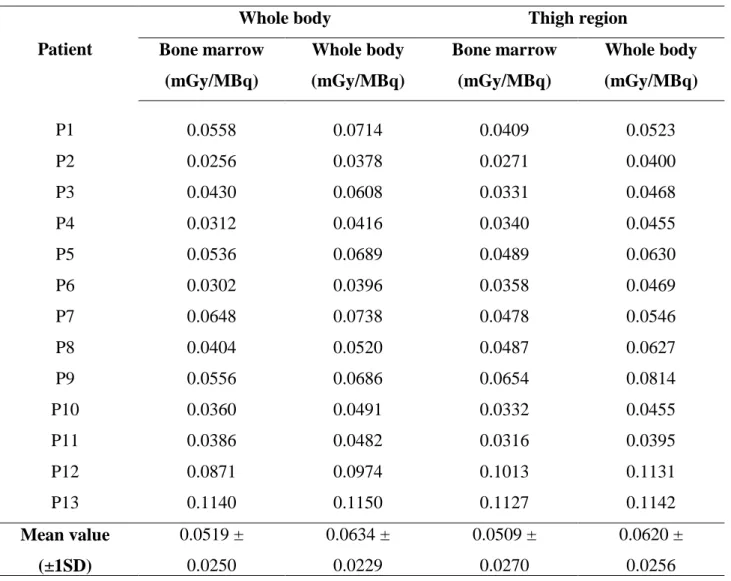
The radiation dose received by the bone marrow and the whole body is presented in Table 3, where the values are according to the dosimetric method employed. The dosimetry based on whole body ROI indicated the average absorbed doses by the bone marrow and whole body of 0.0519 ± 0.0250 mGy/MBq and 0.0634 ± 0.0229 mGy/MBq, respectively. Based on dosimetric method applied in thigh ROI, the average absorbed doses by bone marrow and whole body were 0.0450 ± 0.0239 mGy/MBq and 0.0548 ± 0.0226 mGy/MBq, respectively. It has been observed that the absorbed dose values provided by thigh region dosimetry represent 87% of the absorbed dose value provided by whole body dosimetry. When taking the percentage difference between the values obtained by both dosimetric methods into consideration, it was possible to find an average
correction factor that can be applied to the dosimetry data based on ROI in the thigh region, making absorbed dose values to become similar in the two dosimetric methods. Thus, by adding the correction factor of 13%, which represents the existing difference, in the values provided by thigh region dosimetry, the average absorbed dose by bone marrow and whole body were 0.0509 ± 0.0270 mGy/MBq and 0.0620 ± 0.0256 mGy/MBq, respectively.
Table 3: Bone-marrow and whole-body dosimetry considering both ROI delimitation methods.
Patient
Whole body Thigh region Bone marrow (mGy/MBq) Whole body (mGy/MBq) Bone marrow (mGy/MBq) Whole body (mGy/MBq) P1 0.0558 0.0714 0.0409 0.0523 P2 0.0256 0.0378 0.0271 0.0400 P3 0.0430 0.0608 0.0331 0.0468 P4 0.0312 0.0416 0.0340 0.0455 P5 0.0536 0.0689 0.0489 0.0630 P6 0.0302 0.0396 0.0358 0.0469 P7 0.0648 0.0738 0.0478 0.0546 P8 0.0404 0.0520 0.0487 0.0627 P9 0.0556 0.0686 0.0654 0.0814 P10 0.0360 0.0491 0.0332 0.0455 P11 0.0386 0.0482 0.0316 0.0395 P12 0.0871 0.0974 0.1013 0.1131 P13 0.1140 0.1150 0.1127 0.1142 Mean value 0.0519 ± 0.0634 ± 0.0509 ± 0.0620 ± (±1SD) 0.0250 0.0229 0.0270 0.0256
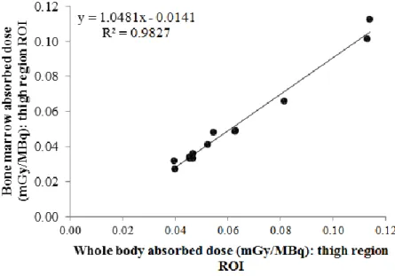
The Figures 2 and 3 illustrate the correlation between doses calculated by both dosimetric methods. Unlike the effective half-life and residence time, the difference between the dose values found was not statistically significant (after correction applied), since the p-value for bone marrow and whole
body absorbed dose was 0.67. The Figures 4 and 5 show the correlation between whole body and bone marrow absorbed dose by whole body and thigh region dosimetry, respectively.
Figure 2: Correlation between bone marrow absorbed dose (mGy/MBq) obtained by whole body
internal dosimetry and thigh region method after the correction factor applied.
Figure 3: Correlation between whole body absorbed dose (mGy/MBq) obtained by whole body
Figure 4: Correlation between bone marrow and whole body absorbed dose (mGy/MBq) by whole
body dosimetry.
Figure 5: Correlation between bone marrow and whole body absorbed dose (mGy/MBq) by thigh
4. DISCUSSION
Since the introduction of internal dosimetry in Nuclear Medicine, a series of studies have been conducted to evaluate the best technique for estimating the dose absorbed by the body of patients undergoing examinations and therapies in the modality[7,8]. Concerning the internal dosimetry for radionuclide therapy, it was observed that the empirically defined activities, sometimes, give more dose than the maximum recommended for bone marrow in elderly patients[9] ; however, this protocol continues to be widely used in Nuclear Medicine Services worldwide.
The internal dosimetry performed using the OLINDA/EXM software was defined as the least invasive method to estimate the absorbed dose by bone marrow and other internal organs, however, this procedure requires patient availability as images are collected at 4, 24, 48, 72, and 96 h after administration of a 131I tracer activity. In addition to this difficulty, there is the need for a trained
professional to collect and analyze the radiometric data. These factors may explain the low attainment of individual internal dosimetry for each therapeutic procedure performed.
During the study, a series of comparisons between the two dosimetry methods were made to verify wheter both provided similar data and what would be the advantage of restricting the region of interest for dosimetric data collection. The choice of a thigh region was based on the idea that to estimate bone marrow irradiation from the blood, a soft tissue compartment would be sufficient to estimate circulating activity. The selected patients had already been submitted to the maximum-safe-dose procedure, so their imaging history was available in the database of our Nuclear Medicine Service, making this retrospective study possible. All patients included in the present study had metastases in the cervical region while no distant metastases were observed. This feature may affect dosimetric data, as the presence of metastases may increase the counts/pixel proportion in the region where the ROI is drawn.
This high number of counts/pixel is a factor to be considered when performing the dosimetric protocol with a thigh region as ROI, since the absorbed dose in bone marrow and whole body based on dosimetry of this region was similar to the absorbed dose obtained by whole body dosimetry only by applying an adjustment factor that took into account the percentage difference between the two. The correction factor applied (13%) is within the uncertainty value already established in the internal dosimetry procedures, whose coefficient of variation reported in the literature is 20%[10]. In
consequently, the estimated absorbed dose, it is suggested that further studies be performed with patients who do not have metastases. Increasing the number of patients is also suggested, as it may bring more satisfactory statistical results.
Radiometric data collection time - insertion and measurement of whole body ROIs in the five images - took an average of 7.2 minutes; when compared to the time taken to perform the same procedure, however, with rectangular ROI measurement only in the thigh region, the time spent was 24 seconds, representing a reduction of almost 95% in the procedure time. By adding the time required to enter dosimetric data into the OLINDA/EXM software (1.5 min), the percentage difference between the two methods continues to represent a significant decrease in the total procedure time, about 80%. The optimization of the time to perform the procedure may represent an encouraging factor for the implementation of dosimetric protocol in the clinical routine.
5. CONCLUSION
Analyzing the dosimetry data obtained through internal dosimetry with whole body ROI and ROI in the thigh region, it was possible to identify that the internal dosimetry by ROI in the thigh provides a dose estimate (mGy/MBq) similar to that estimated with the delimitation of whole body ROI when a correction factor is applied to the data obtained from the thigh region, that is within the uncertainties associated with internal dosimetry in Nuclear Medicine. Even presenting correlation between the data obtained through the two ROIs, we suggest further studies with a larger group of patients, which could increase the level of reliability of the dosimetric method using ROI in the thigh region.
REFERENCES
[1] WILLEGAIGNON J, Princípios básicos da terapia com radionuclídeos. In: Hironaka F, Ono C, Buchpiguel C, Sapienza M, et al. Medicina Nuclear – princípios e aplicações. São Paulo: Atheneu, 2017. Cap 21:557-566.
[2] SAPIENZA M, ENDO I, NETO G, TAVARES M, et al. Tratamento do carcinoma diferenciado da tireóide com iodo-131: intervenções para aumentar a dose absorvida de radiação. Arq Bras
Endocrinol Metab. 2005; 49:341-349.
[3] LUSTER M, CLARKE S, DIETLEIN M, LASSMANN M, et al. Guidelines for radioiodine therapy of differentiated thyroid cancer. Eur J Nucl Med Mol Imaging. 2008; 35:1941–1959.
[4] STABIN M. Fundamentals of Nuclear Medicine Dosimetry. Nova Iorque: Springer; 2008.
[5] WILLEGAIGNON J, PELISSONI R, LIMA B, SAPIENZA M, et al. Estimating 131I biokinetics and radiation doses to the red marrow and whole body in thyroid cancer patients: probe detection versus image quantification. Radiol Bras. 2016; 49: 150-157.
[6] STABIN M, SPARKS R, CROWE E. OLINDA/EXM: the second-generation personal computer software for internal dose assessment in nuclear medicine. J Nucl Med. 2005; 46:1023-1027.
[7] WILLEGAIGNON J, SAPIENZA M, BUCHPIGUEL C. Comparison of different dosimetric methods for red marrow absorved dose calculation in thyroid cancer therapy. Radiation Protection
Dosimetry. 2012; 149:138-146.
[8] CHIESA C, CASTELLANI M, VELLANI C, ORUNESU E, et al. Individualized dosimetry in the management of metastatic differentiated thyroid cancer. QJ Nucl Med Mol Imaging. 2009; 53:546-561.
[9] TUTTLE R, LEBOEUF R, ROBBINS R, QUALEY R, et al. Empiric radioactive iodine dosing regimens frequently exceed maximum tolerated activity levels in elderly patients with thyroid cancer. J Nucl Med. 2006; 47:1587-1591.
[10] SPIELMANN V, LI B, ZANKL M, OEH U, HOESCHEN C. Uncertainty quantification in internal dose calculations for seven selected radiopharmaceuticals. J Nucl Med. 2016; 57:122-128.