Original Article
Preliminary Dosimetry Study of
67Ga-AATS for Human Based on
Biodistribution Data in Rats
Hassan Yousefnia1*, Samaneh Zolghadri1
, Amir Reza Jalilian1
Abstract
Introduction
Gallium-67 (67Ga) has been used as a radionuclide for imaging a variety of solid tumors since 1969. Since then use of various gallium-based radiotracers has been reported. Recently, 67Ga-labeled acetylacetate bis(thiosemicarbazones) (67Ga-AATS) complex with significant tumor accumulation and fast blood clearance has been employed.
Materials and Methods
In this study, the absorbed dose of 67Ga-AATS in each human organ was evaluated and compared with 67 Ga-citrate as the most commonly used form of 67Ga in nuclear medicine. 67Ga was produced via 68Zn(p,2n)67Ga
reaction at 30 MeV cyclotron. Moreover, 67Ga-AATS was produced by adding 50 µl of AATS to absolute
ethanol (1 mg/ml) in a gallium-containing vial at 80-90 °C. The absorbed dose of each human organ was calculated, using RADAR method, based on biodistribution data in Wistar rats.
Results
According to the results, 67Ga-AATS was produced with radionuclidic and radiochemical purity higher than
99% and 93%, respectively. The highest absorbed dose was reported in the bone surface (0.401 mGy/MBq), whereas the whole-body absorbed dose was 0.092 mGy/MBq.
Conclusion
The absorbed dose of each human organ was comparable with the absorbed dose received by each organ after 67Ga-citrate injection. Considering this interesting finding and the significant tumor uptake, it seems
that 67Ga-AATS can be used as an appropriate SPECT tracer.
Keywords: Gallium-67, Radiation Dose, Dosimetry
1- Nuclear Sciences and Technology Research Institute (NSTRI), Tehran, Iran
1. Introduction
Over the past few decades, a large number of experimental studies have evaluated medical radioisotope production [1]. Today, with significant advances in radioisotope production, radiopharmaceuticals play an important role in diagnostic and therapeutic measures for various cancers [2]. However, the sensitivity of tumor-imaging procedures mainly depends on the considerable affinity of radiopharmaceuticals for malignant tissues, compared to normal tissues. Also, the physical properties of radionuclides are regarded as the first parameter which should be considered. The suitable physical properties and availability of gallium-67 (67Ga) contribute to its significance in radiopharmaceutical research [3]. While this radionuclide has been used for imaging a variety of solid tumors since 1969, it has shown great applicability in the management of patients with lymphoma, as well. So far, various gallium-based radiotracers have been reported [4-6] including acetoacetate gallium-67 complex as a potential radiopharmaceutical [7].
Thiosemicarbazone gallium complexes have shown interesting in vitro and in vivo anti-proliferative activities [8]. Generally, traditional bis-thiosemicarbazones such as diacetyl-bis(N4-methylthiosemicarbazone) (ATSM) and pyruvaldehyde-bis(N4-methylthiosemicarbazone) (PTSM) do not form complexes with gallium due to various chemical and molecular orbital considerations. However, the use of 67Ga-labeled acetylacetate bis(thiosemicarbazones) (67Ga-AATS) complex has been recently reported in imaging procedures [9]. This complex with significant tumor accumulation and fast blood clearance can be considered as a potential SPECT radiotracer for imaging malignancies.
One of the important parameters in developing new radiopharmaceuticals is the dose delivered to non-target organs. In fact, any extra radiation dose can damage healthy tissues and result in serious complications. It is generally accepted that even a 10% reduction in patient dose is of high significance [10].
The main aim of patient dosimetry is the evaluation of risks associated with the administration of radiopharmaceuticals and maximum measures which need to be considered [11]. Today, in nuclear medicine, the most commonly used method for the calculation of internal dose estimates is the Radiation Dose Assessment Resource (RADAR) method [12].
While 67Ga-AATS with significant tumor accumulation has been successfully produced, evaluating the absorbed dose of this complex by each human organ is the next step for introducing this complex as a new radiopharmaceutical for diagnostic purposes. Therefore, in this study, the absorbed dose of 67Ga-AATS in each human organ was
evaluated, based on biodistribution data in rats, using RADAR method. Furthermore, since 67Ga citrate is the most commonly used form
of 67Ga in nuclear medicine and is known as a good diagnostic agent in patients with lymphoma (or other malignancies), the absorbed dose of 67Ga-AATS was compared with 67Ga-citrate absorption.
2. Materials and Methods
20*20 cm, TLC Ready Foil, Schleicher & Schuell, Germany).
Normal saline and sodium acetate used for labeling had high purity and were passed through 0.22 mm Cativex filters. Instant TLC (ITLC) was performed by counting Whatman No. 2 papers, using a TLC scanner (Bioscan AR2000, Bioscan Europe Ltd., France). Analytical high-performance liquid chromatography (HPLC), used to determine the specific activity of the samples, was performed by Shimadzu LC-10AT, armed with two detector systems, a flow scintillation analyzer (Packard-150 TR) and a UV-visible spectrophotometer (Shimadzu), using a Whatman Partisphere C-18 column, 250 × 4.6 mm (Whatman, NJ, USA).
Biodistribution data were acquired by counting normal saline-washed tissues after weighing them on a Canberra High-purity Germanium (HPGe) detector (model GC1020, 7500SL); radionuclidic purity was evaluated by the same detector. For the activity measurement of the samples, CRC Capintec Radiometer (NJ, USA) was employed.
All calculations and ITLC counts were based on 184 keV peak. Animal studies were performed in accordance with the guidelines proposed by the United Kingdom Biological Council on the Use of Living Animals in Scientific Investigations (2nd edition).
1.1. Production and quality control of 67
GaCl3
67Ga was produced, using 30 MeV cyclotron
by the Nuclear Medicine Research Group. Also, 68Zn(p,2n)67Ga was used as the best nuclear reaction for the production of 67Ga. Other impurities were removed during the radiochemical separation process. The final 67Ga was assessed in terms of any possible
radionuclidic or radiochemical impurity.
1.2.Production of 67Ga-AATS
AATS was prepared by making slight modifications in the reported method [13]. The labeling procedure was performed according to the previously mentioned method [9]. Briefly, 50 µl of AATS was added to absolute ethanol (1 mg/ml) in a gallium-containing vial and vortexed at 80-90 °C for 30 minutes. The
active solution was evaluated in terms of radiochemical purity by ITLC and HPLC. The content of the vial with maximum radioactivity was diluted to a 5% solution by adding normal saline, followed by passing through a 0.22 mm filter; also, pH was adjusted to 5.5-7.
1.3. Biodistribution of 67Ga-AATS in wild-type rats
For this purpose, 35 µCi of 67Ga-AATS, measured by counting the activity-included syringe before and after the injection in a dose calibrator (with fixed geometry), was injected in each rat. The animals were sacrificed by CO2 asphyxiation at selected time intervals after injection (0.5, 1, 2, 24, and 48 hours). The tissues (blood, heart, lung, brain, intestine, faeces, skin, stomach, kidney, liver, muscle and bone tissues) were weighed and rinsed with normal saline. The specific activities of the tissues were determined as the percentage of the injected dose per gram of tissue, using a high-purity germanium (HPGe) detector, equipped with a sample-holder device.
1.4. Dosimetric studies
The absorbed dose by each human organ was calculated by RADAR method, based on biodistribution data in rats. The accumulated activity in animals was extrapolated to the accumulated activity in humans, using the method proposed by Sparks and colleagues (equation 1) [14]:
Ahuman organ= Aanimal organ
human human
animal animal
OrganMass / BodyMass
OrganMass / BodyMass (1) where à is the accumulated activity in the source organ, which can be calculated by equation 2:
à = ∫ �∞1 A t dt (2) It should be noticed that A(t) is the activity of each organ at time (t).
The curves were extrapolated to infinity by fitting the tail of each curve to a mono-exponential curve with an mono-exponential coefficient equal to the physical decay constant of 67Ga. Afterwards, the area under the curve was calculated. In order to extrapolate the accumulated activity in humans, the mean weight of each organ in a normal human was calculated (Table 1) [12].
Table 1. The mean weight of each organ in an individual with standard weight
Organ Weight (g)
Bone 5500
Heart 330
Stomach 150 Kidneys 310 Small intestine 650
Spleen 150
Muscle 29000
Liver 1800
Lung 500
Brain 1420
Whole body 73000
The radiation absorbed dose was calculated by RADAR formula (12):
= � × where N is the number of disintegrations occurring in a source organ and DF is:
=� ∑� � � ��
DF represents the physical decay characteristics of the radionuclide, the range of emitted radiations and the organ size and configuration, expressed in mGy/MBq.s [15]. It should be mentioned that the DFs were obtained, using OLINDA/EXM software (Vanderbilt University, USA) [16].
3. Results
1.1. Production and quality control of 67
GaCl3
Gallium-67 was prepared as 67GaCl3 by proton bombardment (24 MeV) of 68Zn target at a current intensity of 170 mA and charge of 1400 mAh. The chemical separation process was based on a no-carrier-added method. Radiochemical separation was performed by two-step ion exchange chromatography, with a yield higher than 95%. Radionuclidic purity was evaluated by HPGe detector (higher than 99%).
1.2. Preparation and quality control of 67
Ga-AATS
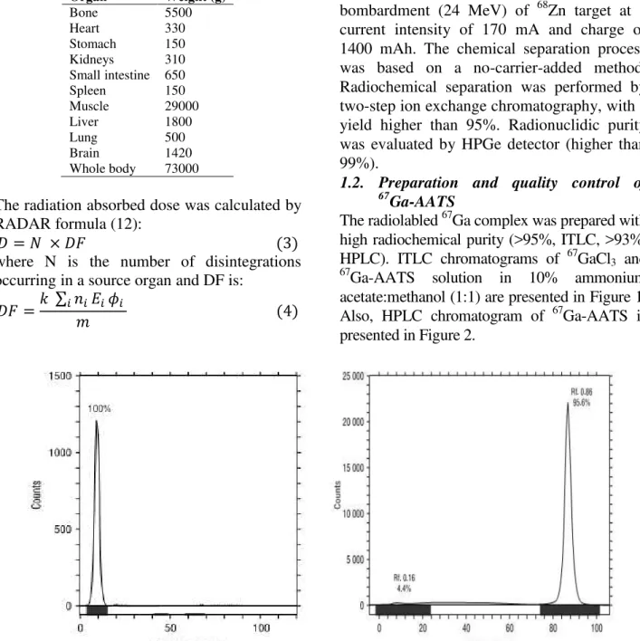
The radiolabled 67Ga complex was prepared with high radiochemical purity (>95%, ITLC, >93%, HPLC). ITLC chromatograms of 67GaCl3 and 67Ga-AATS solution in 10% ammonium
acetate:methanol (1:1) are presented in Figure 1. Also, HPLC chromatogram of 67Ga-AATS is presented in Figure 2.
Figure 1. ITLC of [67Ga]GaCl
3 (left) and [67Ga]-ATSS (right) on Whatman No. 2 paper, using 10% ammonium
Figure 2: HPLC chromatograms of [67Ga]AATS solution on a reversed-phase column, using acetonitrile + 0.1% TFA/water + 0.1% TFA, 90:10
1.3.Biodistribution of 67Ga-AATS in wild-type rats
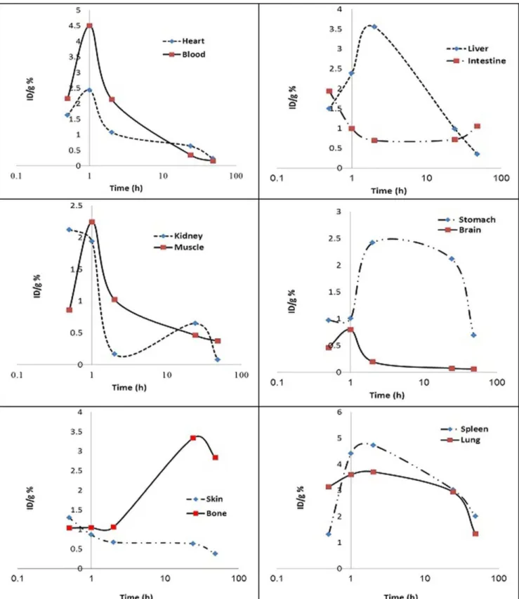
The animals were sacrificed by CO2 asphyxiation at selected time intervals after injection (0.5, 1, 2, 24, and 48h). Dissection
started by drawing blood from the aorta, followed by removing the heart, spleen, muscle, bone, kidney, liver, intestine, stomach, lung and skin samples. The tissue uptake was calculated as the percentage of the area under the curve (of the related photo peak) per gram of the tissue (%ID/g).
1.4. Dosimetric studies
Dosimetric evaluation in human organs was performed by RADAR method, based on biodistribution data in rat organs. The clearance curves of each organ of the rats are shown in Figure 3. Also, the absorbed dose in each human organ after 67Ga-AATS injection is presented in Table 2. Since 67Ga-citrate is a common Ga-67 imaging agent, the calculated dose received by various human organs after 67Ga-citrate injection - is provided in this table,
as well [17].
Table 2. The absorbed dose by each human organ after the injection of 67Ga-AATS and 67Ga-citrate
Tissues
Absorbed dose (mGy/MBq)
67
Ga-AATS
Absorbed dose (mGy/MBq)
67
Ga-citrate
Tissue
Absorbed dose (mGy/MBq)
67
Ga-AATS
Absorbed dose (mGy/MBq)
67
Ga-citrate
Adrenals 0.167 0.13 Muscles 0.081 0.060
Brain 0.073 0.057 Ovaries 0.043 0.082
Breasts 0.039 0.047 Skin 0.067 0.045
Gallbladder wall 0.110 0.082 Pancreas 0.062 0.081
Small intestine 0.073 0.069 Bone surfaces 0.401 0.63
Stomach 0.089 0.059 Spleen 0.117 0.14
Red marrow 0.170 0.21 Testes 0.092 0.056
Cardiac wall 0.054 0.069 Thymus 0.037 0.061
Kidneys 0.112 0.12 Thyroid 0.058 0.062
Liver 0.125 0.12 Uterus 0.086 0.076
Lungs 0.058 0.063 Total body 0.092 0.10
Reference The present study
[16] Reference The present
study
4. Discussion
In this study, 67Ga was produced with a radionuclidic purity of higher than 99%. Total labeling procedure for 67Ga -AATS was performed for only 40 minutes and the radiochemical purity of the complex was higher than 93%. The absorbed dose of 67Ga -AATS has not been reported, so far, despite the use of this complex and the direct relationship between the absorbed dose and the effect of radiopharmaceuticals on disease management.
In this study, the absorbed dose of 67Ga-AATS in each human organ was evaluated for the first time and was compared with 67Ga-citrate as the most commonly used form of 67Ga in nuclear medicine. The dosimetric evaluation of the complex, which was performed based on biodistribution data in Wistar rats, demonstrated the highest absorbed dose in bone surfaces (0.401 mGy/MBq). Also, the maximum absorbed dose for 67Ga-citrate was obtained in bone surfaces (0.63 mGy/MBq). Other organs with considerable absorbed doses included the red marrow, kidneys, liver and spleen with 0.170, 0.112, 0.125 and 0.117 mGy/MBq, respectively. The whole-body absorbed dose after the injection of 67 Ga-AATS was 0.092 mGy/MBq, which is comparable to 0.10 mGy/MBq for 67Ga-citrate.
As presented in Table 2, the absorbed dose of each human organ after the injection of 67Ga -AATS was lower than the maximum absorbed dose, proposed by Food and Drug Administration (FDA) [18] and was comparable with the absorbed dose, received by each organ after the injection of 67 Ga-citrate as a common radiopharmaceutical. Considering this interesting finding and the significant tumor uptake, which has been demonstrated in previous literature, it seems that 67Ga -AATS can be used as an appropriate SPECT tracer.
5. Conclusion
Calculation of the absorbed dose of the tracer showed that human organs absorbed dose was comparable with the absorbed dose received by each organ after 67Ga-citrate injection. Considering this interesting finding and the significant tumor uptake of the radiolabelled compound, it seems that 67Ga-AATS can be used as an appropriate SPECT tracer.
Acknowledgments
The authors wish to thank Nuclear Science and Technology Research Institute (NSTRI) for its financial support.
References
1. Gul K, Hermanne A, Mustafa M, Nortier F, Oblozinsky P, Qaim S, et al. Charged particle cross-section database for medical radioisotope production: diagnostic radioisotopes and monitor reactions, IAEA-TECDOC-1211. Vienna: IAEA; 2001.
2. Therapeutic applications of radiopharmaceuticals, IAEA-TECDOC-1228. Vienna: IAEA; 1999. 3. Firestone RB, Shirley VS, Baglin CM, Zipkin J. Table of isotopes. 8th ed. New York: John Wiley and
Sons; 1996.
4. Tsang BW, Mathias CJ, Fanwick PE, Green MA. Structure-distribution relationships for metal-labeled myocardial imaging agents: comparison of a series of cationic gallium (III) complexes with hexadentate bis(salicylaldimine) ligands. J Med Chem. 1994;37(25):4400–6.
5. Green MA, Mathias CJ, Neumann WL, Janik M, Deutsch EA. Potential gallium-68 tracers for imaging the heart with PET: evaluation of four gallium complexes with functionalized tripodal tris(salicylaldimine) ligands. J Nucl Med. 1993; 34(2):228–33.
6. Tsang BW, Mathias CJ, Green MA. A gallium-68 radiopharmaceutical that is retained in myocardium: 68Ga[(4,6-MeO2sal)2BAPEN]. J Nucl Med. 1993; 34(7):1127–31.
8. Arion VB, Jakupec MA, Galanski M, Unfried P, Keppler BK. Synthesis, structure, spectroscopic and in vitro antitumour studies of a novel gallium(III) complex with 2-acetylpyridine (4)N-dimethylthiosemicarbazone. J Inorg Biochem. 2002; 91(1):298-305.
9. Jalilian AR, Yousefnia H, Garousi J, Novinrouz A, Rajamand AA, Shafaee K. The development of radiogallium-acetylacetonate bis(thiosemicarbazone) complex for tumour imaging. Nucl Med Rev Cent East Eur. 2009; 12(2):65–71.
10. Pernicka F, McLean ID. Dosimetry in diagnostic radiology: an international code of practice, technical report series No.457. Vienna: IAEA; 2007.
11. Stabin MG, Tagesson M, Thomas SR, Ljungberg M, Strand SE. Radiation dosimetry in nuclear medicine. Appl Radiat Isot. 1996; 50(1):73-87.
12. Stabin MG, Siegel JA. Physical models and dose factors for use in internal dose assessment. Health Phys. 2003; 85(3):294-310.
13. Gingras BA, Suprunchuk T, Bayley CH. The preparation of some thiosemicarbazones and their copper complexes, Part III. Can J Chem. 1962; 40(6):1053–7.
14. Sparks RB, Aydogan B. Comparison of the effectiveness of some common animal data scaling techniques in estimating human radiation dose. Sixth International Radiopharmaceutical Dosimetry Symposium. Oak Ridge, TN: Oak Ridge Associated Universities; 1996. P. 705–716.
15. Bevelacqua JJ. Internal dosimetry primer. Radiat Prot Manage. 2005; 22(5): 7-17.
16. Stabin MG, Sparks RB, Crowe E. OLINDA/EXM: the second-generation personal computer software for internal dose assessment in nuclear medicine. J Nucl Med. 2005; 46(6):1023-7.
17. OLINDA - Organ Level Internal Dose Assessment Code (Version 1.1) (2007), copyright Vanderbilt University.
18. Radiation dose to patients from radiopharmaceuticals (addendum 2 to ICRP publication 53). Ann ICRP. 1998;28(3):1-126.