O
RIGINALA
RTICLE Revista Brasileira de FisioterapiaAnti-inflammatory action of the
Ovis aries
lipidic fraction associated to therapeutic
ultrasound in an experimental model
of tendinitis in rats (
Rattus norvegicus
)
Ação anti-inflamatória da fração lipídica do Ovis aries associado ao ultrassom
terapêutico em modelo experimental de tendinite em ratos (Rattus norvegicus)
Marcelino Martins1,2, Antonio L. M. Maia Filho1,2, Charllyton L. S. Costa1, Nayana P. M. F. Coelho1,2, Maricilia S. Costa3, Regiane A. Carvalho4
Abstract
Background: Studies have demonstrated the beneficial effects of topical application of fatty acids as healing agents. The lipid fraction of Ovis aries have an anti-inflammatory action that accelerates the healing process. Ultrasound increases blood flow and the extensibility of collagen structures and tendons. Objectives: To assess the anti-inflammatory action of the Ovis aries lipid fraction associated to pulsed therapeutic ultrasound and friction in an induced tendinitis model. Methods: Fifty Wistar rats were divided into four groups: control that consisted of
Ovis aries gel for topical use; pulsed ultrasound plus oil free sterile lotion; pulsed ultrasound plus Ovis aries gel; and oil free sterile lotion for topical use alone. To induce tendinitis a 10μL intratendinous injection of collagenase was injected into the right Achilles tendon of rats. Treatment consisted of daily applications of ultrasound using the following parameters: 10% pulsed mode, 10% pulsed frequency of 1 MHz and intensity of 0.5 W/cm² for seven or fourteen days. Results: After 7 days of treatment, only the Ovis aries plus ultrasound group showed statistically significant difference when compared to the control group.The variation in the number of inflammatory cells on animals treated for fourteen days for the control, ultrasound plus oil free, ultrasound plus Ovis aries, Ovis aries plus massage and massage plus oil free groups were statistically significant different, p<0.01. It was observed in animals treated for seven days that the ultrasound plus Ovis aries group was statistically significant better than the control group, p<0.05. Conclusion: It can be concluded that treatment using ultrasound plus Ovis aries
is more effective than other treatments as it produces significantly better reduction on the number of inflammatory cells at 7 and 14 days.
Keywords: physicaltherapy; rehabilitation; movement; ultrasound; inflammation; tendinitis.
Resumo
Contextualização: Estudos demonstram o efeito benéfico da aplicação tópica de ácidos graxos como agentes cicatrizantes. A fração lipídica do Ovis aries apresenta uma ação anti-inflamatória que acelera o processo de cicatrização. O ultrassom aumenta o fluxo sanguíneo bem como a extensibilidade das estruturas de colágeno e tendões. Objetivos: Analisar a ação anti-inflamatória da fração lipídica do Ovis aries associado ao ultrassom terapêutico (UST) pulsado e à fricção em modelo de tendinite induzida. Métodos: Cinquenta ratos Wistar foram distribuídos nos seguintes grupos: controle, gel Ovis aries – uso tópico – UST pulsátil + loção estéril (oilfree), UST pulsátil + gel Ovis aries, loção estéril (oil free) – uso tópico. Para induzir a tendinite, utilizou-se uma injeção intratendínea de 10µL de colagenase no tendão do calcâneo direito. O tratamento consistiu em aplicações diárias de ultrassom, com os seguintes parâmetros: modo pulsado 10%, frequência de 1 MHz, pulsátil a 10% com intensidade de 0,5W/cm2, durante sete ou 14 dias. Resultados: A variação do número de células inflamatórias, para os animais tratados por 14 dias, com relação aos grupos controle, UST + oil free e UST +
Ovis aries, apresentou resultados significativos p<0,001. O grupo Ovis aries + massagem e o grupo massagem + oilfree apresentaram resultados significativos, p<0,01. Nos animais tratados por sete dias, observou-se que o grupo UST + Ovis aries, em relação ao controle, é estatisticamente significativo, p<0,05. Conclusão: Pode-se concluir que o tratamento com UST + Ovisaries é mais efetivo que os outros tratamentos, visto que consegue reduzir o número de células inflamatórias no tempo de sete e 14 dias.
Palavras-chave: fisioterapia; reabilitação; movimento; ultrassom; inflamação; tendinite.
Received: 09/06/2010 – Revised: 07/12/2010 – Accepted: 26/04/2011
1 Collegiate of Physical Therapy, Faculdade Integral Diferencial (FACID), Teresina, PI, Brazil
2 Universidade Estadual do Piauí (UESPI), Teresina, PI, Brazil
3 Universidade Vale do Paraíba (UNIVAP), São José dos Campos, SP, Brazil
4 Universidade Nove de Julho (UNINOVE), São Paulo, SP, Brazil
Correspondence to: Marcelino Martins, Coordenação de Fisioterapia, FACID, Av. Rio Poty, 2381, Horto Florestal, CEP 64049-410, Teresina, PI, Brasil, e-mail: [email protected]
Introduction
Tendon injuries are common in sports practice and it is assumed that it corresponds from 30 to 50% of total injuries1.
Such injuries are major health problem in industrialized coun-tries because current occupations often demand a continuous series of repetitive movements2. Tendons are ibrous structures
with cylindroid edges or edges shaped as resistant tapes and made of dense connective tissue3. Tendon injury can occur due
to various factors, such as overload, when the athlete makes an efort beyond his/her capacity, or repetitive efort of the same movement, that can cause an inlammatory process. Tendon overuse may lead to degenerative changes afecting the prac-tice of activities because of the development of calciications on the tendon sheath4.
Fatty acids are compounds containing a long hydrocarbon chain and a carboxyl terminal cluster and have three main func-tions on the human body. hey are structural component of bio-logical membranes; they play the role of intracellular precursors of messages and, when oxidized, they generate energy - ATP (adenosine triphosphate)5,6. here are several studies
demon-strating the beneicial efects of topical application of fatty acids in the treatment of wounds. hey have low cost and are widely used as healing agents as part of the popular culture of diferent countries; and have the property to serve as a protective barrier against micro-organisms, preventing tissue dehydration, beyond the important immunomodulator character7.
herapeutic ultrasound (US) is a non-invasive treatment acting on the repair of tissue injury. Pulsed US is the US mo-dality most commonly chosen by researchers because of the beneicial efects, especially, of its low intensities8. he thermal
efect of ultrasound generated by tissue intermolecular friction occurs by agitation of the interstitial luids’ electrolyte environ-ment that is composed of water and solutes. his thermal efect generated by continuous ultrasound wave is contraindicated in acute inlammatory processes, recent traumas, ischemic areas or on sensitivity disorders. he ultrasound response on the pulse mode have a decreased thermal efects and can be used in acute and subacute inlammation, neuropathic pain and swelling9.
he ultrasound favors the transcutaneous penetration of various substances in animals such as corticosteroids, dexam-ethasone, hydrocortisone and indomethacin anti-inlamma-tory drug; and in healthy humans, such as methyl nicotinate vasodilator10,11. he phonophoresis is a favorable alternative
since it is a non-invasive technique. he thermal and mechani-cal efects of the US lead to physimechani-cal and chemimechani-cal changes of biological tissues, favoring the penetration of active principles present in topical substances. he heating of the area be-ing treated may increase the absorption of the drug with the
increase in blood low, dilation of hair follicles, decrease in skin resistance and increase in the kinetic energy of the drug. he mechanical efects of US are present even if the parameters are set to produce heating12.
Some authors have shown that US is capable of increasing the penetration of some drugs applied topically. his action of the US associated with drugs has promoted researches in various ields of Medicine13. Researches conducted in recent
decades have demonstrated that tendon injuries are a major cause of sufering of manual workers and of workers compen-sation claims2. he high incidence of this condition is due to
the current work environment high productivity and quality demands that overcome the workers health demands. In most cases, the condition develop as consequence of a lack of control of rhythm and speed of movements associated with machinery and furniture ergonomically incorrect14.
he aim of the study was to histologically analyze the anti-inlammatory action of the Ovis aries lipid fraction applied us-ing US on an experimental model of tendinitis in rats.
Methods
Fifty adult male rats, of the Rattus norvegicus specie, Wistar variety (200-250g), aged over 30 days, were included in this study. All rats were allowed to eat and drink water as desired. Half rats (25) received treatment for 7 days and the other half received treatment for 14 days. he experiment was conducted at the Laboratory of Physiology of the Faculdade Integral Diferencial (FACID), Teresina, PI, Brazil. he study was approved by the Ethics Committee of the FACID under the protocol nº 492/2008, on 11/11/2008, according to resolution nº 196/96, of 10 October 1996, of the National Health Council (CNS).
Experimental groups
he animals from the 7 and 14 treatment groups were ran-domly divided into ive subgroups, with a total of ten animals per subgroup:
• Group 1: control;
• Group 2: Ovis aries gel, topical use (massage);
• Group 3: US + sterile lotion (oil free); • Group 4: US + Ovis aries gel;
• Group 5: sterile lotion (oil free), topical use (massage).
Tendinitis induction
at 10%, associated with the same dose of hydrochloride and xylazine at 2%, intramuscularly, with the help of two collabora-tors to hold the animal.
To induce tendinitis experimentally, an intratendineous
injection of 10 µL of collagenase (10mg/ml; SIGMA; C6885) was applied in the right Achilles tendon using a 30G needle.
Collagenase was dissolved in a bufered sterile saline solution of phosphate15.
Preparation of the extract of the
Ovis aries
lipid
fraction
he preparation procedure of the Ovis aries extract de-scribed below was performed at the Laboratory of Natural Products of the Department of Chemistry of the Universidade Federal do Piauí (UFPI), Teresina, PI, Brazil. he fat/protein material obtained from Ovis aries (sheep) was submitted to mechanical manual trituration, yielding 1 kg of triturated material and debris with dimensions not to exceed 2 cm. he fat/protein material was transferred to an extraction bottle and 2 liters of hexane P.A., Vetec brand were added to it. Every two days the material was iltered through preparative ilter paper and a new solvent was added to the material. A total of three extractions were performed and each were kept in room temperature and with low luminosity. At the end of each step of extraction, the iltrate was concentrated under reduced pres-sure at 55° C, in a rotary evaporator. At the end of the solvent concentration process the lipid extract of Ovis aries used in this study was obtained16.
Preparation of the lotion at 5% of
Ovis aries
lipids
A lotion loaded with 5% of lipid extract of Ovis aries was prepared in order to allow a homogeneous application on the injured sites. he oil free lotion was purchased from a com-pounding pharmacy and lipid extract of Ovis aries was added to the oil to establish a concentration of 5% mass/mass. he consistency of the preparation was adjusted by adding sui-cient amount of distilled water. he material was stored in an opaque bottle under refrigeration until use.
Treatment of tendinitis
he ultrasound (Ibramed brand, sonopulse model) was used with the following parameters: 10% pulsed mode, fre-quency of 1 MHz, 10% pulsed with an intensity of 0.5 W/cm2,
direct method of coupling with oscillatory movements in 1 cm2
ERA for 2 minutes. his protocol is in line with recommended parameters for the use of US17,18. he device was properly
calibrated by a specialized company, before and after the pro-posed treatment, in order to verify the maintenance of intensity during treatment19. All animals were treated daily, respecting
the period of seven and 14 days. Treatment was initiated 24 hours after tendinitis induction.
Histological analysis
he animals were sacriiced after treatment respecting the healing period of seven and 14 days. Tendons were dehydrated after ixation, included in parain and then150 ields were pre-pared to be cut in microtome, in a semi-serial manner, with sections of 5 µm in thickness, ive sections per animal, to be stained with hematoxylin-eosin (H&E) and Masson’s trichrome (MT). Histomorphometry was performed using a binocular optical microscope, with the acquisition of photos in a 40X objective, for cellular diferentiation of the total number of in-lammatory cells of the observation ield, intratendineous, us-ing the Image J®
computer program in its cell counter function. It is worth considering that all photos presented in the results had the same increase. Cell diferentiation was not performed but total cells were counted.
Statistical analysis
he collected data were assessed in regards to the coef-icient of variation and the sample distribution to determine the statistical test, considering the signiicance level of 5% (p<0.05)20. A Kolmogorov-Smirnov test demonstrated that data
was normally distributed. Statistical analysis of the variation of the number of inlammatory cells obtained in the treated and untreated groups was conducted, using ANOVA, with Tukey’s Post Hoc Test on the statistical program GraphPad Prism®
, version 3.0. Standard error of measures were used to construct graph error bars.
Results
he results presented on Figure 1for the assessment after 7 days of treatment, demonstrated that the number of inlam-matory cells was statistically signiicant diferent between the group treated with US and Ovis aries when compared to the control group. However, there was no signiicant diference between the other treatment groups and the control group.
he results presented on Figure 2for the assessment con-ducted after 14 days of treatment, showed that the number of inlammatory cells was signiicantly reduced when comparing all groups to the control group.
Figure 1. Graph comparing the variation (Δ) of the number of inflammatory cells after seven days of treatment.
G4 *p<0.05 compared to control group.
Control
Ovis aries + massage UST + Oll free UST + Ovis aries Massage + Oll free
0 50
Number of cells (He-und 7 days)
100 150 200
Figure 2. Graph comparing the variation (Δ) of the number of inflammatory cells after fourteen days of treatment.
Control
Ovis aries + massage UST + Oll free UST + Ovis aries Massage + Oll free
**p< 0.01 in the G1 comparable; ***p< 0.001 in the G1 comparable. 0
50
Number of cells (He-und 14 days)
100 150 200
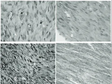
Figure 3. Microscopy of the rat Achilles tendon. Group (3) treated with
pulsed US+ sterile Lotion (Oil free). Photo (A) intratendineous 7 days, stained with H&E and a 40X objective Photo (B) intratendineous 14 days, stained with H&E and a 40X objective, showing large cellularity at the expense of inflammatory cells (1), fibroblasts (2) and edema (3), demonstrating that it was a newly formed granulated tissue, showing a decrease in cellularity due to the decrease of inflammatory cells. The fibroblasts now are predominant and are more ordered (4). Photo (C) intratendineous 7 days, stained with Trichrome to Massom using a 40X objective. Photo (D) intratendineous 14 days, stained with Trichrome to Massom using a 40X objective. The last two photos show the maturation of granulation tissue with increased deposition of extracellular matrix, as seen on photo D compared with C.
Figure 4. Microscopy of the Achilles tendon of rats, group (4) treated
with US + gel Ovis aries. Photo (A) intratendineous 7 days, stained
with H&E and a 40X objective, viewing capillaries (5). Photo (B) intratendineous 14 days, stained with H & E and a 40X objective. Photo (C) intratendineous 7 days, stained with Trichrome to Massom using a 40X objective, viewing the extracellular matrix - elastic tissue (6). Photo (D) intratendineous 14 days, stained with Trichrome to Massom using a 40X objective.
Figure 3, Photo A, stained with H&E shows that for animals treated for seven days with US and sterile lotion there was the presence of maturing granulation tissue, swelling, and newly formed vessels, but in less quantity than in the newly formed granulation tissue, with more organized vessels and prolifera-tion of ibroblasts with deposiprolifera-tion of extracellular matrix (EM). At 14 days of treatment, as shown on Photo B, stained with H&E, there was organizing ibrosis, proliferation of ibroblasts and more organized vessels and in less quantity than in the granulation tissue, but in even greater numbers than in the mature ibrous tissue.
In animals treated for seven days with US and sterile lotion, Photo C stained with TM demonstrated that there was the deposit of EM in a disorganized way, with thinner ibers than in normal conjunctive tissue. In Photo Dstained with TM, for the same patient population after 14 days of treatment there were areas of ibrosis with blue areas (deposited EM) in greater quantities than in the red area (proliferated ibroblasts). he ibrosis was in process of organization.
granulation tissue, but still in less quantity than in mature tis-sue.Photo Cstained with TM of patients treated for seven days
showed thinner and disorganized ibers than in normal con-junctive tissue. For rats treated for 14 days, Photo D stained with TM, showed areas of ibrosis and red areas (proliferated ibroblasts) and organizing ibrosis.
Discussion
herapy with US and Ovis aries showed signiicant better inlammatory process than the control group after treatment for seven days. At 14 days of treatment, it was observed a larger presence of ibroblasts on the treatment groups when com-pared to the control group, with more organized collagen tissue, suggesting the anti-inlammatory action of these therapies.
It has been reported that oleic and linoleic acids present in the Ovis aries lipid fraction can be used as anti-inlammatory agents during the irst phase of the healing process, thus ef-fectively accelerating tissue repair7.
Other studies have reported the positive inluence of topi-cal administration of α-linolenic (n-3), linoleic (n-6) and oleic acids (n-9) in the process of wound healing in rats. It has been observed that from the 5th day of topical treatment with oleic and linoleic acids, there was a signiicant reduction of the wound. In the irst 48 hours there was inhibition of nitric oxide in the site21.
In this study, an important healing action of linoleic and oleic acids, found in the lipid fraction of Ovis aries22,23 was
observed. his healing action could be observed through the number of inlammatory cells as presented on Figure 1. he re-sults obtained showed that there was a decrease in the number of inlammatory cells and an acceleration of the inlammatory process, which is consistent with the literature22,23.
Fatty acids have been shown to accelerate the healing process, acting as chemotactic agents for leukocytes promot-ing angiogenesis as a wound moisturizer24,25.
he efects of US depend on many physical and biological factors, such as: intensity, application time, physiological state of the area to be treated and spatial and temporal structure of the ultrasonic ield26. he identiication of these variables are
necessary for the understanding of the US mechanisms of ac-tion on the biological tissue. Ultrasonic irradiaac-tions have an important role in the cutaneous healing process, accelerating tissue repair and promoting an accelerated healing and scar-ing of the tissue in regards to tissue quality29. hese changes
include increase in the synthesis of protein, mastocytes,
granulation, calcium absorption and mobility of ibroblasts which according to several studies could accelerate the heal-ing process27-30.
he results of the present study showed that a 7 day ultra-sound therapy associated with Ovis aries have signiicantly better anti-inlammatory efect on the acute phase of the in-lammatory process (Figure 1) when compared to the control group. During this period of acute inlammation a vascular response occurs initially, with production of vasoconstriction by the action of norepinephrine and contraction of the en-dothelium followed by vasodilatation and migration of inlam-matory cells (leukocytes and neutrophils) to the injured area. At this time, macrophages remove cellular debris and extracel-lular changed components and ibroblasts initiate the collagen synthesis28-30.
Studies have been conducted to provide a better under-standing of polyunsaturated fatty acids on the immune system and the dynamics of eicosanoids derived from arachidonic acid in the modulation of inlammatory responses and immunity, important for the organism and for the cells31.
he author of a previously published study32 reported the
positive efects of US phonophoresis compared with the efects of topical application of hydrocortisone in the repair of rat’s Achilles tendon after tenotomy. US phonophoresis was found to be the most eicient treatment, and authors concluded that the US stimulates the acceleration of tissue repair and induces transcutaneous penetration of hydrocortisone.
his study assessed the application of the Ovis aries gel with pulsed ultrasound in the acceleration of the inlamma-tion process. Concomitant use of topical anti-inlammatory gel and US is becoming a common practice in rehabilitation services because it facilitates the penetration of substances transcutaneouly13.
Conclusion
he model used on the present study showed that the therapeutic efect of the Ovis aries lipid fraction associated with pulsed ultrasound and friction positively interfered on the healing process of the tendon. here was a statistically signiicant decrease in the number of inlammatory cells for the animals treated for 7 and 14 days when compared to the control group.
Further studies are needed to explain the mechanism of action of ultra sound and phonophoresis and to validate the parameters used in this study.
References
1. Salate ACB. Síndromes por overuse em tendão calcâneo. Fisioter Bras. 2002;3(6):351-5.
2. Regis Filho GI, Michels G, Sell I. Lesões por esforços repetitivos/distúrbios osteomusculares relacionados ao trabalho em cirurgiões-dentistas. Rev Bras Epidemiol. 2006;9(3):346-59.
3. Dangelo JG, Fattini CA. Anatomia humana – sistêmica e segmentar. 3ª Ed. Rio de Janeiro: Atheneu; 2007.
4. Lesic A, Bumbasirevic M. Disorders of the Achilles tendon. Current Orthopaedics. 2004;18:63-75.
5. Curi R, Pompéia C, Mayasaka CK, Procopio J. Entendendo a Gordura: Os ácidos graxos. São Paulo: Manole; 2002.
6. Brasileiro Filho G. Bogliolo: patologia. 7ª ed. São Paulo: Guanabara Koogan; 2006.
7. Hatanaka E, Curi R. Ácidos graxos e cicatrização: uma revisão. Rev Bras Farmacol. 2007;88(2):53-8.
8. Olsson DC, Martins VMV, Pippi NL, Mazzanti A, Tognoli GK. Ultra-som terapêutico na cicatrização tecidual. Ciênc Rural. 2008;38(4):1199-207.
9. Agne JE. Eu sei eletroterapia. 2ª Ed. Santa Maria: Pallotti; 2009.
10. Järvinen TAH, Kääriäinen M, Järvinen M, Kalimo H. Muscle strain injuries. Curr Opin Rheumatol. 2000;12(2):155-61.
11. Polacow MLO, Dib-Giusti HHK, Leonardi GR, Vieira CEC, Guirado GN, Zague V, et al. Efeito do ultra-som e do d-pantenol na regeneração tegumentar. Rev Bras Fisioter. 2005;9(3):365-71.
12. Rosim GC. Análise da Influência do Ultra-Som Terapêutico na Penetração Transcutânea de Diclofenaco Sódico em Humanos Sadios [dissertação]. São Carlos (SP): Universidade de São Paulo; 2003.
13. Jesus GS, Ferreira AS, Mendonça AC. Fonoforese X permeação cutânea. Fisioter Mov. 2006;19(4):83-8.
14. Brasil. Ministério da Previdência Social. Instituto Nacional de Seguro Social e Diretoria do Seguro Social. Ler/Dort Norma Técnica de Avaliação de Incapacidade para fins de Benefícios Previdenciários-INSS. Acesso: 10 de abril de 2010. Disponível em http://www.saude em movimento.com.br/conteúdos.
15. Silva EJ. Espectroscopia Raman e Histologia Clássica na Avaliação de Tendinite Induzida por Colagenase em Ratos Wister [dissertação]: Universidade de Franca; 2005.
16. Brum AAS, Arruda LF, Regitano-d Arce MAB. Métodos de extração e qualidade da fração lipídica de matérias-primas de origem vegetal e animal. Quim Nova. 2009;32(4):849-54.
17. Da Cunha A, Parizotto NA, Vidal Bde C. The effect of therapeutic ultrasound on repair of the achilles tendon (tendon calcaneus) of the rat. Ultrasound Med Biol. 2001;27(12):1691-6.
18. Maia Filho AL, Villaverde AB, Munin E, Aimbire F, Albertini R. Comparative study of the topical application of Aloe vera gel, therapeutic ultrasound and phonophoresis on the tissue repair in collagenase-induced rat tendinitis. Ultrasound Med Biol. 2010;36(10):1682-90.
19. Ng GY, Fung DT. The effect of therapeutic ultrasound intensity on the ultrastructural morphology of tendon repair. Ultrasound Med Biol. 2007;33(11):1750-4.
20. Kupeli E, Tatli II, Akdemir ZS, Yesilada E. Bioassay-guided isolation of anti-inflammatory and antinociceptive glycoterpenoids from the flowers of Verbascum lasianthum Boiss. ex Bentham. J Ethnopharmacol. 2007;110(3):444-50.
21. Cardoso CR, Souza MA, Ferro EA, Favoreto S Jr, Pena JD. Influence of topical administration of n-3 and n-6 essential and n-9 nonessential fatty acids on the healing of cutaneous wounds. Wound Repair Regen. 2004;12(2):235-43.
22. Zapata JFF, Nogueira CM, Seabra LMJ, Barros NN, Borges AS. Composição centesimal e lipídica da carne de ovinos do Nordeste brasileiro. Ciênc Rural. 2001;31(4):691-5.
23. Monteiro EM, Shimokomaki M. Influência do genótipo nos lipídeos totais e na fração insaponificável da carne de cordeiros. Ciênc Rural. 1999;29(3):545-8.
24. Prata MB, Haddad CM, Goldenberg S, Simões MJ, Moura LAR, Trabulsi LR. Uso tópico do açúcar em ferida cutânea: estudo experimental em rato. Acta Cir Bras. 1988;3(2):43-8.
25. Nabas F, Contesini FJ, Menin SEA, Antônio MA, Bighetti AE, Araújo CEP, et al. Efeito antiedematogênico de óleos contendo ácidos graxos ômega-3 e 6 em camundongos. RBM Rev Bras Med. 2009;66(4):92-6.
26. Cândido LC. Nova Abordagem no Tratamento de Feridas. São Paulo: SENAC; 2001.
27. Ferreira AS, Mendonça AC. Ultra-Som Terapêutico nas Lesões Cutâneas: Uma Revisão. Revista FAFIBE On Line 2007 Disponível em: http://carefisioterapia.webs.com/apps/blog/ show/1082639-ultra-som-terap-ico-nas-les-cut-as-uma-revis-o
28. Olsson DC, Martins VMV, Martins E, Mazzanti A. Estimulação ultra-sônica pulsada e contínua no processo cicatricial de ratos submetidos à celiotomia. Ciênc Rural. 2006;36(3):865-72.
29. Watson T. Ultrasound in contemporary physiotherapy practice. Ultrsonics. 2008;48(4):321-9.
30. Kitchen S. Eletroterapia prática baseada em evidências. 11ª ed. São Paulo: Manole; 2003.
31. Mendonça AC, Ferreira AS, Barbieri CH, Thomazine JA, Mazzer N. Efeitos do ultra-som pulsado de baixa intensidade sobre a cicatrização por segunda intenção de lesões cutâneas totais em ratos. Acta Ortop Bras. 2006;14(3):152-7.