From the Institute of Orthopedics and Traumatology, Hospital das Clínicas, Faculty of Medicine, University of São Paulo - São Paulo/SP, Brazil.
Received for publication on September 16, 2003.
ORIGINAL RESEARCH
ANATOMIC STUDY OF THE DORSAL ARTERIAL
SYSTEM OF THE HAND
Marcelo Rosa de Rezende, Rames Mattar Júnior, Álvaro Baik Cho, Oswaldo Hideo Hasegawa and Samuel Ribak
REZENDE MR de et al. - Anatomic study of the dorsal arterial system of the hand. Rev. Hosp. Clín. Fac. Med. S. Paulo 59(2):71-76, 2004.
Historically, the dorsal arterial system of the hand received less attention than the palmar system. The studies concerning dorsal arterial anatomy present some controversies regarding the origin and presence of the dorsal metacarpal artery branches. Knowledge of the anatomy of dorsal metacarpal arteries is especially applied in the surgical planning for flaps taken from the dorsum of the hand. The purpose of this study is to analyze the arterial anatomy of the dorsum of the hand, compare our observations with those of previous studies from the literature, and therefore to define parameters for surgical planning for flaps supplied by the dorsal metacarpal arteries.
METHOD: Twenty-six dissections were performed at the dorsum of the right hand of 26 cadavers by making a distal-based U-shaped incision. After catheterization of the radial artery at the wrist level, a plastic dye solution with low viscosity and quick solidification was injected to allow adequate exposure of even small vessels. The radial artery and its branches, the dorsal arterial arch, the dorsal metacarpal arteries, the distal and proximal communicating branches of the palmar system, and the distal cutaneous branches were carefully dissected and identified.
RESULTS: The distal cutaneous branches originating from the dorsal metacarpal arteries were observed in all cases; these were located an average of 1.2 cm proximal from the metacarpophalangeal joint. The first dorsal metacarpal artery presented in 3 different patterns regarding its course: fascial, subfascial, and mixed. The branching pattern of the radial artery at the first intermetacarpal space was its division into 3 branches. We observed the presence of the dorsal arterial arch arising from the radial artery in 100% of the cases. The distance between the dorsal arterial arch and the branching point of the radial artery was an average of 2 cm. The first and second dorsal metacarpal arteries were visualized in all cases. The third and fourth dorsal metacarpal arteries were visualized in 96.2% and 92.3% of cases, respectively. There was proximal and distal communication between the dorsal arterial arch and the palmar system through the communicating branches contributing to the dorsal metacarpal artery formation.
CONCLUSION: At the dorsum of the hand there is a rich arterial net that anastomoses with the palmar arterial system. This anatomical characteristic allows the utilization of the dorsal aspect of the hand as potential donor site for cutaneous flaps.
KEY WORDS: Arterial system. Dorsal. Hand. Anatomy. Fasciocutaneous flaps.
Many kinds of flaps have been pro-posed for covering the dorsal aspect of the hand1-3. Foucher & Braun’s article
(1979) was the first to describe the flap supplied by the first dorsal metacarpal artery (1DMA)4. In reviewing the
litera-ture regarding the anatomy of the dor-sal arterial system of the hand, we no-ticed some controversy about the
fre-quency of the dorsal metacarpal arter-ies (DMAs)4-8, the location of the distal
cutaneous branches5,9-12, and the
amount of contribution of the dorsal and palmar arterial system to the forma-tion of the DMAs13-15. Since that
METHOD
For the study of dorsal arterial sys-tem of the hand, 26 dissections in 26 cadavers were performed in the Obit Verification Service of our University. All specimens were male, with a mean age of 47.2 years (range 29 to 71 years). The cadavers were chosen ran-domly, and no female were used due to legal restrictions.
The radial artery was identified fol-lowing a 3 cm longitudinal incision made between the flexor carpi radialis and the abductor pollicis longus ten-dons in the volar and radial aspect of the wrist. It was dissected and divided 1 cm proximally to the radius styloid process. A catheter was introduced 2 cm into the distal stump of the vessel and fixed to preclude dye solution leakage. The radial artery and its branches were washed with 20 mL of saline solution followed by manual injection of 6 mL of dye solution—a mixture of lead ox-ide, vinyl, latex, and acetone—in puls-ing manner. This blue solution had low density and proved to be quite effective in depicting even very small vessels. This technique is a modification of the technique described by other authors in previous studies16. Before the
begning of the dissection, there was an in-terval of 10 minutes for solidification of the dye solution. All the dissections were performed by the same author un-der loupes magnification (3.5X).
The dissection was made with a dor-sal U-shaped incision, starting at the transition of the dorsal and palmar skin, at the level of fifth metacarpophalangeal (MCP) joint. Then, the incision turned radially, 1 cm proximal to the ulnar sty-loid and continued transversally across the wrist, turning distally again at the level of radius styloid, ending at the level of the second MCP joint. The distal-based flap was elevated between the subcutaneous tissue and the fascia. The dorsal retinaculum of the second, third, and fourth extensor compartments
was opened longitudinally. The exten-sor pollicis longus, extenexten-sor digitorum communis, and extensor indicis proprius tendons were divided proximally and re-tracted distally. The second compartment tendons were separated from their inser-tions and retracted proximally. At this point, the radial artery and its branches, the dorsal arterial arch (DAA), the DMAs, the distal and proximal communicating branches of the palmar system, and the distal cutaneous branches were carefully dissected and identified. The distance between the emergence of the DAA and the branching point of the radial artery and the distance of the distal cutaneous branches from the MCP joints were meas-ured.
RESULTS
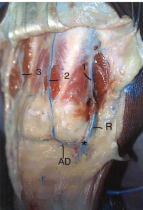
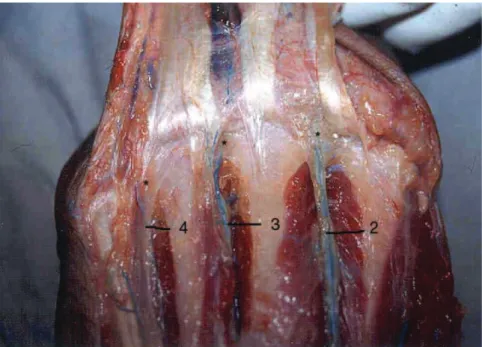
Just distal to the tendinous junc-tures between the extensor tendons, the second, third, and the fourth dorsal metacarpal arteries (DMAs) gave off small branches, the distal cutaneous branches, that perforated the fascia and the subcutaneous plane to supply the dorsal skin of the hand. (Fig. 1). They were identified all DMAs, in 100% of the cases. The distance be-tween the emergence point of these branches (from the DMAs) and the MCP joint of the adjacent ulnar digit averaged 1.2 cm.
The course of the first DMA was fascial in 14 (53.8%) cases and subfascial in 2 (7.7%). It was dupli-cated in 10 cases (38.4%), with one fascial and another subfascial branch in 8 (30.8%) and a double fascial branch in 2 (7.7%). The first DMA was found in the first intermetacarpal space in all specimens, and it arose from the radial artery in all except 1 case, in which it arose from a branch of the ra-dial artery that ran to the dorso-rara-dial aspect of the thumb (Fig. 2).
The radial artery divided into 3 branches between the base of first and
second metacarpals in 22 (84.6 %) specimens: the princeps pollicis artery, the first DMA, and the branch to the deep palmar arterial arch (Fig. 3). In 4 (15.3%) specimens, it divided into 4
Figure 2 - Picture illustrating the typical arterial pattern observed during the dissections. 1: 1DMA; 2: 2DMA; 3: 3DMA; R: radial artery; AD: dorsal arterial arch.
branches, the last one running to the dorso-ulnar aspect of the thumb.
The dorsal arterial arch (DAA) was found at the level of the distal carpal row, underneath the third and the fourth dorsal extensor compartments, and it was present in all specimens. The distance between the origin of
DAA and the distal division of the ra-dial artery was an average of 2.0 cm (range 1.3 to 2.3 cm) (Fig. 3). The DAA arose from the radial artery in 21 (79.8%) cadavers (Fig. 4). In the re-maining 5 (19.2%), it arose from a branch of the radial artery that formed the second DMA and a communicat-ing branch to the palmar system.
The most frequent arterial pattern observed during the dissections was the first DMA (1DMA) arising from the radial artery and the remaining DMAs arising from the DAA (Fig. 2).
The second dorsal metacarpal ar-tery (2DMA) was present in all speci-mens, at the second intermetacarpal space (Fig. 2). It arose from the DAA in 18 specimens (69.2%) and from the radial artery in 6 (23.1%). In one speci-men it arose directly from the first DMA, and in another one it was formed exclusively by a branch of the palmar arterial arch (PAA). In 25 (96.2%) ca-davers a proximal communicating branch arising from the palmar arch contributed to form the 2DMA. The DAA was dominant in its formation in 28% of specimens while the PAA was dominant in 8%.
The third dorsal metacarpal artery (3DMA) arose from the DAA and re-ceived a contribution from a proximal palmar arterial branch (PPAB) in 24 (92.3%) specimens (Fig. 2). In 1 speci-men it arose from the 2DMA (Fig. 4). It was found in the third interme-tacarpal space in all but 1 (3.8%) speci-men. The PAA was dominant in 27% of the specimens.
The fourth dorsal metacarpal artery (4DMA) was formed by the DAA and the PPAB in 22 (80.7%) specimens (Fig .4). In 2 (7.7%) specimens it was formed ex-clusively by the PPAB, and in 1 (3.8%) it was formed solely by the DAA. It was not found in the fourth intermetacarpal space in 2 specimens. The PAA was dominant in 38.4% of cases.
The second, third, and fourth DMAs divided into 2 branches at the level of the metacarpal heads after they had given off the distal cutane-ous branches: one branch was the ex-tension of DMA in the dorsal aspect of proximal phalange and the other was the distal palmar communicating branch (Fig. 5).
Figure 3 - Fourth DMA arising from the dorsal arterial arch and the proximal palmar communicating branch. Third DMA arising from the 2DMA. 2: 2DMA; 3: 3DMA; 4: 4DMA; PP: proximal palmar communicating branch ; RD: branch from the dorsal arterial arch; AD: dorsal arterial arch ; R: radial artery.
Figure 4 - Picture showing the distal cutaneous branches arising from the DMAs. The extensor tendons were cut and retracted distally. 2: 2DMA; 3: 3DMA; 4: 4DMA
CONCLUSION
The literature regarding the arterial anatomy of the dorsal aspect of the hand was reviewed and some diver-gences concerning the presence and the constancy of DMAs and their rela-tionships with the palmar arterial sys-tem were observed9,17-22.
According to previous studies26,27,
the radial artery divides into 3 branches at the level of the anatomic snuffbox: the princeps pollicis artery, the 1DMA, and the branch to the deep palmar arch. This division pattern was confirmed in 84.6% of our cases, but in 15.3% of cases, a fourth branch run-ning to the dorso-ulnar aspect of the thumb was observed.
The dissection of the DAA was very difficult due to its close relation-ship to the local capsuloligamentar structures and the presence of the ex-tensor digitorum tendons.
Youssif ’s28 description of the
1DMA arising from the dorsal and pal-mar arterial arches was not confirmed by us nor by other authors15,17,20,25,29,30.
Hamdy20 observed the division of
1DMA into 3 branches. In contrast, our study showed the 1DMA as a single artery or in a bifurcation pattern. Based on the variations of its course
and branching pattern, we strongly suggest that to be certain to include the 1DMA, the fasciocutaneous pedi-cled flap based on the 1DMA must be raised in the subfascial plane, includ-ing a strip of the interosseous muscle fascia with the pedicle.
The first and second DMAs were anatomically constant, making them very safe as a source of pedicle flaps. In the other hand, the third and fourth DMAs were not found in 3.8% and 7.7% of cases, respectively. Therefore, it would be wiser to confirm their pres-ence with Doppler or even arteriogra-phy prior to surgery. We found the third and fourth DMAs with a higher frequency than has been reported in the literature. Perhaps this finding could be explained by the different dye solution used by us; or by the dif-ferent amount of significance given to smaller vessels.
Coleman14, Yousif28, and Olave15
emphasized the great importance of the palmar arterial system throughout its communicating branches in the forma-tion of the DMAs. Our findings were more objective than those because we took into account the vessel caliber in the evaluation of the dominance of the arterial system. The PAA was dominant in the third and fourth DMAs. In the
2DMA, the DAA was dominant in the majority of cases, although a proximal palmar communicating branch was present in the majority of cases.
The characteristics of the dye so-lution used in this study—good penetrance, low viscosity, and quick solidification—allowed us to perform precise dissection of even very small vessels. Arteriography or contrast-en-hanced magnetic resonance could be employed as alternative methods for the study of the arterial anatomy of the hand of living humans. However, the arteriography would not give a tri-dimensional view of the vessels, and both exams would not be able to visu-alize very small vessels with the pre-cision possible in the dissections of cadavers.
A very important aspect of this study was the identification and pre-cise location of the distal cutaneous branches in all DMAs. Their constant anatomy allows us to plan the reversed flow dorsal metacarpal artery flap with greater accuracy and safety, since they represent its pivoting point. Therefore, we conclude that the flaps described by Quaba12 can be safely employed
af-ter the presence of the corresponding DMA is confirmed.
RESUMO
REZENDE MR de e col. - Estudo ana-tômico do sistema arterial dorsal da mão. Rev. Hosp. Clín. Fac. Med. S. Paulo 59(2):71-76, 2004.
Historicamente o sistema arterial dorsal da mão recebeu menos atenção em relação ao palmar. Os trabalhos que abordam a anatomia arterial dorsal apresentam pontos divergentes no que se refere a origem, a freqüência e a pre-sença de ramos das artérias metacarpais dorsais. Este conhecimento se aplica, em especial, no planejamento
cirúrgi-co de retalhos que tenham cirúrgi-como área doadora o dorso da mão. O objetivo deste trabalho é o de estudar a anato-mia do sistema arterial dorsal da mão, confrontando estes achados com os da literatura e desta maneira, definir parâmetros para o planejamento dos retalhos supridos pelas artérias meta-carpais dorsais da mão.
CASUÍSTICA E MÉTODO: Fo-ram realizadas 26 dissecções na região dorsal da mão direita de 26 cadáveres, através de uma incisão em forma de U de base distal. Após a cateterização da
artéria radial a nível do punho, foi in-jetado um corante plástico de baixa viscosidade e rápida solidificação que permitiu adequada visibilização até mesmo de pequenos vasos. A artéria radial e seus ramos, o arco dorsal, as artérias metacarpais dorsais, os ramos comunicantes distais e proximais do sistema palmar e os ramos cutâneos distais, foram cuidadosamente disseca-dos e identificadisseca-dos.
to-dos os casos, em média, a 1,2 cm proximal a articulação metacarpo-falangeana. A primeira artéria metacar-pal dorsal apresentou três padrões dife-rentes em relação ao seu trajeto no pri-meiro espaço intermetacarpal: fascial, subfascial e misto. O padrão de ramifi-cação da artéria radial, no primeiro es-paço intermetacarpal, foi o de sua divi-são em três ramos. Observamos a pre-sença do arco arterial dorsal em 100% dos casos, com sua origem na artéria
ra-dial. A distância entre a emergência do arco dorsal e o ponto de ramificação da artéria radial foi em média de 2 cm. As artérias primeira e segunda metacarpais dorsais estiveram presentes em todos os casos. As artérias terceira e quarta metacarpais dorsais estiveram presentes em 96,2% e 92,3% dos casos, respecti-vamente. Constatamos que houve uma comunicação proximal e distal do arco dorsal com o sistema palmar, através de ramos comunicantes que contribuíram
para a formação das artérias metacarpais dorsais.
CONCLUSÃO: Existe uma rica rede arterial no dorso da mão, que apresenta um grande número de anastomoses com o sistema arterial palmar, permitindo a utilização desta região como uma fonte potencial de retalhos cutâneos.
UNITERMOS: Sistema arterial. Dorsal. Mão. Anatomia. Retalhos fasciocutâneos.
REFERENCES
1. Sapp J, Allen RJ, Dupin C. A reversed digital artery island flap for the treatment of fingertip injuries. J Hand Surg (Am) 1983; 18:528.
2. Smith PJ. A sliding flap to cover dorsal skin defects over the proximal interphalangeal joint. Hand 1982; 14: 271. 3. Vilain R, Dupuis JFE. Use of the flag flap for coverage of a small
area on a finger or the palm. 20 years experience. Plastic Reconstr Surg 1973; 51(4): 397-401.
4. Foucher GD, Braun JB. A new island flap transfer from the dorsum of the index to the thumb. Plastic Reconstr Surg 1979; 63(3): 344-349.
5. Levame JH, Otero C, Berdugo G. Vascularization artérielle des tégumenets de la face dorsale de la main et des doigts. Ann Chir Plast 1967; 12:316-24.
6. Murakami T, Takaya K, Outi H. The origin, course and distribution of arteries to the thumb, with special reference to the so-called a. princeps pollicis. Okajimas Fol Anat Jap 1969; 46:123-137. 7. Ikeda A, Ugawa A, Kazihara Y, et al. Arterial patterns in the hand based on a three-dimensional analysis of 220 cadaver hands. J Hand Surg 1988; 13(A):501-9.
8. Rezende MR, Mattar RJ, Azze RJ, et al. Estudo clínico do retalho das artérias metacárpicas dorsais. Rev Bras Ortop 1997; 31:231-236.
9. MARUYAMA Y - The reverse dorsal metacarpal flap. Br J Plast Surg 1990; 43:24 – 27.
10. Karacalar SA, Akin S, Özcan M. The second dorsal metacarpal flap with pivot points. Br J Plast Surg 1996; 49:97-102. 11. Pelissier P, Casoli V, Bakahach J, et al. Reverse dorsal digital and
metacarpal flaps. A review of 27 cases. Plast Recontr Surg 1999;.103:159-165.
12. Quaba AA, Davison PM. The distally-based dorsal hand flap. Br J Plast Surg 1990; 43:28-39.
13. Earley MJ. The arterial supply of the thumb, first web and index finger and its surgical application. J Hand Surg 1969; 11B(2): 163-174.
14. Coleman SS, Anson BJ. Arterial patterns in the hand based upon a study of 650 specimens. Surg Gynecol Obstet 1961; 113(4):409-424.
15. Olave E, Prates JC, Grabrielle C, et al. Perforating branches: Important contribution to the formation of the dorsal metacarpal arteries. Scand J Plast Recontr Hand Surg 1997; 32:221-7.
16. Rees MJW, Taylor I. A simplified oxide cadaver injection technique. Plastic Reconstr Surg 1985, 77:141-5.
17. Dautel G, Merle M, Borreily J, et al. Variations anatorniques du réseau vasculaire de Ia prernière cominissure dorsale. Applications larribeau cerf-volant. Ann Chir Main 1989; 8:53-59.
18. Earley MJ. The arterial supply of the thumb, first web and index finger and its surgical application. Br J Hand Surg 1986; 11:163-172
19. Earley MJ, Milner RH. Dorsal metacarpal flaps. Br J Plast Surg 1987; 40:333-341.
20. Hamdy A, El-Khatib HA. Clinical experiences with the extended first dorsal metacarpal artery island flap for thumb reconstruction. J Hand Surg (Am) 1998; 23:647.
21. Pierer G, Steffen J, Hoflemer H. The vascular blood supply of the second metacarpal bone: anatomic basis for a new vascularized bone graft in hand surgery. Surg Radiolog Anat 1992; 14:103-112.
22. Khan K, Riaz M, Small JO. The use of the second dorsal metacarpal artery for vascularized bone graft. J Hand Surg 1995; 23(B):308 –310.
24. Joshi BB. A local dorsolateral island flap for restorations of sensation after avulsion injury of fingertip pulp. Plast Reconstr Surg 1974; 54:175.
25. Sherif MM. First dorsal metacarpal artery flap in hand reconstruction. Anatomical study. J Hand Surg [Am] 1994; 19:32-8.
26. Gray DJ. Some variations appearing in the dissecting room. Stanford M BuII 1945, 3:120-127.
27. Iselin F. The flag flap. Plast Reconstr Surg 1973; 52:374-7.
28. Yousif JN, Ye Z, Sanger JR, et al. The versatile metacarpal and reverse metacarpal artery flaps in hand surgery. Ann Plast Surg 1992; 29:523-31.
29. Bertelli JA, Paglieri A, Lasssau JP. Role of the first dorsal metacarpal artery in the construction of pedicled bone graft. Surg Radiol Anat 1992; 14:275-77.

