L
I/_
LIFE AT HIGH ALTITUDES
PAN AMERICAN HEALTH ORGANIZATION Pan American Sanitary Bureau, Regional Office of the
/0
LIFE AT
HIGH ALTITUDES
Proceedings of the Special Session
held during the Fifth Meeting of the
PAHO Advisory Committee
on Medical Research
15 June 1966
I w 5'N-'
Scientific Publication No. 140
PAN AMERICAN HEALTH ORGANIZATION Pan American Sanitary Bureau, Regional Office of the
WORLD HEALTH ORGANIZATION
525 Twenty-third Street, N.W. Washington, D.C., 20037
PAN
AMN RtC:Ak .,.~. TARY BURiAUSeptember 1966
' Urx,
NOTE
At each meeting of the Pan American Health Organization Advisory Committee on Medical Research, a special one-day session is held on a topic chosen by the Committee as being of particular interest. Experts in the field under discussion are invited to participate. At the Fifth Meeting, which convened in June 1966 in Washington, D.C., the session
PAHO ADVISORY COMMITTEE ON MEDICAL RESEARCH
Dr. Hernán Alessandri
Ex-Decano, Facultad de Medicina Universidad de Chile
Santiago, Chile
Dr. Otto Bier
Departamento de Microbiologia e Imunologia
Escola Paulista de Medicina Sáo Paulo, Brasil
Dr. Roberto Caldeyro-Barcia Jefe, Servicio de Fisiología Obstétrica Facultad de Medicina
Montevideo, Uruguay
Dr. Carlos Chagas
Chief, Brazilian Delegation to UNESCO Paris, France
Dr. Ignacio Chávez
Ex-Rector, Universidad Nacional Autónoma de México México, D.F., México
Dr. René Dubos Professor and Member The Rockefeller University New York, New York, U.S.A.
Dr. Bernardo A. Houssay Director, Instituto de Biología y
Medicina Experimental Buenos Aires, Argentina
Dr. Alberto Hurtado
Decano, Facultad de Medicina Cayetano Heredia
Lima, Perú
Dr. Walsh McDermott
Chairman, Department of Public Health Cornell University Medical College New York, New York, U.S.A.
Dr. James V. Neel
Department of Human Genetics University of Michigan School of
Medicine
Ann Arbor, Michigan, U.S.A.
Dr. Anthony M.-M. Payne
Chairman, Department of Epidemiology and Public Health
Yale University School of Medicine New Haven, Connecticut, U.S.A.
Dr. Marcel Roche
Director, Instituto Venezolano de Investigaciones Científicas Caracas, Venezuela
Dr. James A. Shannon
Director, National Institutes of Health Bethesda, Maryland, U.S.A.
Dr. J. C. Waterlow
Tropical Metabolism Research Unit University of the West Indies Kingston, Jamaica
Professor Abel Wolman Emeritus Professor of Sanitary
Engineering and Water Resources The Johns Hopkins University Baltimore, Maryland, U.S.A.
SECRETARIAT
Office of Research Coordination
Dr. Mauricio Martins da Silva
Chief
Mr. Louis Munan
Research Scientist
PAN AMERICAN HEALTH ORGANIZATION Pan American Sanitary Bureau
Special Session on
LIFE AT HIGH ALTITUDES
Moderator: Dr. Alberto Hurtado
PARTICIPANTS
Dr. Javier Arias-Stella
Departamento de Anatomía Patológica Facultad de Medicina Cayetano
Heredia Lima, Perú
Dr. Hugo P. Chiodi
Department of Physical Medicine and Rehabilitation
College of Physicians and Surgeons Columbia University
New York, New York, U.S.A.
Dr. José Donayre
Instituto de Investigaciones de la Altura
Facultad de Medicina Cayetano Heredia
Lima, Perú
Dr. Donald A. Heath Department of Pathology University of Birmingham Birmingham, England
Dr. Ralph H. Kellogg Department of Physiology
University of California Medical Center
San Francisco, California, U.S.A.
Dr. John C. Mithoefer Cardio-Pulmonary Laboratory Mary Imogene Bassett Hospital Cooperstown, New York, U.S.A.
Dr. Federico Moncloa
Instituto de Investigaciones de la Altura
Facultad de Medicina Cayetano Heredia
Lima, Perú
Dr. Carlos Monge, Jr.
Instituto de Investigaciones de la Altura
Facultad de Medicina Cayetano Heredia
Lima, Perú
Dr. Dante Peñaloza
Instituto de Investigaciones de la Altura
Facultad de Medicina Cayetano Heredia
Lima, Perú
Dr. Hermann Rahn Department of Physiology School of Medicine
State University of N.Y. at Buffalo Buffalo, New York, U.S.A.
Dr. Baltazar Reynafarje Instituto de Biología Andina Universidad de San Marcos
Lima, Perú
Dr. César Reynafarje Instituto de Biología Andina Universidad de San Marcos Lima, Perú
Dr. Tulio Velásquez Instituto de Biología Andina Universidad de San Marcos Lima, Perú
CONTENTS
Introduction to the Study of Man at High Altitudes: Conductance of 02 from
the Environment to the Tissues Hermann Rahn 2
Natural Acclimatization to High Altitudes
Review of Concepts Alberto Hurtado 7
Morphological Patterns
Mechanism of Pulmonary Arterial Hypertension Javier Arias-Stella 9
The Structure, Composition, and Extensibility of the Pulmonary Trunk at Sea Level and High Altitude in Peru Donald A. Heath 13
Physiological Patterns
The Respiration of Andean Natives John C. Mithoefer 21 Cardiovascular Characteristics of Healthy Man Dante Peñaloza 27
Hematological Aspects César Reynafarje 32
Endocrine Factors Federico Moncloa 36
Enzymatic Changes Baltazar Reynafarje 40
Clinical Conditions Carlos Monge C. 46
Discussion 49
Acquired Acclimatization
To High Altitudes Alexander von Muralt 53
To Sea Level Tulio Velásquez 58
Regulation of Breathing Ralph H. Kellogg 64
Discussion Hugo Chiodi and others 67
Population Growth and Fertility at High Altitude José Donayre 74
Needs for Further Research Alberto Hurtado 80
LIFE
AT HIGH ALTITUDES
Moderator: Last year's decision by the Ad-visory Committee on Medical Research of the Pan American Health Organization to hold this special session on life at high altitudes was taken because several million people in this hemi-sphere, especially in Peru and Bolivia, live per-manently at over ten thousand feet. Other popu-lations, in Argentina, Colombia, and Mexico, live just a little lower. As we shall see later, this environmental factor modifies many char-acteristics, both functional and organic, of people subjected to it. We are beginning to rec-ognize that such factors also have implications
for disease incidence and evolution, and
conse-quently for sanitary and public health matters. I feel certain that today's session will demon-strate that high altitude is an excellent laboratory in which to study how the human body is able to respond and adapt to the environment in which it lives provided that physiological re-serves and limitations are not exceeded. There are yet many problems to be investigated and answers to be found.
I am happy to propose Dr. Hermann Rahn of the United States and Dr. Carlos Monge of Peru as rapporteurs.
Dr. Rahn is also our first speaker.
INTRODUCTION TO THE STUDY OF MAN AT HIGH ALTITUDES:
CONDUCTANCE OF
02FROM THE ENVIRONMENT TO THE TISSUES
Hermann Rahn
Normal function in man requires a continu-ous delivery of oxygen to the tissues. At rest
this is about 300 ml 02 per minute. However,
it must also be delivered above the "critical" 02 pressure for optimal oxidative enzyme reactions. If we choose a value of 3.5 mm Hg pO2 for this pressure (10), then oxygen delivery below that
value at the site of the mitochondria is synono-mous with some degree of hypoxia. One might predict that the average 02 tension in the cytoplasm surrounding the mitochondria (tissue or cellular 0,2 tension) would be considerably higher. This 02 tension of a cell must not only
provide an adequate 02 gradient between the
cytoplasm and its own mitochondria but must also serve neighboring cells further removed from the capillary. In addition, one might ex-pect a slight reserve or cushion in case of
inter-ruption in the 02 transport. Even so, some
tissues, such as the retina, will begin to cease functioning in as little as three seconds when the blood supply is interrupted (2).
Experimental data suggest that the "critical"
pO2 of the venous blood below which tissue
function is impaired is 18 mm for cat brain (13), 14 for contracting dog soleus muscle (14), 15 for cerebral cortex (4), and 28 and 29 for cat and dog muscles at rest (12, 14). Thus one might say that the normal tissue 02
tensions are at least 15 and may be as high as or higher than venous blood tensions (5). The particular value, however, will also depend upon such factors as the diffusion distance be-tween the capillary and the cells and the meta-bolic rate of the particular tissue.
If we assume these to be the normal values, then we must ask what the values are at high altitude and during the adaptation to high alti-tude. What can the over-all 02 transport prob-lem do to maintain the minimal tissue 0,2 ten-sion requirements? What are the immediate
responses of man to preserve the necessary 02 pressure? What are the long-term adaptations?
I should like to contrast the problems of 02 transport at sea level, where the inspired pO2 is 150 mm Hg, with those that exist at eighteen thousand feet, where the inspired value is less than half-namely, 70. This is about the highest altitude to which man can become acclimatized-at which he can live and work for months (16) and years (6).
Man at sea level
If the tissue must have an average
environ-ment of about 10 mm pO2, then 150-10, or
140 mm, can be used in the over-all transport. Figure 1 indicates the pressure levels at different distances along the transport chain. The oxygen flow is constant at any level. It is delivered from the infinite reservoir of the atmosphere and cascades down the transport chain to disappear
-Sea leve l- Ventilotion Circuloion Tissue
1i...I
1-140
r120
k ... ... ..:... _
-40
-20
Figure 1. THE 02 TENSIONS IN MAN LIVING AT SEA LEVEL FROM THE INSPIRED GAS TO THE TISSUE LEVEL
nll
in the "hungry mouths" of the mitochondria. So long as the 02 flow is delivered at the proper pressure, optimal oxidative enzyme reactions oc-cur; if, on the other hand, the pressure falls below this critical level (ca. 4 mm pO2) somce degree of hypoxia is imminent.
In Table 1, I have given approximate values for the 02 pressure drop at various levels. If we assume a vO2 of 300 ml/min we can describe the 02 conductance at each level. Thus the 02 conductance for ventilation is 6 ml of 02 for
O, Conductance of.
Ventilation = k VA .lveolar ventilation
Lungs = k DL ... diffusion capacity
= k Distr ... A / distribution
02 Conductance
°02
Ventilation
Lung
Circulation
Tissue Diffusion
02 2 Po?
A po
02 Cond.
300 50 6
300 l0 30
300 50 6
300 30 10
= k 1/Shunt ... venous admixture Total Conductance 300 . 140 = 2.14 Circulation = k Q ... cardiac output
= k Hb ... Hb concentration = k S ... siope of diss. curve
Tissues = k a'd ... constant
= k 1/diff. dist. no. ofcopill./mmt
every mm pO2 drop. The 02 conductance for the circulation is the same. The tissue conduct-ance is 10 if we assume a 30 mm pO2 drop be-tween the venous value and the mitochondria, and that for the lung is very high-namely, 30. (The over-all conductance is 2.14 and is equal to the reciprocals of each individual conductance or equal to the oxygen uptake divided by the total pressure drop.)
In Table 2 I have broken down the four major conductances into their various component parts and defined them. These will be discussed in more detail below. Suffice it here to point out that there are many factors indeed that play a role in the conductance of 02, some of them more important than others in man's adaptation to high altitude.
Man at high altitude
When man goes to altitude and the inspired 0,2 falls, the 0,2 conductance must obviously
in-crease (Figure 2). At eighteen thousand feet, where the inspired 02 is 70, the over-all con-ductance must at least double if the tissue ten-sion is to remain the same, since the vO2 is
un-altered. The question, then, is, Do all the con-ductances increase equally or are some more efficient than others in adapting to the increased demand? In Table 3 I have given some com-parative estimates for man at sea level and at eighteen thousand feet. Most of the values for
Vent iltlion irculion Tissue
-Seo level- .'
140
-120
0
.""" :""- 4,0
L "l".",,S ' : vsnous , |
Ventilotion
Lungs
Circulotion
Tissues
altitude data of 1
Venti,
tude re< increases
02/mm Lungs
somewh. the conc
affects tl pected t of the a of the < reasons . in prodt Altogeth arterial ( the conc altitude. arterial conducta
It is the incr
altitude distribut
consequc ence. H gained ii
look for this hypc
Circul
02 cond By doub Howeve approacl
.Po. Cond. ments of the myocardium. The evidence
sug-i. iso Ititude sea a.titude gests (9, 1) that the cardiac output is not
ap-Accbm. ocIeel lel
preciably altered at rest and changes with
exer-k
~VI
50 32 6 9.5 cise in much the same fashion as in sea-levelk DL /man (1). The increase in Hb, however, is
im-k Distr h lo 3 30 100 portant. A doubling in Hb should double the
k 1/Shunt 4
conductance. However, the change in the slope
0k
0 6 2 of the oxygen dissociation curve is of equal
k Hb V 50 15 6 20
k S v importance. The steeper the slope the higher
the conductance, and combined with an increase k .d
i~ /diftwdist. 4 30 15 10 20 in Hb it provides one of the major contributions
/40 65 2.14 4.62 to the increased 02 conductance at altitude
(from a value of 6 to 20 ml/mm PO2). are based upon or calculated from the
Tissues. We have finally to consider the
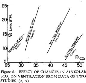
dif-Dill et al. (6) and Hurtado ill e al. (6) and Hurtado e al. (8, et al. (8, 9).9). fusion conductance of the tissues. The diffusion lation. The increased ventilation at
alti-coefficient and a for 02 (a.d) is independent luces the ApuO, froim 50 to 32 and of altitude. However, one can reduce the dif-; the conductance from 6 to 9.5 mril fusion distance between the capillary and the
P2 .cells furthest removed-the intracapillary
s. The diffusion capacity may increase distance-by increasing the number of
capil-at capil-at altitude (15). This would increase
uctane. The distribution fator as it laries/mm2. By doubling the number of capil-ductance. The distribution factor as it
laries one can reduce by about half the 02
he arterial-alveolar O= difference is ex- diffusion gradient from the capillary to the o decrease (7) as a result of the shift periphery of its tissue cylinder. With this as-alveolar gases down on the steep part sumption the ApO, for the tissues in Table 3 ,xygen dissociation curve. For similar is reduced from 30 at sea level to 15 at altitude a shunt would also become less effective
and thus O2 conductance is doubled. icing an alveolar-arterial 02 difference.
her, I have assumed a total alveolar- The total O2 pressure drop as shown in 'er2 di.e 1 of3m m. Thi incrl avelases Table 3 is 140 at sea level and 65 at altitude. 02 difference of 3 mm. This increases
0uc. ,ifferene of m 3* mm.a ee This incr Since the vO2 is 300 in both cases, the over-all
During exercise, how30 at sea lever, the alveo 100 at- conductance is 300/140, or 2.14, at sea level and 02 may increase at altitude (1) ; the 300/65, or 4.62, at altitude. The various fac-' an e tors that contributed to the increase in con-ance change would then be smaller.
ductances at altitude have been checked under of interest here to speculate whether two headings in Table 3. One column has been rease in pulmonary artery pressure ates inmpulmonary , aproerg prmssre , labeled
Dissoc. Since the physiological range of
is simply a way of providing more even the 02 dissociation curve has now been displaced ion of pulmonary blood flow and as a to its steep part, there is an automatic reduction ence a smaller alveolar-arterial ce a smaller alveolar-arterial O2 0 differ- differ- in the 02 differences that normally arise from
lowever, it seems that so little is actuallyowever, it sees that so little is actually the presence of the distribution factor, the shunt n terms of 02 conductance that we must
other explanations for the occurrence of factor, and the oxygen dissociation ertension. curve. This shift on the 02 dissociation curve
!ation. One obvious way to increase the therefore increases the 02 conductances and
uctance is to increase the cardiac output. takes place automatically upon exposure to low ling it we can double the conductance. 02 without requiring any active response. These r, in the long run this is a rather costly effects are well known.
active responses that come into play to increase the 02 conductance. The increase in ventilation is well known. It is of interest to note, how-ever, that the increase in conductance is rela-tively small-from 6 to 9.5. The 02 conductance of the lung is increased about threefold. Being very high to start with, it does not contribute very much by increasing further. The changes in the diffusion capacity of the lung are probably small at rest, and the better distribution of the vA/q achieved by the increased pulmonary artery pressure is probably also negligible at rest. However, both of these must be considered at least contributory factors that are achieved slowly during the acclimation process.
The increase in hemoglobin is also achieved gradually as well as the reduction in the diffu-sion distance. Recent evidence presented by Cassin et al. (3) suggests that the increase in
capillarization at altitude represents not simply an opening up of previously closed vessels but an actual growth of new vessels. Both the changes in Hb and the new blood vessels are important contributors to the increased 02 con-ductance. However, they require time for their ultimate expression. Thus other conductances must take over during the acute exposure. An increase in cardiac output and a greater alveolar ventilation are frequently seen in newcomers; they may represent compensation for the more slowly acquired compensatory devices, such as the changes in diffusion capacity, pulmonary hypertension, increased hemoglobin, and num-ber of capillaries. In general it will be noted that by far the biggest change in 02 conduct-ance is contributed by the circulation and the tissues.
Tissue Diffusion. Finally, we must look more
closely at the 02 diffusion process from the capillary to the tissues. This is a much neglected area that deserves closer scrutiny, since the 02 tensions are not easily measured. We must therefore still depend upon various models such as were proposed originally by Krogh (11) and later by many others. The most recent model, by Diemer (5), provides a new approach based upon the observation that in adjacent capillaries
of the brain the blood flows in a countercurrent fashion. I should like to use the old Krogh-Erlang equation and adapt it from the excellent analysis recently provided by Landis and Pap-penheimer (12).
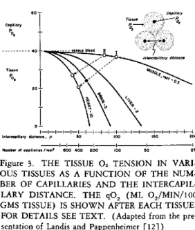
In Figure 3 are shown the tissue 02 tensions for various tissues as a function of their inter-capillary distances, and also the number of capil-laries/mm2. I have assumed that the capillary PO2 was 40 for all tissues. The number follow-ing each tissue indicates the qO2, ml O2/min/ 100 gm of tissue. For example, brain tissue has
approximately 300 capillaries per mm2, which
corresponds to an intercapillary distance of about
58 /¿ (5). If qO2 is 5, the pO, at the
periphery of each tissue cylinder surrounding the capillary is 22 (see solid circle).
On the other hand, the resting gracilis muscle of the rat (3) has about 100 capillaries/mm2. If the resting qO2 is only 0.3 its tissue PO2 is
37, as is shown by the open circle marked I. One may now predict the changes in tissue ten-sion during work. If the 02 uptake increases
thirty-three-fold (qO2= 10) and the number
of capillaries increases four-fold, from 100 to
400/mm2, the new 02 tension is now reduced to
20, as is shown by the new open circle. Cassin
et al. (3) have also determined the increase
Copillory Po.
P,
--.' ' -- p
0,01
- .MWOOP,04,
-In...cop llor diatone, , 50 100 150 200
i-rII--i--l --1 I
«ih07
o- cWlleri· /.H .1 ' 00 0OO 200 100 50 25
Figure 3. THE TISSUE 02 TENSION IN
VARI-OUS TISSUES AS A FUNCTION OF THE NUM-BER OF CAPILLARIES AND THE INTERCAPIL-LARY DISTANCE. THE qO2 (ML 02/MIN/100 GMS TISSUE) IS SHOWN AFTER EACH TISSUE. FOR DETAILS SEE TEXT. (Adapted from the pre-sentation of Landis and Pappenheimer [12])
60-in capillaries of the same muscle after a thirty-six:day period of acclimation to an altitude of twenty thousand feet. In this case the resting muscle had 150 capillaries/mm2 and its resting tissue 02 tension is shown by the open circle marked II. If it now changes its 0, uptake and capillarization during contraction in the same manner as the unacclimatized muscle, its P02 is
reduced only to 27.
These examples serve simply to emphasize the importance of capillarization in the 02 con-ductance through tissues. Thus we see that the increase in the number of open capillaries plays a most important role not only in daily life at sea level but particularly during acclimation to high altitude by changing the 02 conductance. Unfortunately, our models are still very crude
and possibly misleading. Until we have more exact measurements of actual pO2 values in the tissue, this approach must suffice.
The description of the 02 transport from the inspired gas to the venous blood has now been well established for man at sea level and at altitude. Our understanding of the last step from the capillaries to the mitochondria is in-complete, based largely upon models. It is this last link in the description that may turn out to be the most exciting, particularly if we can tie it in to the changes of the tissue enzyme system during acclimation to altitude.
NATURAL ACCLIMATIZATION TO HIGH ALTITUDES:
REVIEW OF CONCEPTS
Alberto Hurtado
The term acclimatization, in reference to a high-altitude environment, does not have a precise and uniform significance among in-vestigators and in the related medical literature. Very frequently, it has been applied to designate the changes found after a brief exposure, of days or even hours, in low-pressure chambers or in elevated places. In my opinion, according to experience obtained in many years of observa-tion, this common interpretation is not justified. When subjected to a constant condition of hypoxia, or oxygen deficiency, the human body needs a long time to develop fully adequate adaptive mechanisms, functional and organic. And even when such tolerance is apparently present, there are always, or nearly always, some qualitative differences between a man born and reared in the hypoxic environment and one who has been subjected to it temporarily. It seems logical, therefore, to make a distinction between these two experimental subjects. There is a tendency, I believe a proper one, to use the terms natural acclimatization for the character-istics of native high-altitude dwellers and
acquired acclimatization for the adaptation
reached after more or less prolonged exposure. It also seems appropriate to rate the effectiveness of acclimatization in terms of similarity to what is found in the native inhabitant, who is the man best adapted, and not exclusively from the usual standpoint of deviations from sea-level physiology.
Another source of variability in the findings reported, and in their interpretation, refers to the level of the given or simulated altitude in-vestigated and the consequent degree of hypoxia. Quite frequently-especially when low-pressure chambers are used-the level of altitude is far beyond the possibilities of tolerance or adapta-tion by the human or animal subjects. What is being investigated in that case is not exactly
ac-climatization or tolerance but rather deteriora-tion. I could mention many examples, but I shall limit myself to one. We know that hypoxia stimulates erythropoietic activity, and one well-known adaptive response is an increase in the circulating red blood cells and hemoglobin. Some evidence, however, suggests that when the hypoxia is very severe its effect becomes depressive.
On the other hand, not infrequently a level of altitude is used at which the decrease in the partial pressure of oxygen and the saturation of the blood with it are extremely moderate or in-significant, and so there is not a real and ac-tive need for the development of compensating mechanisms. The degree of hypoxia, like the duration of the exposure, is a very important factor in determining the nature and the degree of responses. These considerations are not al-ways taken properly into account. Because of the peculiar shape of the oxygen dissociation curve, the relationship between level of altitude and degree of hypoxia is not a linear one. As-cending five hundred meters at an altitude of two to three thousand meters does not have the same consequences and significance as a similar displacement at four to five thousand meters.
look for new physiological patterns in relation to the environment, to have a correct view as to what constitutes normality, and to be able to estimate the changes that take place when patho-logical processes set in.
Another factor that has added special interest to altitude research is the awareness that hypoxia is not exclusive to low-pressure environments. In many diseases affecting the respiratory and circulatory systems, the circulating blood, and the metabolic and chemical activities of the tissue cells, difficulties may arise in the acquisi-tion of oxygen at lung level or in its transport and adequate distribution or in its utilization in body chemistry. It has therefore become of interest to the clinician and the clinical in-vestigator to know how the body may develop mechanisms that will compensate for a de-ficiency responsible for signs, symptoms, and disability. High-altitude research activities are not, in fact, remote from everyday clinical implications.
Finally, I may call attention to one of the great scientific events of the present century-the struggle and century-the determination to conquer space. This endeavor poses great and numerous difficulties, and one has to do with the need for providing a constant oxygen supply to main-tain life. Conditions at high altitudes, where men are living or can live, are not quite like those in space; all the same, it has become
im-portant to know how man may best compensate for oxygen deficiency and how he can be helped toward longer tolerance of it.
MORPHOLOGICAL PATTERNS:
MECHANISM OF PULMONARY ARTERIAL HYPERTENSION*
Javier Arias-Stella
Early in the study of the biology of the Andean regions Peruvian investigators proved that high-altitude natives and residents showed an enlarged cardiac silhouette compared with what was seen in sea-level subjects (1, 2, 3, 4). In line with these observations electrocardio-graphic differences were also demonstrated. Rotta (5) and later Peñaloza et al. (6, 7)
pointed out that the electrocardiographic changes indicated preponderance of the right ventricle. In 1956 the existence of a moderate degree of pulmonary hypertension in high-altitude natives was described (8). The interesting work of Peñaloza et al. (9, 10, 11) has defined this form
of pulmonary hypertension. Thus, it has been established that its level is higher in children than in adults (12), that it increases with alti-tude (13), that it reaches very high values during exercise (14), and so on. It has been demonstrated in other species (15, 16).
Although the hypertension is moderate it is accompanied by definite anatomic changes in the heart, in the pulmonary trunk, and in the lungs (17-22). Studies carried out in our labora-tory have shown that from the fourth month of life there is right ventricular hypertrophy in the high-altitude native. This hypertrophy is greater at the base of the ventricle (17, 18)-a finding that goes along well with the char-acteristics of the electrocardiogram. The pul-monary trunk is thicker and richer in elastic fibers than at sea level (21, 22) and has a greater elastine content, behaving like a resistant vessel to physical deformations (25).
Although Campos and Iglesias (26), in rou-tine histologic studies, found no differences in the small pulmonary vessels of a group of
sub-* This differs from the author's presentation at the Special Session under the heading "Natural Acclimati-zation to High Altitudes: Morphological Patterns."
jects autopsied at La Oroya, the investigations performed in our laboratory leave no doubt that definite differences do exist in the small pul-monary arterial branches.
Using a quantitative histologic procedure, we have shown that after the first month of life there is a greater muscularization in the small pulmonary arterial branches in people born and living at high altitudes (23, 24). Our findings correlate with the report by Pefaloza et al. (10)
of increased pulmonary vascular resistance in high-altitude natives. We therefore can now locate the anatomical point of origin of the pul-monary hypertension at the level of the periph-eral pulmonary arterial branches. In the genesis of the hypertension augmented blood flow and capillary engorgement have, on sound physio-logic bases, been ruled out as possible factors (8, 10, 24). However, the contributing role played by increased blood viscosity is debatable (27). The first problem that should be elucidated is whether the anatomical differences are present in high-altitude subjects from birth or before, or whether the changes take place later.
Our anatomical investigations have demon-strated that up to the seventh postnatal day there are no differences between high-altitude and sea-level subjects in the weight of the right ventricle
(17). This finding agrees with the observations of Pefaloza et al. (12), who have shown that at
and sea-level humans at the end of intrauterine life, no differences will be found in the levels of pulmonary arterial pressures.
From what has been said it can be concluded that the changes we described in the pulmonary vasculature of high-altitude people dating from the first month of life (24) are an acquired post-natal phenomenon. Now we need an explana-tion to the quesexplana-tion, Why are the peripheral pulmonary arterial branches structurally differ-ent at high altitudes? And what is the mecha-nism that gives rise to this difference?
Since we are dealing with an acquired post-natal difference it is reasonable to think of the role played by environmental factors as causal agents. Beyond any doubt, the level of oxygen tension in the atmosphere is the fundamental physical factor of biological importance in high-altitude environments.
Abundant information has been accumulated indicating that diminished oxygen tension can affect the physiology and the structure of the pulmonary vascular system (16, 29, 30, 31, 32).
The experiments of Von Euler and Liljen-strand (29) were the first to show that an in-crement in pulmonary arterial pressure due to vasoconstriction followed a fall in the tension level of breathed oxygen. A similar observation has been made in sea-level animals placed at high altitudes (16j. The vasoconstrictive effect is demonstrated by the fact that, in these experi-ments, when oxygen is administered the elevated pulmonary arterial pression is immediately re-duced to the original normal levels. When the hypoxic situation is maintained for subacute or subchronic periods, as in the experiments of Grover and Reeves, the pulmonary hypertension is found to be associated with vasoconstrictive effects and with anatomic changes in the form of increased arterial muscularization (16). In this case the oxygen administration reduces the ele-vated blood pressure, but to a level above the original normal sea-level value. This demon-strates that the oxygen acts on the vasoconstric-tive physiologic phenomena but has no effect on the anatomic changes.
Our findings in high-altitude natives after
birth (24) accord with the experiments of Grover and Reeves (16). We have found sug-gestive evidence of vasoconstriction in the peripheral pulmonary arterial branches in high-altitude infants. On the other hand, Pefaloza
et al. (12) reported an increased pulmonary
vascular resistance at high altitudes, which is greater in children under five years than in those from six to fourteen.
Taking into account (a) the proved effect of the level of oxygen tension on the vascular "tone" of the small pulmonary arterial branches, (b) the fact that at birth there are no differences in the weight of the, right ventricle and in the characteristics of the pulmonary arterial branches at sea level and high altitudes, (c) the observations showing that the anatomic differ-ences described at high altitudes begin after the first week of life, (d) the demonstration that the physiologic and anatomic changes bear a close correlation with altitude levels, and (e) Can-non's basic principle of homeostasis, one can attempt an explanation of the probable mecha-nism through which the high-altitude pulmonary hypertension occurs.
During intrauterine life the fetus develops in a markedly hypoxic environment, one that is not comparable to any in which normal human life exists. Mithoefer (33) has emphasized the fact that during intrauterine life the arterial oxygen tension in the fetus is 20, which would corre-spond to an atmospheric oxygen tension of ap-proximately 61-that is, to an altitude of about eight thousand meters above sea level. There-fore, at birth, wherever it occurs, the fetus passes from a hypoxic to a better-oxygenated environ-ment. Even at fourteen or fifteen thousand feet above sea level the newborn is a "lowland" new-comer. The chain of events that takes place in the newborn after birth are related to the equi-librium established with the environment. Those environmental factors that are able to exert bio-logical actions are the ones that have to be taken into account.
signifi-cant environmental factor is the level of oxygen tension.
At birth the newborn enters for the first time into contact with what is going to be its per-manent environment, and a homeostatic equi-librium with it takes place. The elevated pul-monary arterial pressure due to the increased pulmonary vascular resistance characteristic of fetal life is suddenly reduced at birth, mainly as a result of the alveolar expansion (34). Once this mechanical readjustment has occurred, the pulmonary vasculature enters into equilibrium with the atmospheric factor that influences it-that is, the atmospheric oxygen tension level. The figure below gives an idea of the sequence of physiologic and anatomic events during this stage of equilibrium.
Since the fetus passes from a less to a more oxygenated medium and the effect on the pul-monary peripheral arterial system is a relaxing one, a relaxation occurs in the small muscular pulmonary arteries. This effect has been well
documented in lambs (32).
If vascular tone is defined as the state of
tensile equilibrium of a vessel with its
environ-ment, and a greater postnatal pulmonary arte-rial relaxation corresponds to higher oxygen ten-sion, we have here a diminished "vascular tone." To lower oxygen tension levels corresponds a lesser degree of arterial relaxation or, in other words, a greater vascular tone. The figure de-picts the changes thought to occur in the pul-monary vascular tone according to the different altitude levels at which birth takes place. The vascular tone and its consequence, the degree of pulmonary arterial resistance, in the immediate postnatal stage are thus determined by the par-ticular level of the atmospheric oxygen tension. The anatomic changes in the pulmonary vas-culature during the postnatal period, which in-clude among other things an involution in the muscularization of the peripheral pulmonary arterial branches and in the mass of the right ventricle, evolve up to a point corresponding to the level of pulmonary arterial pressure that has been determined by the pulmonary vascular resistance.
The figure illustrates the anatomic changes in the pulmonary vasculature at various altitude levels. As the anatomic modifications evolve
INTRAUTERINE
LIFE
(at any altitude level)
8.0001
-Un
w
r-w
z
2
1-
F-¡_
Parcial atmospheric oxygen tension
PULMONARY PERIPHERAL ARTERIAL MUSCLE
Vascular Tone at distal level
Pa 02
20-
45-90
o
Post-NATAL
7
©
0i0000
after 6 months of life
7
00000
FETAL PULMONARY VASCULAR STRUCTURE AND POST ALTITUDE. SCHEME TO EXPLAIN THE MECHANISM OF MONARY HYPERTENSION.
PUL-slowly, compared to the physiologic changes, the fetal vestiges persist for a while after birth. This explains why the pulmonary arterial pressure is greater during early infancy than thereafter.
In the figure it can be seen that to each level of oxygen tension corresponds a different degree of physiologic and anatomic postnatal readjust-ment. This explains the demonstrated fact that the hemodynamic and anatomic changes differ according to the altitude level (13, 21). It is clear to us that differences exist among people born at different altitude levels, but when the altitude difference is slight the changes are minimal and pass unnoticed. Only when we compare sea-level with high-altitude places, as I have done here, do they become obvious and easily detectable.
The explanation advanced by Grover and Reeves (16) that a moderate increment in pul-monary arterial pressure favors a better per-fusion in those pulmonary areas subjected to the effect of hydrostatic pressures is helpful toward
an understanding of the objective of this type of equilibrium.
In the same way that the body surface changes with external temperature to augment or diminish heat irradiation and thus maintain an optimal internal temperature, so the level of atmospheric oxygen tension determines the anatomic and physiologic changes described, which together with some others tend to assure
better utilization of the atmospheric oxygen. The cardiovascular phenomena of high alti-tudes are therefore not a special characteristic of life in rarefied atmospheres but another example of a homeostatic adjustment.
As with any homeostatic mechanism, the ca-pacity for physiologic equilibrium has some limits. We are gathering evidence, based on the magnitude of the anatomic changes and the fre-quency of deaths due to right ventricular failure, that for the variables atmospheric oxygen
ten-sion/pulmonary arterial pressure, this limit is
MORPHOLOGICAL PATTERNS: THE STRUCTURE, COMPOSITION,
AND EXTENSIBILITY OF THE PULMONARY TRUNK AT
SEA
LEVEL
AND HIGH ALTITUDE IN PERU
Donald
A. Heath
Last year members of the High Altitude Research Unit of the Cayetano Heredia Medical School, Peru, and of the Faculty of Medicine of Birmingham University, England, undertook a joint study of the pulmonary trunk of people indigenous to high altitudes (1). We investi-gated its extensibility in people living at sea level in Lima and at a high altitude in' the Andes. At the same time we carried out histo-logical studies of the elastic tissue pattern of its media and chemical determinations of the pro-portions of elastin and collagen within it. We were stimulated to undertake this study by the fascinating observation that the configuration of the elastic tissue in the media of the adult pul-monary trunk of those indigenous to high alti-tudes may resemble that of the aorta (2). This is in striking contrast to the state of affairs in persons living at sea level.
In fetal life the magnitude of the pulmonary arterial pressure is comparable to the aortic and the configuration of the elastic tissue in the media of the pulmonary trunk is very similar to that of the aorta. In both arteries there is a dense network of long, parallel elastic fibrils. By the end of the first month of extrauterine life in those living at sea level or low altitude the blood pressure in the pulmonary arteries falls to the low level maintained for the rest of adult life. A group of us working at the Mayo Clinic have shown that normally, during the first two years of extrauterine life, the elastic tissue in the wall of the pulmonary trunk undergoes involution, becoming fragmented and subse-quently forming an open network of branched fibrils (3).
The tissues of the media of the normal pul-monary trunk of an adult at sea level are more extensible than those of the aorta (4, 5)
-prob-ably because they include a lower proportion of elastin, although differences in the proportion of collagen may also play a part. With increasing age there is a progressive decrease in extensi-bility, and we have previously ascribed this to the apparent increases in the proportion of col-lagen visible in histological sections (5).
When pulmonary hypertension has persisted from birth, as a result of a large congenital "post-tricuspid shunt" (4), the pattern of the elastic tissue of the media of the pulmonary trunk retains its fetal appearance and is thus similar to that of the aortic media (3). In such patients the media of the pulmonary trunk is less extensible than normal (6). In contrast, when pulmonary hypertension arises later in life as a result of a "pretricuspid shunt" (4) or mitral stenosis, the elastic tissue of the pul-monary trunk remains of the normal adult type (3) and the extensibility of the wall is normal (6). It has been shown that the Peru-vian Indian native to high altitude sustains physiological pulmonary hypertension (7) and in such people the appearance of this tissue may resemble that of the aorta (2).
During the course of our studies in Peru we investigated the extensibility of the pulmonary trunk in people living at sea level and at a high altitude and compared this physical property of the media with its histological pattern of elastic tissue and with its chemically determined pro-portions of elastin and collagen. We examined rings of pulmonary trunk from seventy-three subjects. Sixty-six had died in Lima, which is at sea level, but some of these had spent most of their lives at a high altitude and had died during transit through Lima or else had lived in Lima for only a short time before their death.
Seven had lived and died in the vicinity of Cerro de Pasco, which is at 14,200 feet.
Three portions of pulmonary trunk were ex-amined in each case. After the adventitia had been stripped off the media, the extensibility of a circumferential strip was measured in the manner we have described previously (5). The volume of the arterial strip was assessed by weighing it and assuming its specific gravity to be unity. The cross-sectional area was calculated by dividing the volume by the length. A series of extensile loads up to 100 gm were used, and the results were expressed as the percentage extension for a given extensile force (dynes/
mm2 cross-sectional area).
A second adjacent portion of pulmonary trunk was subsequently examined histologically to de-termine the type of elastic tissue pattern in the media. The sections were stained by the Lawson modification of the Weigert-Sheridan method. The remainder of the pulmonary trunk, free of adventitia, was cut into small pieces and put in a vacuum desiccator. After the material was dry it was ground with a mortar and pestle and placed in the desiccator once again for subse-quent chemical analysis. Collagen and elastin were estimated in this dried material by the method of Lawry, Gilligan, and Katersky (8), with the modifications that the first extraction with sodium hydroxide was prolonged to forty-eight hours and that autoclaving was carried out at 15 lb/sq. in. for fifty-four hours. Measure-ments of collagen were made on thirty-one specimens and of elastin on forty-five specimens. The results of our studies were as follows.
Histology
The elastic tissue pattern of the media of the
pulmonary trunk was classified as one of the three types that we have designated previously: "adult pulmonary," "aortic," and "transitional" (3). Sixty-three of the sixty-six cases from Lima and four of the seven who had lived all their lives at Cerro de Pasco showed an adult pul-monary configuration consisting of an open net-work of branched, irregularly shaped elastic fibrils (Figure 1). There was much variation in
Figure 1. TRANSVERSE SECTION OF PULMO-NARY TRUNK FROM PERUVIAN INDIAN AGED 14, SHOWING ADULT PULMONARY PATTERN OF ELASTIC TISSUE. Such a configuration of elastica is characteristic of persons living at low altitude (Elastic/Van Gieson; X 150)
the amount of elastic tissue present, a feature that we have noted previously in low-altitude cases (9). The appearances were similar to those found in a series of cases in Birmingham, England (5). It was frequently possible to determine, from examination of the elastic tis-sue of the media, that a subject was over forty years of age-the fibrils showed a fuzziness of outline, a background elastosis of very fine con-necting fibrils, and they tended to take up stains for elastin less well than those of the younger age group.
10,000ooo
cJ
w
z
In
5,000
IO i a 0-4
1
/..//
... //
dc,/,
uO 20 40 60
*/o EXTENSION.
Figure 3. AVERAGE VALUES FOR EXTENSIBIL-ITY OF PULMONARY TRUNK IN DIFFERENT AGE GROUPS
Figure 2. TRANSVERSE SECTION OF THE PUL-MONARY TRUNK FROM A PERUVIAN IN-DIAN AGED 18 YEARS, SHOWING AN AORTIC PATTERN OF ELASTIC TISSUE. Such a configura. tion of elastica is characteristic of persons living at high altitude (Elastic/Van Gieson; X 150)
pattern more closely resembled the adult pul-monary than the aortic type and for the pur-poses of statistical analysis has been included in the adult pulmonary group.
The mean thickness of the fresh media of the pulmonary trunk in this series was 1121/, (S.D. = 58/t). The range of thickness of the medias
showing an aortic pattern of elastic tissue was 960 to 1220,.
Extensibility
The relation between length and tension was a curved one similar to that found in previous studies (5, 6). In the subjects with an adult pulmonary configuration, extensibility decreased as age increased (Figure 3). The average values of extension for a series of common extensile forces shown in Figure 3 were obtained by drawing separate length-force curves for each case and estimating the degree of extension for a given extensile force by interpolation.
The relation between the age and the loga-rithm of the degree of extension approximated to a straight line, the slope and intercept of which varied with the extensile force. Thus, for a particular extensile force, the relation
be-tween age and extension could be given by
Al = a 10Ib g
where 1l is the percentage increase in length and a and b are constants. The values of a and b varied according to the extensile force and a
number of them were calculated from regres-sion equations relating log,, Al to age. There was no simple formula relating these constants and the magnitude of the extensile force as in our previous study (5), so the relations be-tween the two constants and the extensile force are shown graphically (Figures 4 and 5).
It thus proved possible to assess the expected degree of extension for any given extensile
10,000
5,000'
>-u) w z 0
1
u10 1;2 1:4 1'6
COEFFICIENT "a"
Figure 4. RELATION BETWEEN FORCE AND COEFFICIENT 'A'
1'8 2'0
EXTENSILE
ro -
1oPooo.
5,0oo
oo01 -0D002 -0o003 -0004
COEFFICIENT "b"
Figure 5. RELATION BETWEEN EXTENSILE FORCE AND COEFFICIENT 'B'
force by substituting the obtained values of a
and b together with the age of the subject in the
formula. In Figure 6 we see the degree of ex-tension predicted in this manner, plotted against the observed degree of extension in forty-two specimens with a normal adult pulmonary con-figuration of elastica in the media of the pul-monary trunk.
It proved possible, in addition, to plot the observed degree of extension against that pre-dicted from our equation in the five specimens with an aortic pattern of elastic tissue in the
80.
601
40j
20
c
Figure 6 AND O EXTEN!
* . **.
. . .
t.
·
$ , .
.,:¢,.*
·
1* +~
media (Figure 7). Also included in this graph is the line of the normal regression equation with lines at a distance of twice the standard error. It will be noted that the extensibility of these five specimens is less than normal.
Chemical Analyses
Collagen was estimated in twenty-eight speci-mens with an adult pulmonary pattern of elastic tissue, and the average content was 31.0 per cent (S.D.=4.0 per cent) ; the slight tendency for the content to decrease with age was readily attributable statistically to chance (Figure 8). Collagen was also estimated in three specimens with an aortic pattern of elastica, and the con-tents were 28.3, 31.7 and 37.9 per cent, which did not differ appreciably from the group with the normal adult pulmonary pattern (Figure 8).
Elastin was estimated in forty-one specimens with an adult pulmonary pattern of elastic tis-sue, and the average content was 27.2 per cent (S.D.=4.8 per cent). There was a tendency for the content to increase with age (Figure 9) that was statistically unlikely to have arisen by chance. In four specimens with an aortic
pat-z
Xoo
tu
1-tu
f-80'
60
40.
20-rle / . . .
-2'0
4'0
6-0
8'0
J
1
OBSERVED
0/EXTENSION
o 20 40 60 80 Figure 7. RELATION BETWEEN OBSERVED
DEGREE OF EXTENSION AND THAT
PRE-OBSERVED /lo EXTENSION. DICTED FROM EQUATION 1 IN FIVE SUB-JECTS WITH AORTIC PATTERN OF ELASTIC 5. RELATION BETWEEN PREDICTED TISSUE. "Normal" regression line of equation 2 )BSERVED VALUES FOR PERCENTAGE is shown, together with lines at a distance of twice
SION the standard error of the estimate
:E
z
o
z o
(n
z
I-X Lto
u
1-
·
.
e o e**
·
* :
40
AGE
Collagen and elastin were both estimated in twenty-seven specimens with an adult pulmo-nary pattern of elastic tissue (Figure 10). There was a positive relation between the ratio of elas-tin to collagen and the age of the subject. Col-lagen and elastin were both estimated in three specimens with an aortic pattern of elastic tis-sue; in these three subjects the ratio was inap-propriately high for the age (Figure 10), and the difference was statistically unlikely to have arisen by chance more often than once in a thousand times.
Discussion of Results
* = PULMONARY PATTERN
o = AORTIC PATTERN.
Figure 8. RELATION BETWEEN AGE AND CONTENT OF COLLAGEN (% DRY WEIGHT)
tern of elastica, the elastin contents were 31.0, 31.4, 37.9 and 40.0 percent. The average age of these four subjects was only 22.5 years com-pared to an average age of 45.4 years for the low-altitude group. These figures hence confirm that there is a higher proportion of elastin in the pulmonary trunk with an aortic configura-tion of elastica.
These results show that in Peruvians living at sea level in and around Lima the pulmonary trunk has a normal adult pulmonary pattern and its extensibility is similar to that which we have previously demonstrated in a group of subjects in Birmingham, England (5). Our previous observation that this extensibility decreases with age (5) has also been confirmed in Peruvians. The curved shape of the force-extension dia-gram of the walls of arteries has been ascribed to the shorter relaxed length of the elastic fibrils relative to that of collagen (10). The more horizontal initial part of the curve was
o 12,
o e
.
* *
* .+o ·
.
10' * - e
V) _i'n-J -JO0
w U e o
o o
"v
2'0
4'0
6b
8s0
AGE
o =AORTIC PATTERN
* = PULMONARY PATTERN.
Figure 9. RELATION BETWEEN AGE AND THE CONTENT OF ELASTIN (% DRY WEIGHT)
08'
06
u4¿ .4
e
.
e . le
e *
e
. *
e e
.
e O
*
~~~e
e
:
e
) 2'0 40
AGE
ec0 80
* =PULMONARY PATTERN.
o = AORTIC PATTERN
Figure 10. RELATION BETWEEN THE AGE AND THE RATIO OF ELASTIN TO COLLAGEN
· 0 o
:2:
LIÚ (D 4
4
o
O~ioi
0
40
30
20
1 ,
-o
.
considered to be due mainly to tl the elastic tissue, while the deve more vertical later part of the c with the gradual involvement of
If this view is accepted, the slo should be related to the propor present. Plotting the degree of ex age at a small extensile force o]
mm2 reveals that in specimens
pulmonary pattern the degree of with age (Figure 11). This fin tent with the increasing propor found chemically in our investi the degree of extension producec force is plotted against the propo there is a negative relation (Figu with adult pulmonary and aort elastica.
The slope of the initial part extension curves becomes steepe. ing age (Figure 3), but the incre of the later part of the curves even greater rate. An elementarí the curvilinearity of the curve is culating the ratio of the degree o 10,000 dynes/mm2 to that at 5,0( This ratio would decrease from in the presence of a rectilinear theoretical minimum of 1.0 as th increased. When the ratio is 1
40-Z
z
30-0
z
20-z
1o
.n
.
O
0o 4'o 0
AGE. .. PULMONARY PATTERN .* AORTIC PATTERN
Figure 11. RELATION BETWEEI THE DEGREE OF EXTENSION AT
MM2
he extension of lopment of the curve coincided collagen fibres. ,pe of the curve rtion of elastin :tension against f 2,500 dynes/ with an adult extension falls ding is consis-tion of elastin gation. When d by this small irtion of elastin re 12) in cases tic patterns of
40-z -J il, 30,
20-0o o o
e ®
o . .
10i
10 20 30 40
°/o EXTENSION AT 2,500 DYNES/MM2
o= AORTIC PATTERN. .= PULMONARY PATTERN
Figure 12. RELATION BETWEEN DEGREE OF
EXTENSION AT 2,500 DYNES/MM2
AND CON-TENT OF ELASTIN (% DRY WEIGHT)
age there is a negative correlation when there r with increas- is an adult pulmonary pattern of elastic tissue. The progressive decrease in exensibility with asing steepness age in the later steep portion of the
force-proceeds at an extension curve has been ascribed to an
in-y expressibon of creased proportion of collagen in both systemic s given by
cal-of extension at (11) and pulmonary arteries (5). Our reason
30
dyneS/MM2. for supposing this to be the case was the ap-a vap-alue of 2.0 parent increase in collagen with age in sections relation to a stained with Van Gieson's stain. The present investigation, however, has shown that chemical e curvilinearity
e crttvid ineit estimations do not support the idea of an in-crease with age in the amount of collagen in the pulmonary trunk. A similar discrepancy is found in Fallot's tetrad, where there is an in-crease in collagen seen in histological sections of the pulmonary trunk (3) but not confirmed chemically (12). It may be that collagen be-comes less extensible with age; there have cer-tainly been reports that the bulk modulus of collagen increases with age (13, 14). Alter-natively, it may be that the collagen fibers be-come straighter with increasing age so that they become stretched earlier. However, we must bear in mind that the geometry of the pattern 60 80 of the elastic and collagen fibers in the
pulmo-nary trunk will also influence its extensibility, and any minor modifications of this pattern with N AGE AND increasing age might also play a part in
dimin-2,500 DYNES/ ishing extensibility.
Figur RATI
MM-In tern chem amou fitted The disea high sibili
.16 planation for the abnormally high ratios of the
ratio of extension at 10,000 dynes/mm2 to that
at 5,000 dynes/mm2 (Figure 13).
1 .4 . When the degree of extension is shown as a
percentage of that predicted it is seen that four out of the five pulmonary trunks with an aortic
.2 % pattern in these subjects indigenous to high
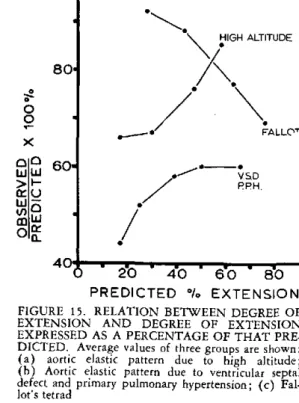
altitudes show a maximal decrease in extensi-bility at low degrees of extension (Figure 14). 20 40 6o o In Figure 15 the average for these five mens is compared with the average of two
speci-AGE
re 13. RELATION BETWEEN AGE AND mens with an aortic pattern of elastic tissue in 10 OF EXTENSIBILITIES AT io,000 DYNES,/ the pulmonary trunk due to congenital heart
AND 5,00( DYNES/MM'
disease (6). Both groups show a maximal the Peruvian Indians with an aortic pat- diminution in extensibility at low degrees of of elastic tissue in the pulmonary trunk, extension. These observations may further be
iical studies showed an abnormally high contrasted with a group of subjects with Fallot's unt of elastin for the person's age. This tetrad (6) (Figure 15). In such persons there in well with the histological appearances. is an abnormally small proportion of elastic same is true in cases of congenital heart tissue and an abnormally high proportion of ,se (12). These subjects indigenous to collagen visible histologically. In Figure 15 we altitudes showed an abnormally low exten- note that in these specimens the extensibility ty of their pulmonary trunk at a low ex- becomes lower with increasing extension. This tensile torce ot 2,500 dynes/mm2, and this is
consistent with the presence of excess elastin (Figure 2). These Indians also showed a nor-mal proportion of collagen in the pulmonary trunk, so that the ratio of elastin to collagen was abnormally high (Figure 10). This is an
ex-AORTIC PATTERN.
PREDICTED /. EXTENSION.
Figure 14. RELATION BETWEEN DEGREE OF EXTENSION AND DEGREE OF EXTENSION EXPRESSED AS PERCENTAGE OF THAT PRE-DICTED IN SUBJECTS WITH AORTIC PAT-TERN OF ELASTIC TISSUE
80'
o
0 X
aC 60.
VSD
PPH
/
40+ m
--_ - 2-0 - 4'0 - 6'0' 80
PREDICTED 0% EXTENSION.
FIGURE 15. RELATION BETWEEN DEGREE OF EXTENSION AND DEGREE OF EXTENSION
EXPRESSED AS A PERCENTAGE OF THAT PRE-DICTED. Average values of three groups are shown: (a) aortic elastic pattern due to high altitude; (b) Aortic elastic pattern due to ventricular septal defect and primary pulmonary hypertension; (c)
Fal-lot's tetrad a
w
z
8 o
4
zo 'o
1
0
bJ z
z z
w
1
is the opposite groups with an i
It is clear fror a considerable U
chemical compos of the pulmonar: high altitudes. \ simple descriptio of the terminal a pulmonary capil lung. Such obs importance to an' and, furthermore siderable accurac lung morphomet The application will enable us t the internal surf numbers of alve healthy subjects and in the unfo veloped chronic disease).
Internal surface ar in the lungs in 7 which there was n
NU CONDITION
STI
Normal
Bronchiolar Emphysema
Alveolar Emphysema
situation to that of the two It is important for us to be frank and admit ncreased proportion of elastin. that at the moment we have no inkling about n these data that we now have the basic morbid anatomy of Monge's disease. nderstanding of the structure, During my visit to the Andes I was intrigued ition, and biophysical behavior by the similarity of the clinical picture of y trunk of those indigenous to Monge's disease to that of cor pulmonale com-XWe still lack, however, even a plicating bronchiolar emphysema that we see not n of the microscopical anatomy uncommonly in the industrial areas of Great tir passages and the state of the Britain. Hypoxia, hypercarbia, polycythemia, lary bed in the high-altitude and certain pulmonary vascular changes are ervations are of fundamental common to both. I think we should make every y understanding of the problem effort to ascertain whether the reduction of e, can now be made with con- internal surface area of pulmonary capillary bed y by use of the techniques of found in bronchiolar emphysema (16) is also ry introduced by Weibel (15). seen in Monge's disease. Our studies in lung
of morphometric techniques morphometry in Birmingham, England, have o determine the magnitude of revealed a considerable reduction in this area face area of the lung and the in cases of bronchiolar and alveolar emphysema eolar spaces within it both in (see table) (16). Is it possible that cases of so-indigenous to high altitudes called chronic mountain sickness are in fact ,rtunate minority who have de- simply people with emphysema, especially of mountain sickness (Monge's the bronchiolar type, who happen to be living on mountaintops rather than persons who are showing a pathological, exaggerated response rea and numbers of alveolar spaces
cases of emphysema and 3 cases in to the hypoxia of high altitude?
o evidence of heart or lung disease I am glad to be able to report that we are
NUMBERS making plans to resolve this problem. Dr.
OF ALVEOLI Kruger of the High Altitude Research Unit
sURFACE AREA
MBER (V2) PER 6,000 IN UNITS OF at Lima has just spent two months with us at DIED
ML LUNG 10 PER Birmingham to learn these methods of lung
ML OF
VOLUME MLUNG morphometry and has now returned to the
Andes to collect these much-needed data on 3 58.5 to 83.4 106 to 127 the internal surface area of the lung. We hope that we shall soon be getting some idea as to 3 25.0 to 55.1 20 to 80 the nature of the basic morbid anatomy of
Monge's disease.
Moderator: We shall now hear from Dr. 4 17.2 to 40.3 19 to 34