Review
Printed in Brazil - ©2014 Sociedade Brasileira de Química0103 - 5053 $6.00+0.00*e-mail: [email protected]
New Trends in Sample Preparation in Brazil: an Overview of Bioanalytical
Applications by Liquid Chromatography
Neila M. Cassiano, Juliana C. Barreiro and Quezia B. Cass*
Departamento de Química, Universidade Federal de São Carlos, 13565-905 São Carlos-SP, Brazil
Esta revisão versa sobre o uso de novas tecnologias no preparo de amostras biológicas, com ênfase nos trabalhos reportados por pesquisadores brasileiros. A miniaturização, automação e injeção direta de amostra com o intuito de atender a demanda por métodos mais eficientes e com menor geração de resíduos, apresentam-se criticamente discutidos.
This paper reviews the new technologies used for bioanalytical sample preparation as developed and explored by Brazilian researchers. Off-line, at-line and on-line sample clean-up approaches, as well microextraction systems, have been critically discussed. The main gains in these applications are enhanced sensitivity with cleaner samples and/or high-throughput capabilities.
Keywords: miniaturization, 2D LC, sorptive phases, microextraction, micro-LLE-based
techniques
1. Introduction
Sample preparation is an integral part of the analytical process and it is by far the highest problem in bioanalysis, furthermore is considered the most environmental pollutant step of the analysis. All the talk of reducing the tedious sample preparation step by liquid chromatography coupled to tandem mass spectrometry has proved to be no more
than a wild dream of fancy.1 On the other hand, sample
clean-up procedures yielding higher analytical turnover with low limits of quantification are of utmost importance.
The goal of the analysis influences the procedure used in sample preparation. In bioanalysis, it usually involves fractionation, isolation and enrichment of the target analytes from the matrix. Most of these protocols are performed off-line affecting time and the cost of the analysis.
Green analytical chemistry is a growing concept in
separation science.2 Thus, considered as a priority, new
approaches to decrease risk contamination and waste have been pursued, with new technologies for direct sample injection or miniaturization of well settled approaches. Automated sample preparation has also been widespread used, and scores of reviews have covered this important
topic.3-8 We shall henceforth outline the Brazilian
contribution to this subject throughout the last decade.
2. Restrict Access Media (RAM)
RAM phases have been explored for direct sample injection since their commercial introduction at the end of eighties. The unique feature of these columns is that they exclude the macromolecules of the matrix while selectively
retaining the small molecules.5,8,9 In 1985, Hagestam
and Pinkerton introduced the internal surface reversed phase (ISRP) support by Regis Technologies, and later
on Boos et al. prepared the alkyl-diol silica (ADS) phase,
commercialized as LiChrospher® ADS by Merck.9 Among
the variety of RAM sorbents, proteins-based phases were one of the first type, being introduced by Yoshida and
co-workers.10 Avidin and α
1-acid glycoprotein RAM columns
were commercialized later on.9 The RAM bovine serum
albumin (BSA) phases were initially criticized, for they had
not great stability. The work of Menezes and Felix11 changed
it all by cross-linking the protein with glutaraldehyde. The RAM-BSA columns have demonstrated high efficiency for
protein depletion using only water as mobile phase.12 The
RAM-BSA columns prepared in accordance with Menezes
and Felix protocol13 has no need of buffer for salting out
the sample proteins.14,15 Their smooth and relatively easy
preparation14,16 with high reproducibility makes them an
excellent choice for direct sample injection. The RAM-BSA columns were successfully explored for assessing, with a
of the proton pump inhibitors (PPIs) pantoprazole,16
omeprazole18 and lansoprazole.19 For that, RAM-BSA-C
8
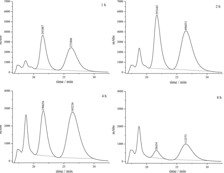
columns were coupled to polysaccharide chiral columns, in a two multidimensional liquid chromatography (2D LC) configuration. Figure 1 illustrates the quality of samples analyses. In spite of the higher number of work using RAM columns for protein depletion, few works deals with chiral
separation, the PPIs works were among the first.9 To explore
other RAM hydrophobic phases BSA-C18, BSA-phenyl
and BSA-CN phases were prepared and their capacity for
depletion of plasma and bovine milk protein evaluated.12,20
Protein depletion was obtained with high percentage with all columns using only water as mobile phase.
The RAM-BSA-C18 column coupled to a C18 analytical
column was used for quantification of amoxicillin in human
plasma.21 To extract amoxicillin with high precision and
high recovery, it was necessary ion pairing (by addition of a cationic counter-ion) in the sample, and the use of
0.01 mol L-1 phosphate buffer, pH of 7.2, as mobile phase,
at protein depletion stage.
A RAM-BSA-phenyl column coupled to a C18
analytical column was efficiently used for quantification of
cefoperazone in bovine milk.20 For quantifying cephalexin,
a RAM-BSA-C8 was used in the first dimension.22
To further explore12,20 the RAM-BSA columns retention
capability of small hydrophilic molecules in samples with high protein content, 2D LC methods were employed for the simultaneously quantifying sulfamethoxazole and
trimethoprim in bovine milk23 and in whole eggs24 samples.
The on-line sample clean-up procedure was carried out also for quantification of these bacteriostatic drugs in bovine milk while employing amperometric detection at a
boron-doped diamond electrode.25
The efficient coupling of RAM-BSA columns to polysaccharide columns granted their use for simultaneous quantification of enantiomeric mixtures and their metabolite
in human14,26 or bovine27 plasma. For modafinil enantiomers
and its two major metabolites, the small RAM-BSA-C8
column (10 × 4.6 mm i.d., 10 µm, 100 Å) was coupled to
an amylose tris[(S)-1-phenylethylcarbamate] chiral column.
The quality of the performance of both columns was
maintained with over 280 plasma injections of 100 µL, each.26
Meanwhile, Santos-Neto et al.28,29 demonstrated the
viability of using RAM columns in a capillary approach. For that, they explored a 25 cm fused silica capillary tubing of
50 µm i.d. coupled to a YMC® ODS-AQ capillary analytical
column (5 µm, 120 Å; YMC Europe) which is coupled to an electrospray interface tandem mass spectrometry
(ESI-MS/MS). As RAM phases, they used a LiChrospher®
ADS-C18 (25 µm, 60 Å; Merck), SPS® C18 (Semi-Permeable
Surface, 5 µm, 100 Å; Regis Technologies), and the in-house
prepared bovine serum albumin-C18 (BSA-C18, 10 µm,
120 Å). Throughout these approaches, the back-flush mode was preferred for coupling the columns in the 2D LC
system.28,30,31 Lower sample dilution and consumption of
mobile phase are the main attractive of the capillary approach
procedure.28 Furthermore, the use of ESI-MS/MS furnished
limits of quantification of 1.0 ng mL-1 for five antidepressant
drugs, with injection of 1 µL of sample.30
The RAM columns are usually used in the LC-LC 2D
configuration, due to lower selectivity.5,9 Menezes and
co-workers, however, have used long RAM-BSA or -HSA (human serum albumin) columns, as single column mode,
for protein depletion and analysis of small molecules.32-35
A RAM-BSA-C18 (50 × 4.6 mm i.d., 10 µm, 100 Å)
column was used in the single mode coupled to an ion trap mass spectrometry for measuring both carbamazepine and its active metabolite carbamazepine 10,11-epoxide, in
human milk.36 Lipids and proteins are the main components
of breast milk interfering in drug recovery and their detection by MS/MS. The molecular mass cut-off of the
RAM-BSA-C18 column for the human milk proteins were
evaluated by the elution profile of β-casein (24,000 Da) and α-lactalbumin (14,178 Da), at concentrations of 5.0 g L-1,
examined at the same conditions used for the human milk samples. Extraction efficiency, accuracy, and precision were achieved employing 100 µL of the sample.
The use of ultra high performance liquid chromatography (UHPLC) tandem mass spectrometry for the fast analysis of small molecules within complex matrices, with lower quantification and detection limits, is already well
established.8,37 The methods for sample clean-up, however,
have remained a drawback for such procedures. It has been recently reported on the use of an injection/column-switching
system for coupling a RAM-BSA-C8, (50 × 2.1 mm, 10 µm,
100 Å) to an Acquity UPLC® BEH C
18 (50 × 2.1 mm, 1.7 µm;
Waters) column.38 The method requires a 14 min analysis,
and the RAM-BSA columns have once more proven to be capable of fast on-line protein depletion.
A recent review about 2D LC-MS,8 has drawn attention
to procedures such as turbulent flow injection that usually
requires pretreatment, while with RAM columns this is not necessary.
In spite of published works on environmental
samples,39,40 no papers on 2D LC-MS chiral bioanalytical
applications have been published by the Brazilian researchers. Considering the expertise in the field, we expect to see further applications on chiral separation.
3. Molecularly Imprinted Solid-Phase Extraction (MISPE)
Molecularly imprinted polymers (MIPs) are synthetic polymers with high specific molecular recognition capability. MIPs preparation is usually carried out by polymerization of a monomer around a template using a cross-linker in the presence of an initiator. Afterwards, the template molecule is removed, yielding a polymeric matrix with specific cavities, which are complementary to the template in size, shape, and position of the functional groups.41-43
The use of solid-phase extraction based on MIPs has been explored for a variety of analytes within different
biological matrices.42,43 In this respect, Vieira et al.44
reported an off-line MISPE procedure for the selective
extraction of trans,trans-muconic acid from urine samples
followed by LC-UV analysis. Their method exhibited good sensitivity in applications within occupational and environmental toxicology. A cotinine-imprinted polymer was also developed for the off-line quantification of cotinine in saliva samples by LC with diode array detection
(LC-DAD).45 The method was successfully applied for
extracting cotinine from smoker’s samples. Recently,
Melo and Queiroz46 developed an off-line MISPE to
preconcentrate parabens from human milk samples. The MISPE⁄LC-UV was adequated to determine methyl, ethyl and propyl parabens and compared with the classical extraction methods, the miniaturized approach minimized the volumes of organic solvent and biological fluid.
4. At-Line Solid-Phase Extraction (SPE)
The at-line approach is usually mixed-up with the on-line one. The difference between them is, however, well settled. In the first case, there is the automation of the SPE process, while in the second case there is a multimodal LC-LC system.
At-line SPE47 is an important approach for labor-intense
analysis, furnishing high throughput with high precision and sensitivity. In this context, it has been used for clinical
analysis and in bioequivalence studies. Carvalho et al.48
attention to the quality of such technique as an alternative to immunoaffinity assays and gas chromatography-mass spectrometry (GC-MS) for steroids clinical profiling. Works have demonstrated that the SPE at-line with a LC-MS/MS is an important tool for bioequivalence studies, whereby thousands of samples are processed with minimum of
manual operation.49-53 In Brazil, this sample preparation
approach has been explored also for pharmacokinetics
studies, as in the quantification of gatifloxacin in rat plasma54
and of etoricoxib in human plasma.55
5. Solid-Phase Microextraction (SPME)
SPME was first introduced in the early 1990s by Pawliszyn’s group as a new and effective sample preparation method to solve problems commonly associated to solid phase extraction, such as: high blank values, variability among the cartridges from different manufactures, and interferences due to adsorption of analytes on the SPE
cartridges.7,56 In the SPME approach, fused silica capillary
are coated on the outside with an appropriate stationary phase, and the analytes are adsorbed by simple exposure to fiber. For gaseous samples, headspace is used, while direct
immersion is employed for liquid samples.4,6,7 To meet this
end, analytes’ partition and desorption are involved.57
Initially, the SPME was developed for the analysis of organic compounds from aqueous sample matrices using
GC.56 Later it was applied for preconcentration of drugs
from biological fluids by GC or LC.58 While in GC analysis
the analytes are thermally desorbed by a heat chamber,56
without solvent, in LC the desorption is carried out either off-line or on-line mode in a suitable volume of selected solvent.6,7
The success of the SPME is determined by the physicochemical properties and the thickness of fiber
coatings.6 The variety of commercially available coatings
has contributed to the number of classes of analytes that can be successfully analyzed in different matrices. For example: polydimethylsiloxane (PDMS) for extraction of non-polar analytes; polyacrylate (PA) and polydimethylsiloxane-divinylbenzene (PDMS-DVB) for extraction of polar analytes, especially phenols and amines, respectively; carboxen-polydimethylsiloxane (CAR-PDMS) for extraction of volatile/low molar mass analytes; carbowax-divinylbenzene (CW-DVB) for extraction of polar analytes (especially alcohols); carbowax-templated resin (CW-TPR) for extraction of polar analytes; and divinylbenzene-carboxen-polydimethylsiloxane (DVB-CAR-PDMS) for extraction of broad range of analytes. Other less frequently used coatings include carbon nanotubes, several crown
ethers, MIPs, anodized metals, and ionic liquids.4,6
In Brazil, the first work using off-line SPME for LC bioanalysis was reported on the determination of lamotrigine, carbamazepine and carbamazepine
10,11-epoxide in human plasma.59 Silva et al.60 and
Cantú et al.61 reported the use of off-line SPME-LC on
the determination of antidepressants and anticonvulsants in plasma samples. Both works showed high sensitivity and reproducibility on the quantification of these drugs in human plasma with no special interface. A comparison between commercial (PA, PDMS-DVB, CW-TPR) and in-house fibers (polyurethane, octadecylsilane, aminosilane)
was carried out by Queiroz and co-workers.61 The in-house
fibers gave higher quantification limits than the commercial ones; the authors stressed, however, the benefits of in-house fibers, such as, easy preparation, good mechanical strength and low cost. Regardless of all desorption inconvenient in the off-line mode, and the increase in solid residue, an advantage is that the use of multiple SPME fibers, improves
the throughput analysis.7
Based on the Pawliszyn’s work,62 Lanças and
co-workers63 developed a new heated interface with lower
inner volume for on-line coupling in order to minimize common problems related to the commercial SPME-LC interface. Improved sensitivity, qualitative and quantitative results were obtained for fluoxetine analysis when compared either to an interface without heating or to SPME extraction in the off-line mode.
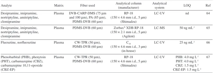
Table 1 summarizes the SPME Brazilian methods based on the use of heated interface for drug analysis in biological matrix.
Queiroz and co-workers68,69 evaluated the use of new
phases coating, polypyrrole (PPY) and polythiophene (PTh) prepared on a stainless-steel wire by electrochemical polymerization. The PPY fiber offered higher sensitivity (in order of 10 times more) due to the matrix effect observed in the quantification of common antidepressants (citalopram, paroxetine, fluoxetine and sertraline).
Another important application of SPME is reported
by Bonato and co-workers.70-72 These works deal with
the quantification of chiral drugs and their metabolites in human urine, by off-line approach, using CW-TPR, PDMS-DVB and PA fibers. Good sensitivity, selectivity and less consume of organic solvents were achieved.
6. In-tube Solid-Phase Microextraction (in-tube SPME)
extraction capillary column, which is placed in an automatic six-port valve. In the draw/eject extraction systems, the capillary column can be installed between the injection loop and the injection needle of the LC autosampler. It can also
be placed in the injection loop.7,73-75
Capillaries prepared with selective materials, such as immunosorbent, PPY polymer and RAM sorbent, have been developed to improve both extraction efficiency and selectivity. The immunoaffinity capillaries SPME devices
were used for quantifying fluoxetine in serum samples76
and interferon alpha2a in plasma sample.77 The drug were
extracted using draw/eject procedure and the separation was
carried out at a C18 column.
An in-tube SPME PPY-coated capillary was prepared in-house in order to analyze fluoxetine and norfluoxetine enantiomers in plasma samples. The capillary was used
in a draw/eject system using a Chiralcel OD-R as the
analytical column.78 The developed method was employed
for monitoring patients under fluoxetine therapy (Prozac,
20 mg day-1).
The same research group has also prepared a
RAM-BSA-C18 in-tube-SPME capillary for the determination
of interferon alpha2a in human plasma.79 A single draw/eject
cycle was used for sample extraction while the analysis was
carried out at a LichroCART RP-18 column. The developed
method has adequate sensitivity and selectivity for therapeutic
monitoring of interferon alpha2a in human plasma samples.
The main drawback of in-tube SPME is the requirement of very clean samples, since the capillary column can be easily blocked. Therefore, previous sample pretreatment, such as filtration or protein precipitation, is usually required to extend the lifetime of the capillary and to prevent
clogging of the flow line system.7,73-75 The biocompatibility
of the RAM-BSA support79 allowed the direct injection
of plasma samples with no sample manipulation other than dilution, reducing, thus, the total analysis time and in accordance with the green analytical chemistry aim.
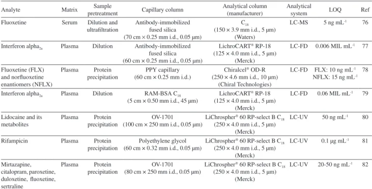
The works carried out in Brazil using on-line in-tube SPME system are summarized in Table 2.
7. Stir Bar Sorptive Extraction (SBSE)
The SBSE was introduced in 1999 by Baltussen et al.83
as a solventless sample preparation method. Magnetic
coated stirs bars are the main attraction of this technique.84
In SPME, the maximum volume of coated PDMS on to the fiber is of 0.5 µL (film thickness 100 µm), whereas, about
25-125 µL of PDMS are used for the stir bars in SBSE,
increasing the extraction efficiency.6,85,86
One limitation of SBSE, when compared to SPME, is that there is only one commercial available extraction
sorbent, PDMS, although in different length and thickness.84
Aiming to increase the applicability of SBSE, in-house sorptive phases have been developed. Accordingly,
Melo et al.87 produced a dual-phase polymeric coating
consisting of PDMS and PPY for antidepressants extraction (mirtazapine, citalopram, paroxetine, duloxetine, fluoxetine and sertraline) from human plasma. The PDMS-PPY coated stir bar showed high extraction efficiency (sensitivity and selectivity) toward the targets analytes.
The quantification of ivermectine in bovine plasma was carried out using a PDMS modified bars with 5% of polydimethylphenylsiloxane and 10% of polydiethyleneglycol succinate. They showed higher efficiency when compared
with the commercial PDMS bars.85
In Brazil, SBSE has been used mostly for analyzing
antidepressants,88,89 anticonvulsants90 and antituberculous
drugs in biological matrices.91
Table 1. Bionalytical methods employing heated SPME-LC interface
Analyte Matrix Fiber used Analytical column (manufacturer) Analytical system LOQ Ref
Desipramine, imipramine, nortriptyline, amitriptyline, clomipramine
Plasma DVB-CARP-DMS (75 µm and 100 µm), PA (85 µm),
PDMS-DVB (60 µm)
RP-18 (150 × 4.6 mm i.d., 5 µm)
(Shimadzu)
LC-UV nd 64
Desipramine, imipramine, nortriptyline, amitriptyline, clomipramine
Plasma PDMS-DVB (60 µm) Zorbax XDB RP-18
(150 × 2.1 mm i.d., 5 µm) (Agilent)
LC-MS 50 ng mL-1 65
Fluoxetine, norfluoxetine Plasma CW-TPR (50 µm), PDMS-DVB (60 µm)
C18
(150 × 4.6 mm i.d., 3 µm) (in-house)
LC-UV 25 ng mL-1 66
Phenobarbital (PHB), phenytoin (PHT), carbamazepine (CBZ), carbamazepine 10,11-epoxide (CBZ-EP)
Plasma CW-TPR (50 µm), PDMS-DVB (60 µm)
RP-18 (150 × 4.6 mm i.d., 5 µm)
(Shimadzu)
LC-UV PHB: 4.0 mg L-1
PHT: 4.0 mg L-1
CBZ: 1.5 mg L-1
CBZ-EP: 1.5 mg L-1
67
8. Microextraction by Packed Sorbent (MEPS)
MEPS are the miniaturization of SPE and works with reduced sorbent bed volume, and they are suitable for a large sample volume range (10-1000 µL). Since they are integrated in the syringe, they diminish the number of steps typically involved in the conventional SPE, and are easily
automatized.6 Sorbent materials such as silica based (C
2,
C8, C18), strong cation exchanger (SCX), RAM, hydrophilic
materials, carbon, polystyrene-divinylbenzene copolymers,
and MIPs can be used.92,93
The main advantage of the MEPS, when compared to conventional SPE, is that the packed syringe is used several times, more than 100 times for plasma or urine samples, without loss of performance. A protocol for guaranteeing the performance of MEPS cartridges is, however, necessary, which includes dilution, centrifugation, and precipitation
steps.93 Chaves et al.94 reported the use of MEPS based on
C8/SCX for the extraction of sertraline, mirtazapine,
fluoxetine, citalopram, and paroxetine in human plasma. Effects on the extraction efficiency were examined for sample volumes, pH, number of extraction cycles, and desorption conditions. Different sample pre-treatment was also evaluated, such as precipitation, centrifugation and dilution. Dilute samples (1:1, v/v) led to sensitive, selectivity, and accurate quantification of the selected drugs. Furthermore, the cartridge might be reused more than 50 times.
MEPS cartridge (C8/SCX), as sample clean-up
procedure, was used for the first time by Salami et al.95
for the simultaneous determination of sulfonamides in whole egg samples. Precipitation and centrifugation were, however, used as sample pre-treatment. The cartridges were reused more than 60 times with minimum loss of extraction efficiency.
A C18 cartridge was used by Bordin et al.96 for
determining voriconazole in oral fluids and plasma. The extraction variables parameters such as, pH of the buffer used in the sample, number and flow rate of extraction cycles were examined. For the analysis, the pH was the most important factor. Extraction time was
of 4 min per sample. The cartridge was used for about
40 extractions.
An important issue to be noticed is that MEPS efficiently reduces the organic solvent volume used and the amount of solid residue. Thus, it can be considered as a green chemistry approach.
9. Hollow Fiber Liquid-Phase Microextraction (HF-LPME)
HF-LPME developed in 1999 by Pedersen-Bjergaard
and Rasmussen97 uses a water immiscible organic solvent
immobilized as a thin supported liquid membrane (SLM) in the pores of a hollow polypropylene fiber. For extraction,
Table 2. Use of in-tube SPME in biological sample
Analyte Matrix pretreatment Sample Capillary column Analytical column(manufacturer) Analytical system LOQ Ref
Fluoxetine Serum Dilution and ultrafiltration
Antibody-immobilized fused silica (70 cm × 0.25 mm i.d., 0.05 µm)
C18
(150 × 3.9 mm i.d., 5 µm) (Waters)
LC-MS 5 ng mL-1 76
Interferon alpha2a Plasma Dilution Antibody-immobilized
fused silica
(60 cm × 0.25 mm i.d., 0.05 µm)
LichroCART RP-18
(125 × 4.0 mm i.d., 5 µm) (Merck)
LC-FD 0.006 MIL mL-1 77
Fluoxetine (FLX) and norfluoxetine enantiomers (NFLX)
Plasma Protein precipitation
PPY capillary (60 cm × 0.25 mm i.d.)
Chiralcel OD-R
(250 × 4.6 mm i.d., 10 µm) (Chiral Technologies)
LC-FD FLX: 10 ng mL-1
NFLX: 15 ng mL-1
78
Interferon alpha2a Plasma Dilution RAM-BSA C18 (5 cm × 0.50 mm i.d., 45 µm)
LichroCART RP-18
(125 × 4.0 mm i.d., 5 µm) (Merck)
LC-FD 0.06 MIL mL-1 79
Lidocaine and its metabolites
Plasma Protein precipitation
OV-1701
(100 cm × 250 mm i.d., 0.05 µm)
LiChrospher 60 RP-select B C
18
(250 × 4.0 mm i.d., 5 µm) (Merck)
LC-UV 50 ng mL-1 80
Rifampicin Plasma Protein precipitation
Polyethylene glycol (60 cm × 0.32 mm i.d., 0.05 µm)
LiChrospher 60 RP-select B C
18
(250 × 4.0 mm i.d., 5 µm) (Merck)
LC-UV 0.1 µg mL-1 81
Mirtazapine, citalopram, paroxetine, duloxetine, fluoxetine, sertraline
Plasma Protein precipitation
OV-1701
(80 cm × 250 mm i.d., 0.05 µm)
LiChrospher 60 RP-select B C
18
(250 × 4.0 mm i.d., 5 µm) (Merck)
LC-UV 20-50 ng mL-1 82
the lumen of the hollow fiber is filled with a small volume of an acceptor phase (usually in the range 2-30 µL), and the whole assembly is placed in the matrix sample (typically within 100 µL to 4 mL). The analytes are extracted from the aqueous sample (donor phase), through the organic SLM, and further into the acceptor phase (aqueous or organic) inside its lumen. After extraction, the acceptor solution is
collected and analyzed.98,99
HF-LPME can be performed in either two or three-phase mode. In the first mode, the acceptor solution and the immobilized organic solvent are the same. They may
be employed for compounds with high solubility in non-polar organic solvents, and acidic/basic analytes. In the three-phase mode, the acceptor phase is an acidic or alkaline aqueous solution and, thus, it is used for extraction of acids
and bases.98-101
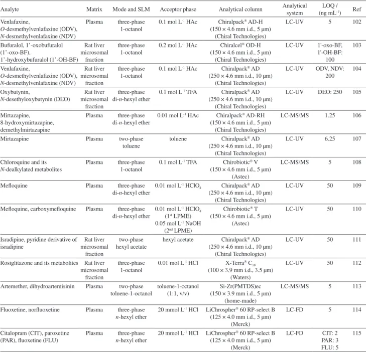
Different research groups in Brazil have employed two and three-phase HF-PLME for the determination of drugs and their metabolites in biological fluids, as the HF-LPME technique allows high analyte enrichments, despite UV detection being known to provide relatively poor sensitivity (Table 3).
Table 3. Application of HF-LPME in biological sample
Analyte Matrix Mode and SLM Acceptor phase Analytical column Analytical system (ng mLLOQ / -1) Ref
Venlafaxine,
O-desmethylvenlafaxine (ODV),
N-desmethylvenlafaxine (NDV)
Plasma three-phase 1-octanol
0.1 mol L-1 HAc Chiralpack AD-H
(150 × 4.6 mm i.d., 5 µm) (Chiral Technologies)
LC-UV 5 102
Bufuralol, 1’-oxobufuralol (1’-oxo-BF), 1’-hydroxybufuralol (1’-OH-BF) Rat liver microsomal fraction three-phase 1-octanol
0.2 mol L-1 HAc Chiralcel OD-H
(150 × 4.6 mm i.d., 5 µm) (Chiral Technologies) LC-UV 1'-oxo-BF, 1'-OH-BF: 100 103 Venlafaxine,
O-desmethylvenlafaxine (ODV), N-desmethylvenlafaxine (NDV)
Rat liver microsomal
fraction
three-phase 1-octanol
0.1 mol L-1 HAc Chiralpack AD
(250 × 4.6 mm i.d., 10 µm) (Chiral Technologies)
LC-UV ODV, NDV: 200
104
Oxybutynin,
N-desethyloxybutynin (DEO)
Rat liver microsomal
fraction
three-phase di-n-hexyl ether
0.1 mol L-1 TFA Chiralpack AD
(250 × 4.6 mm i.d., 10 µm) (Chiral Technologies)
LC-UV DEO: 250 105
Mirtazapine, 8-hydroxymirtazapine, demethylmirtazapine
Plasma three-phase di-n-hexyl ether
0.01 mol L-1 HAc Chiralpack AD-RH
(150 × 4.6 mm i.d., 5 µm) (Chiral Technologies)
LC-MS/MS 1.25 106
Mirtazapine Plasma two-phase
toluene
toluene Chiralpack AD
(250 × 4.6 mm i.d., 10 µm) (Chiral Technologies)
LC-UV 6.25 107
Chloroquine and its
N-dealkylated metabolites
Plasma three-phase 1-octanol
0.1 mol L-1 TFA Chirobiotic V
(150 × 4.6 mm i.d., 5 µm) (Astec)
LC-MS/MS 5 108
Mefloquine Plasma three-phase
di-n-hexyl ether
0.01 mol L-1 HClO
4 Chiralpack AD
(250 × 4.6 mm i.d., 10 µm) (Chiral Technologies)
LC-UV 50 109
Mefloquine, carboxymefloquine Plasma three-phase di-n-hexyl ether
0.01 mol L-1 HClO 4
(1st LPME)
0.05 mol L-1 NaOH
(2nd LPME)
Chirobiotic T
(150 × 4.6 mm i.d., 5 µm) (Astec)
LC-UV 50 110
Isradipine, pyridine derivative of isradipine Rat liver microsomal fraction two-phase hexyl acetate
hexyl acetate Chiralpack AD
(250 × 4.6 mm i.d., 10 µm) (Chiral Technologies)
LC-UV 50 111
Rosiglitazone and its metabolites Rat liver microsomal
fraction
three-phase 1-octanol
0.01 mol L-1 HCl X-Terra C
18
(100 × 3.9 mm i.d., 3.5 µm) (Waters)
LC-UV 50 112
Artemether, dihydroartemisinin Plasma two-phase toluene-1-octanol
toluene-1-octanol (1:1, v/v)
Si-Zr(PMTDS)ec (150 × 3.9 mm i.d., 5 µm)
(home-made)
LC-MS/MS 5 113
Fluoxetine, norfluoxetine Plasma three-phase
n-hexyl ether
20 mmol L-1 HCl LiChrospher 60 RP-select B
(125 × 4.0 mm i.d., 5 µm) (Merck)
LC-FD 5 114
Citalopram (CIT), paroxetine (PAR), fluoxetine (FLU)
Plasma three-phase
n-hexyl ether
20 mmol L-1 HCl LiChrospher 60 RP-select B
(125 × 4.0 mm i.d., 5 µm) (Merck)
LC-FD CIT: 2 PAR: 3 FLU: 5
115
Bonato and co-workers have used HF-PLME procedures followed by LC analysis for quantification of drugs in different biological matrices using either chiral columns (macrocyclic antibiotic and polysaccharide-based
stationary phases)102-111 or achiral columns.112,113
Siqueira and co-workers114 and Porto et al.115 employed
three-phase HF-LPME, coupled to LC-fluorescence detection, for the analysis of fluoxetine/norfluoxetine, and citalopram, paroxetine and fluoxetine in human plasma, respectively. Both methods showed excellent sample clean-up, selectivity and sensitivity.
High recovery associated with low organic solvent consumption is the main attractive of this sample clean-up procedure. Furthermore, its application in sample clean-up procedures for chiral bioanalysis with UV detection demonstrates the high capability of producing clean samples. Chiral selectors easily lose selectivity, demanding generally cleaner samples.
10. Final Considerations
Greener analytical procedures are characteristic to miniaturization, to at-line and on-line systems, due to reduced generation of waste. 2D LC systems have proven their ability in producing on-line matrix protein depletion, and their versatility has been efficiently explored by Brazilian researches for a variety of bioanalytical applications. The use of capillary columns, in-tube SPME, and the coupling of a RAM to a UHPLC column have demonstrated that the researchers are not only interested in efficient sample enrichment, but in producing less waste.
The trends on sample preparation, as here reported, reflect the effort of achieving green parameters and getting cleaner samples with overall smaller analysis time.
Acknowledgements
The authors acknowledge the financial support from Fundação de Amparo à Pesquisa do Estado de São Paulo FAPESP (Process 2009/17138-0) and Conselho Nacional de Desenvolvimento Científico e Tecnológico (CNPq).
Neila Maria Cassiano received her PhD in Analytical Chemistry from Federal University of São Carlos (UFSCar, São Carlos city, Brazil) in the group of Eduardo F. A. Neves in 1998. She has also worked as post-doctoral fellow in the same institution in 2002. Currently, she is a laboratory technician in the Chemistry
Department at UFSCar and associate researcher on the group of Organic Synthesis and HPLC. She has been working on development/validation of analytical methods for quality control of raw material and products by LC, separation of chiral compounds in both analytical and preparative scale and development/application of restricted access media column for analysis of pharmaceutical compounds in biological fluids by direct injection of samples.
Ju l i a n a C r i s t i n a B a r r e i r o
received her PhD in Chemistry from Federal University of São Carlos (UFSCar, São Carlos city, Brazil) in the group of Milton D. Capelato in collaboration with EMBRAPA-Agricultural Instrumentation in 2005. Her research interests are development of methods for quantitation of chiral and achiral compounds by LC-MS in wastewater samples and separation of chiral compounds in both analytical and preparative scale. Currently, she is working as post-doctoral fellow in the group of Organic Synthesis and HPLC at UFSCar, where she investigates the mechanisms involved during chiral discrimination of sulfoxide compounds and the chiral stationary phase by Nuclear Magnetic Resonance.
Q u e z i a B e z e r r a C a s s i s a n Associated Professor at the Chemistry Department of the Federal University
of São Carlos (UFSCar, São Carlos
city, Brazil). She received her PhD at
the The City University (London) in the group of Professor P. G. Sammes in 1981, working on chemistry of some vinyl sulfoxides. Her present research interests are development of methods for quantitation/isolation of chiral and achiral compounds by LC, with a list of works on development and use of polysaccharide chiral columns in both analytical and preparative scale. Also, she has been working on development and application of restricted access media for small molecules in complex matrix. New models for ligand screening by the use of immobilized enzymatic reactors are part of her recent researches.
References
1. Carvalho, V. M.; J. Chromatogr., B: Anal. Technol. Biomed. Life Sci. 2012, 883, 50.
2. Tobiszewski, M.; Mechlinska, A.; Zygmunt, B.; Namiesnik, J.;
3. Singleton, C.; Bioanalysis 2012, 4, 1123. 4. Ramos, L.; J. Chromatogr., A 2012, 1221, 84.
5. Cassiano, N. M.; Barreiro, J. C.; Moraes, M. C.; Oliveira, R. V.; Cass, Q. B.; Bioanalysis 2009, 1, 577.
6. Kole, P. L.; Venkatesh, G.; Kotecha, J.; Sheshala, R.; Biomed. Chromatogr. 2011, 25, 199.
7. Kataoka, H.; Saito, K.; J. Pharm. Biomed. Anal. 2011, 54, 926. 8. Cassiano, N. M.; Barreiro, J. C.; Oliveira, R. V.; Cass, Q. B.;
Bioanalysis 2012, 4, 2737.
9. Cassiano, N. M.; Lima, V. V.; Oliveira, R. V.; de Pietro, A. C.; Cass, Q. B.; Anal. Bioanal. Chem. 2006, 384, 1462.
10. Yoshida, H.; Morita, I.; Tamai, G.; Masujima, T.; Tsuru, T.; Takai, N.; Imai, H.; Chromatographia 1984, 19, 466. 11. Menezes, M. L.; Felix, G.; J. Liq. Chromatogr. Rel. Technol.
1998, 21, 2863.
12. de Lima, V. V. ; Cassiano, N. M.; Cass, Q. B.; Quim. Nova 2006, 29, 72.
13. Menezes, M. L.; Felix, G.; J. Liq. Chromatogr. Rel. Technol.
1996, 19, 3221.
14. Cassiano, N. M.; Cass, Q. B.; Degani, A. L. G.; Wainer, I. W.;
Chirality 2002, 14, 731.
15. Tanaka, H.; Takahashi, K.; Ohira, M.; J. Chromatogr., A 2000, 869, 151.
16. Cass, Q. B.; Degani, A. L. G.; Cassiano, N. M.; Pedrazolli, J.;
J. Chromatogr., B: Anal. Technol. Biomed. Life Sci.2002, 766, 153. 17. Cassiano, N. M.; Oliveira, R. V.; Bernasconi, G. C. R.; Cass,
Q. B.; Chirality 2012, 24, 289.
18. Cass, Q. B.; Lima, V. V.; Oliveira, R. V.; Cassiano, N. M.; Degani, A. L. G.; Pedrazzoli, J.; J. Chromatogr., B: Anal. Technol. Biomed. Life Sci. 2003, 798, 275.
19. Gomes, R. F.; Cassiano, N. M.; Pedrazzoli, J.; Cass, Q. B.;
Chirality 2010, 22, 35.
20. Oliveira, R. V.; Cass, Q. B.; J. Agr. Food Chem. 2006, 54, 1180. 21. Cass, Q. B.; Gomes, R. F.; Calafatti, S. A.; Pedrazolli, J.;
J. Chromatogr., A 2003, 987, 235.
22. Oliveira, R. V.; de Pietro, A. C.; Cass, Q. B.; Talanta 2007, 71,
1233.
23. Pereira, A. V.; Cass, Q. B.; J. Chromatogr., B: Anal. Technol. Biomed. Life Sci. 2005, 826, 139.
24. de Paula, F. C. C. R.; de Pietro, A. C.; Cass, Q. B.; J. Chromatogr., A
2008, 1189, 221.
25. Andrade, L. S.; Rocha-Filho, R. C.; Cass, Q. B.; Fatibello-Filho, O.;
Electroanalysis 2009, 21, 1475.
26. Cass, Q. B.; Galatti, T. F.; J. Pharm. Biomed. Anal. 2008, 46,
937.
27. Belaz, K. R. A.; Cass, Q. B.; Oliveira, R. V.; Talanta 2008, 76,
146.
28. Santos-Neto, A. J.; Rodrigues, J. C.; Fernandes, C.; Titato, G. M.; Alves, C.; Lanças, F. M.; J. Chromatogr., A 2006, 1105, 71. 29. Santos-Neto, A. J.; Markides, K. E.; Sjoberg, P. J. R.;
Bergquist, J.; Lanças, F. M. Anal. Chem. 2007, 79, 6359.
30. Santos-Neto, A. J.; Bergquist, J.; Lanças, F. M.; Sjoberg, P. J. R.;
J. Chromatogr., A 2008, 1189, 514.
31. Santos-Neto, A. J.; Fernandes, C.; Rodrigues, J. C.; Lanças, F. M.; J. Sep. Sci. 2008, 31, 78.
32. Sanchez, A.; Toledo-Pinto, E. A.; Menezes, M. L.; Pereira, O. C. M.; Pharmacol. Res. 2004, 50, 481.
33. Menezes, M. L.; Muzardo, G. A.; Chaves, M. S.; J. Liq. Chromatogr. Rel. Technol. 2004, 27, 1799.
34. Menezes, M. L.; Felix, G.; Demarchi, A. C. C. O.;
Chromatographia 1998, 47, 81.
35. Menezes, M. L.; Simionato, E. M. R. S.; Felix, G.; J. Liq. Chromatogr. Rel. Technol. 2008, 31, 2603.
36. Lopes, B. R.; Barreiro, J. C.; Baraldi, P. T.; Cass, Q. B.;
J. Chromatogr., B: Anal. Technol. Biomed. Life Sci. 2012, 889, 17.
37. Guillarme, D.; Schappler, J.; Rudaz, S.; Veuthey, J-L.; TrAC, Trends Anal. Chem. 2010, 29, 15.
38. Moura, F.; de Almeida, F. G.; Lopes, B. R.; Cass, Q. B.; J. Sep. Sci. 2012, 35, 2615.
39. Barreiro, J. C.; Vanzolini, K. L.; Madureira, T. V.; Tiritan, M. E.; Cass, Q. B.; Talanta 2010, 82, 384.
40. Barreiro, J. C.; Vanzoline, K. L.; Cass, Q. B.; J. Chromatogr., A
2011, 1218, 2865.
41. Pichon, V.; J. Chromatogr., A 2007, 1152, 41.
42. He, C. Y.; Long, Y. Y.; Pan, J. L.; Li, K.; Liu, F.; J. Biochem. Biophys. Methods 2007, 70, 133.
43. Haginaka, J.; J. Sep. Sci. 2009, 32, 1548.
44. Vieira, A. C.; Zampieri, R. A.; de Siqueira, M. E. P. B.; Martins, I.; Figueiredo, E. C.; Analyst 2012, 137, 2462.
45. Vitor, R. V.; Martins, M. C. G.; Figueiredo, E. C.; Martins, I.;
Anal. Bioanal. Chem. 2011, 400, 2109.
46. Melo, L. P.; Queiroz, M. E. C.; Anal. Methods 2013, 5, 3538. 47. Hyotylainen, T.; J. Chromatogr., A 2007, 1153, 14.
48. Carvalho, V. M.; Nakamura, O. H.; Vieira, J. G. H.;
J. Chromatogr., B: Anal. Technol. Biomed. Life Sci. 2008, 872,
154.
49. Estrela, R. D. C. E.; Salvadori, M. C.; Suarez-Kurtz, G.; Rapid Commun. Mass Spectrom. 2004, 18, 1147.
50. Estrela, R. D. E.; Salvadori, M. C.; Raices, R. S. L.; Suarez-Kurtz, G.; J. Mass Spectrom. 2003, 38, 378.
51. Gonçalves, J. C. S.; Monteiro, T. M.; Neves, C. S. D.; Gram, K. R. D.; Volpato, N. M.; Silva, V. A.; Caminha, R.; Gonçalves, M. D. B.; dos Santos, F. M.; da Silveira, G. E.; Noel, F.; Ther. Drug Monit. 2005, 27, 601.
52. Raices, R. S. L.; Salvadori, M. C.; Estrela, R. D. E.; Neto, F. R. D.; Suarez-Kurtz, G.; Rapid Commun. Mass Spectrom.
2003, 17, 1611.
53. Suenaga, E. M.; Ifa, D. R.; Cruz, A. C.; Pereira, R.; Abib, E.; Tominaga, M.; Nakaie, C. R.; J. Sep. Sci. 2009, 32, 637.
54. Tasso, L.; Costa, T. D.; J. Pharm. Biomed. Anal. 2007, 44, 205. 55. Dalmora, S. L.; Brum, L.; Ferretto, R. M.; de Oliveira, P. R.;
56. Arthur, C. L.; Pawliszyn, J.; Anal. Chem. 1990, 62, 2145. 57. Zhang, Z. Y.; Yang, M. J.; Pawliszyn, J.; Anal. Chem. 1994, 66,
A844.
58. Chen, J.; Pawliszyn, J.; Anal. Chem. 1995, 67, 2530.
59. Queiroz, M. E. C.; Silva, S. M.; Carvalho, D.; Lanças, F. M.;
J. Sep. Sci. 2002, 25, 91.
60. Silva, B. J. G.; Queiroz, R. H. C.; Queiroz, M. E. C.; J. Anal. Toxicol. 2007, 31, 313.
61. Cantu, M. D.; Toso, D. R.; Lacerda, C. A.; Lanças, F. M.; Carrilho, E.; Queiroz, M. E. C.; Anal. Bioanal. Chem. 2006, 386, 256.
62. Daimon, H.; Pawliszyn, J.; Anal. Commun. 1997, 34, 365.
63. Rodrigues, J. C.; Santos-Neto, A. J.; Fernandes, C.; Alves, C.; Contadori, A. S.; Lanças, F. M.; J. Chromatogr., A 2006, 1105,
208.
64. Alves, C.; Fernandes, C.; Santos-Neto, A. J.; Rodrigues, J. C.; Queiroz, M. E. C.; Lanças, F. M.; J. Chromatogr. Sci. 2006, 44, 340.
65. Alves, C.; Santos-Neto, A. J.; Fernandes, C.; Rodrigues, J. C.; Lanças, F. M.; J. Mass Spectrom. 2007, 42, 1342.
66. Fernandes, C.; Santos-Neto, A. J.; Rodrigues, J. C.; Alves, C.; Lanças, F. M.; J. Chromatogr., B: Anal. Technol. Biomed. Life Sci. 2007, 847, 217.
67. Alves, C.; Gomes, P.; dos Santos-Neto, A. J.; Rodrigues, J. C.; Lanças, F. M.; Anal. Methods 2012, 4, 1519.
68. Chaves, A. R.; Chiericato, G.; Queiroz, M. E. C.; J. Chromatogr., B: Anal. Technol. Biomed. Life Sci. 2009, 877, 587.
69. Caris, J. A.; Chaves, A. R.; Queiroz, M. E. C.; J. Braz. Chem. Soc. 2012, 23, 57.
70. de Oliveira, A. R. M.; Cesarino, E. J.; Bonato, P. S.;
J. Chromatogr., B: Anal. Technol. Biomed. Life Sci. 2005, 818, 285.
71. de Oliveira, A. R. M.; de Santana, F. J. M.; Bonato, P. S.; Anal. Chim. Acta 2005, 538, 25.
72. de Oliveira, A. R. M.; Bonato, P. S.; J. Sep. Sci. 2007, 30, 2351. 73. Kataoka, H.; Ishizaki, A.; Nonaka, Y.; Saito, K.; Anal. Chim.
Acta 2009, 655, 8.
74. Kataoka, H.; Saito, K.; Bioanalysis 2012, 4, 809.
75. Queiroz, M. E. C.; Lanças, F. M.; Quim. Nova 2005, 28, 880. 76. Queiroz, M. E. C.; Oliveira, E. B.; Breton, F.; Pawliszyn, J.;
J. Chromatogr., A 2007, 1174, 72.
77. Chaves, A. R.; Queiroz, M. E. C.; J. Chromatogr., B: Anal. Technol. Biomed. Life Sci. 2013, 928, 37.
78. Silva, B. J. G.; Lanças, F. M.; Queiroz, M. E. C.; J. Chromatogr., A 2009, 1216, 8590.
79. Chaves, A. R.; Silva, B. J. G.; Lanças, F. M.; Queiroz, M. E. C.;
J. Chromatogr., A 2011, 1218, 3376.
80. Caris, J. A.; Silva, B. J. G.; Moises, E. C. D.; Lanchote, V. L.; Queiroz, M. E. C.; J. Sep. Sci. 2012, 35, 734.
81. Melo, L. P.; Queiroz, R. H. C.; Queiroz, M. E. C.; J. Chromatogr., B: Anal. Technol. Biomed. Life Sci. 2011, 879, 2454.
82. Silva, B. J. G.; Lanças, F. M.; Queiroz, M. E. C.; J. Chromatogr., B: Anal. Technol. Biomed. Life Sci. 2008, 862, 181.
83. Baltussen, E.; Sandra, P.; David, F.; Janssen, H. G.; Cramers, C.;
Anal. Chem. 1999, 71, 5213.
84. David, F.; Sandra, P.; J. Chromatogr., A 2007, 1152, 54. 85. Lanças, F. M.; Queiroz, M. E. C.; Grossi, P.; Olivares, I. R. B.;
J. Sep. Sci. 2009, 32, 813.
86. Chaves, A. R.; Queiroz, M. E. C.; Quim. Nova 2008, 31, 1814.
87. Melo, L. R.;, Nogueira, A. M.; Lanças, F. M.; Queiroz, M. E. C.;
Anal. Chim. Acta 2009, 633, 57.
88. Fernandes, C.; Jiayu, P.; Sandra, P.; Lanças, F. M.;
Chromatographia 2006, 64, 517.
89. Chaves, A. R.; Silva, S. M.; Queiroz, R. H. C.; Lanças, F. M.; Queiroz, M. E. C.; J. Chromatogr., B: Anal. Technol. Biomed. Life Sci. 2007, 850, 295.
90. Queiroz, R. H. C.; Bertucci, C.; Malfara, W. R.; Dreossi, S. A. C.; Chaves, A. R.; Valerio, D. A. R.; Queiroz, M. E. C.;
J. Pharm. Biomed. Anal. 2008, 48, 428.
91. Balbao, M. S.; Bertucci, C.; Bergamaschi, M. M.; Queiroz, R. H. C.; Malfara, W. R.; Dreossi, S. A. C.; Mello, L. D.; Queiroz, M. E. C.; J. Pharm. Biomed. Anal. 2010, 51, 1078. 92. Abdel-Rehim, M.; Anal. Chim. Acta 2011, 701, 119.
93. Abdel-Rehim, M.; J. Chromatogr., A 2010, 1217, 2569. 94. Chaves, A. R.; Leandro, F. Z.; Carris, J. A.; Queiroz, M. E. C.;
J. Chromatogr., B: Anal. Technol. Biomed. Life Sci. 2010, 878, 2123.
95. Salami, F. H.; Queiroz, M. E. C.; J. Braz. Chem. Soc. 2011, 22, 1656.
96. Bordin, N. A.; Antunes, M. V.; Spaniol, B.; Andreolla, H. F.; Pasqualotto, A. C.; Linden, R.; Lat. Am. J. Pharm. 2011, 30,
2043.
97. Pedersen-Bjergaard, S.; Rasmussen, K. E.; Anal. Chem. 1999, 71, 2650.
98. Pedersen-Bjergaard, S.; Rasmussen, K. E.; J. Chromatogr., B: Anal. Technol. Biomed. Life Sci. 2005, 817, 3.
99. Pedersen-Bjergaard, S.; Rasmussen, K. E.; J. Chromatogr., A
2008, 1184, 132.
100. Ghambarian, M.; Yamini, Y.; Esrafili, A.; Microchim. Acta
2012, 177, 271.
101. de Oliveira, A. R. M.; Magalhães, I. R. D.; de Santana, F. J. M.; Bonato, P. S.; Quim. Nova 2008, 31, 637.
102. da Fonseca, P.; Bonato, P. S.; Bioanalysis 2013, 5, 721.
103. Barth, T.; Simões, R. A.; Pupo, M. T.; Okano, L. T.; Bonato, P. S.; J. Sep. Sci. 2011, 34, 3578.
104. da Fonseca, P.; Bonato, P. S.; Anal. Bioanal. Chem.2010,
396, 817.
105. da Fonseca, P.; de Freitas, L. A. P.; Pinto, L. F. R.; Pestana, C. R.; Bonato, P. S.; J. Chromatogr., B: Anal. Technol. Biomed. Life Sci. 2008, 875, 161.
107. de Santana, F. J. M.; de Oliveira, A. R. M.; Bonato, P. S.;
Anal. Chim. Acta 2005, 549, 96.
108. Magalhães, I. R. D.; Bonato, P. S.; J. Sep. Sci.2008, 31, 3106.
109. Magalhães, I. R. D.; Bonato, P. S.; J. Pharm. Biomed. Anal.
2008, 46, 929.
110. Magalhães, I. R. D.; Bonato, P. S.; Anal. Bioanal. Chem.
2009, 393, 1805.
111. Simões, R. A.; de Oliveira, A. R. M.; Bonato, P. S.; Anal. Bioanal. Chem. 2011, 399, 2435.
112. Calixto, L. A.; Bonato, P. S.; J. Sep. Sci. 2010, 33, 2872. 113. Magalhães, I. R. S.; Jabor, V. A. P.; Faria, A. M.; Collins,
C. H.; Jardim, I.; Bonato, P. S.; Talanta 2010, 81, 941.
114. de Freitas, D. F.; Porto, C. E. D.; Vieira, E. P.; de Siqueira, M.; J. Pharm. Biomed. Anal. 2010, 51, 170.
115. Porto, C. E. D.; Maia, P. P.; de Freitas, D. F.; Araujo, R. C. C.; de Siqueira, M.; Martins, I.; dos Santos-Neto, A. J.;
Quim. Nova 2012, 35, 72.
Submitted: September 11, 2013
Published online: November 29, 2013