Evaluation of the bending strength of rigid internal fixation with
absorbable and metallic screws in mandibular ramus sagittal split
osteotomy –
in vitro
study
Avaliação da resistência à flexão da fixação interna rígida, com
parafusos absorvíveis e metálicos, na osteotomia sagital do ramo
mandibular – estudo
in vitro
Petrus Pereira Gomes* Rubens Guimarães Filho** Renato Mazzonetto***
ABSTRACT:The aim of this study was to evaluate,in vitro, the bending strength of internal fixation with absorbable and metallic screws in mandibular ramus sagittal split osteotomy in sheep hemimandibles. The screws were inserted as lag screws, with an inverted “L” configuration, and the set was submitted to bending strength tests. The load and displacement of the peak and final load averages were, respectively, 18.45 kgf, 8.19 mm and 14.38 kgf for Group I, and 16.67 kgf, 6.73 mm and 13.98 kgf for Group II. The results were submitted to statistical analysis by Student’sttest and by the Pearson correlation analysis. The groups showed no statistically significant differences, indicating the fea-sibility of both for osteosynthesis in mandibular ramus sagittal split osteotomies.
DESCRIPTORS:Osteotomy; Fracture fixation, internal; Mandibular advancement; Lactic acid.
RESUMO:O objetivo deste estudo foi avaliar,in vitro, a resistência à flexão da fixação interna, através do uso de para-fusos absorvíveis e metálicos, na osteotomia sagital do ramo em hemimandíbulas de carneiros. Os parapara-fusos foram in-seridos como “lag screw”, com configuração em “L” invertido e o conjunto submetido ao teste de resistência à flexão. As médias da carga e do deslocamento do pico e da carga final do Grupo I foram de 18,45 kgf, 8,19 mm e 14,38 kgf, e do Grupo II de 16,67 kgf, 6,73 mm e 13,98 kgf, respectivamente. Os resultados foram submetidos à análise estatística pelo testetde Student e pela análise de correlação de Pearson. Os grupos exibiram diferenças estatisticamente não significativas, indicando a viabilidade de ambos para a osteossíntese em osteotomias sagitais do ramo mandibular. DESCRITORES:Osteotomia; Fixação interna de fraturas; Avanço mandibular; Ácido láctico.
INTRODUCTION
The introduction of mandibular ramus sagittal split osteotomy, by Trauner, Obwegeser26 (1957),
represented a great advance in the correction of dentofacial deformities. Successive modifications of the technique were suggested, mainly for increa-sing the stability of the fixed segments11,28. Spiessl23
(1974) was the first to employ screws for fixing mandibular ramus sagittal osteotomies, using the lag screw technique with three 2.7 mm screws po-sitioned in an inverted “L”9.
A wide variety of osteosynthesis materials and techniques have been employed by intra/extraoral accesses, including plates with mono- and bicor-tical screws, whether or not associated with
maxil-lomandibular fixation (MMF) or skeletal fixation and positional or lag screws9. Osteosynthesis
ma-terials should have some ideal characteristics like ease of use, the ability to fix adequately to bone segments, biocompatibility, and should be able to gradually transfer physiological loads to the bone being repaired. They should also be absorbed after they have completed their function16. Metallic
ma-terials like titanium have excellent properties for use in osteosynthesis. There are, however, some disadvantages described in the literature, like the release of metallic ions, continual mechanical stimulus, osteolysis under the implants21,
interfer-ence in radiotherapy, production of artifacts in computerized tomography and magnetic reso-nance, occurrence of corrosion and allergic
tions, and the palpability and hypersensitivity to cold1.
Kulkarni et al.19(1966) were the first authors to
experimentally evaluate the employment of absor-bable materials. Cutright et al.6(1971) reported the
use of absorbable fixation for stabilizing mandibu-lar fractures in monkeys. Important advances have occurred with the employment of this techni-que, especially with regard to the chemical compo-sition of materials, clinical and biomechanical pro-perties, and the forms of presentation. Polylactic acid (PLA), which is absorbed by hydrolysis, is the most commonly employed material in oral and ma-xillofacial surgery16. Törmälä et al.25 (1988)
intro-duced the self-reinforcement techniques in the production of PLA, thus significantly increasing its mechanical properties. The absorbable materials are outstanding due to the ability of not requiring removal after the bone repair period, thus elimina-ting a second surgical procedure. Furthermore, they do not interfere in the treatment with radiot-herapy and do not generate artifacts in computeri-zed tomography and magnetic resonance exami-nations. Studies indicate that the absorbable osteosynthesis systems gradually lose mechanical strength, a loss that generally becomes significant after about six months, which is sufficient for bone consolidation. The total absorption time of these materials is not yet known, varying according to chemical composition, place of implantation and the amount of material used5.
The aim of this experimental study is to evalua-te,in vitro,the bending strength of an internal
fixa-tion carried out with the use of absorbable and me-tallic screws of equal diameter, inserted as lag screws.
MATERIALS AND METHODS
The bending strength tests were carried out on 20 right hemimandibles of adult sheep supplied by a slaughterhouse. All the soft tissues enveloping them were removed. The mandibular ramus sagit-tal split osteotomy in the hemimandibles was car-ried out by using a fissure cone bur, mounted on a handpiece coupled to an electrical motor, following the precepts of Trauner, Obwegeser26 (1957) with
the modifications of Dal Pont7 (1961) and
Hunsuck15 (1968). After the osteotomy, the distal
segment was advanced by 5 mm. To fix the hemi-mandible segments, screws with 2.0 mm external diameters and 12 mm long were inserted as lag screws. The specimens were divided into two groups, Group I being constituted of 10 hemiman-dibles, each fixed with three absorbable screws of self-reinforced polylactic acid copolimer (SR-PLA, Bionx Implants, Tampere, Finland) 70L:30DL. Group II comprised 10 hemimandibles, each fixed with three commercially available pure titanium screws (Osteomed, Addison, Texas, United States of America).
The screws were inserted 90° with the lateral bone surface throughout threading. Insertion was considered to be complete when the screw head touched the buccal cortical, and the extremity of the screw passed the medial cortical. After being fi-xed the hemimandibles had the posterior edge of
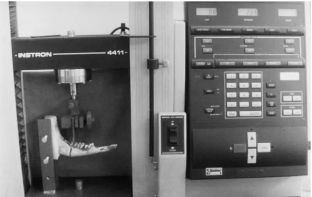
FIGURE 1 -InstronÒmachine during
the ramus and condyle embedded in an acrylic re-sin block.
The hemimandibles, adapted to a fixing device, were taken for bending tests in an InstronÒ
univer-sal test machine (Instron Co., Canton, Massachu-setts, USA) – model 4411 (Figure 1). The speed was set at 1 mm/min, with maximum displacement of 10 mm, for the application of a progressive load to the system. The load was always applied vertically in the area between the last two molar teeth of each hemimandible. The progression of loading and displacement was followed on the machine monitor, and the load data in kilogram-force (kgf), and extent in millimeters (mm) of any peak during the experiment were noted. When the final displa-cement of 10 mm was reached, a reading was made of the value corresponding to the final load, in kilogram-force (kgf).
Statistical analysis of the values obtained was carried out using Student’sttest and by the Pear-son correlation analysis at a significance level of 5%.
RESULTS
Student’sttest was applied to Groups I and II with regard to the mean values of the peak displa-cement (p = 0.23), of the peak load (p = 0.55) and of the final load (p = 0.88) (Table 1). For all the data,
the difference between both groups was not statis-tically significant.
The Pearson correlation analysis was applied to the peak and final loads, independently, for each of the groups. Group I showed a positive correlation between the peak and final loads (p = 0.007), whe-reas in Group II there was no correlation between the respective peak and final loads (p = 0.22).
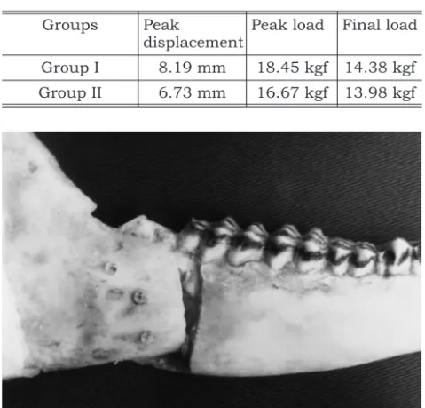
The analysis of Group I showed that the distal segment of the hemimandibles underwent bending throughout the extent of the displacement (10 mm), by means of the area of contact between the osteotomy segments, without evidence of bone fracture in the region where the screws were inser-ted (Figure 2). In Group II, however, the distal seg-ment underwent less bending, which may explain the horizontal bone fractures on the areas where the metallic screws were inserted (Figure 3).
DISCUSSION
The mandibular ramus sagittal split osteotomy is the most widely employed procedure for correc-ting dentoskeletal deformities in the mandible20.
The wide dissemination of this technique is pos-sibly related to its versatility and to the favorable results that have been obtained26.
In this study, all the distal segments of the hemimandibles were advanced by 5 mm, thus keeping a reference standard in conformity with other studies that evaluated the stability and fixa-tion methods of mandibular ramus sagittal osteot-omy10,27.
The sheep mandible has been used for experi-mental studies of osteotomies and rigid internal fi-xation in the ramus area, mainly due to the simila-rities in format, size and structure to the human
TABLE 1 -Mean value of peak displacement, peak load and final load.
Groups Peak
displacement
Peak load Final load
Group I 8.19 mm 18.45 kgf 14.38 kgf
Group II 6.73 mm 16.67 kgf 13.98 kgf
FIGURE 2 -Bend of distal segment, by absorbable screw
seg-mandible27. It is important to emphasize that the
data obtained from biomechanical studies using analogous bones cannot be directly transferred to clinical use in humans, serving only as indicative parameters of the behavior of a certain technique and/or material.
Since Spiessl23 described, in 1974, the
oste-osynthesis technique with screws, investigations have been carried out with regard to whether or not should the bone corticals be compressed22, as
the use of screws, like the lag screw, would permit a greater surface compression area, thus favoring bone tissue stability and primary bone repair wit-hout the development of bone callus. There are no statistically significant differences between the stability provided by positional screws and by the lag screw technique when used for osteosynthesis of segments in sagittal split osteotomy13.
Different numbers and configurations of screws employed for rigid internal fixation have been eva-luated. It is known, however, that the use of one screw is unacceptable and that stabilization with two screws is still a method with few documented reports. The use of three screws seems to be the ideal, as the addition of more screws shows no me-chanical advantage14. In this study, the option was
to use the inverted “L” configuration for fixation of the hemimandibles, as this is the most acceptable pattern and one that provides the greatest resis-tance to displacement of the fixed segments after mandibular ramus sagittal split osteotomy14.
The biomechanical test employed a two-dimen-sional model with a free end lever, due to the faci-lity of obtaining data, lower cost and ease of use. It was, however, not able to simulate the complex be-havior of the human mandible at work. The load peaks generated in some specimens during the bending test probably occurred due to the sudden reductions in biomechanical resistance of the system at some moment during load application, and this could have been the point of failure in the-se hemimandibles. In the other specimens, there was a progression of loading and displacement without the occurrence of peaks, so the final dis-placement (10 mm) was considered as the point of failure. One should consider, however, that a dis-placement of 10 mm is far in excess of the clinical and radiographic limit of what would be conside-red a failure of the fixation system. According to Ardary et al.2(1989), this limit would be 1 mm.
The results indicated that there were no statis-tically significant differences between the groups,
as both attained the final displacement proposed for the study, with similar final load averages, in spite of the individual variation of the values in the samples. This suggests that possibly the absorba-ble osteosynthesis system in this study showed bending strengths comparable to those of the me-tallic system.
However, the elasticity module of the SR-PLA (70L:30DL) screws, 6 GPa, is similar to that of the cortical bone, 5 to 30 GPa, while the metallic mate-rial reaches around 200 GPa17. Perhaps this
justifi-es the finding in some hemimandibljustifi-es after the bending test (Figures 2 and 3). Tate et al.24(1994)
found masticatory forces in the molar area of up to 13.8 kgf, in the first six weeks, compared to forces of 26 kgf after the sixth post-operative week of sur-gical treatment of mandibular angle fractures. The importance of these data is that they are maximum forces obtained during occlusion, and that the amount of force used during functional activities is probably much less. This explains why forms of fi-xation with less rigid materials may be success-fully used. Clinically, the amount of resistance to load necessary for a fixation material to promote adequate bone repair in a functional mandible is unknown18.
Experimental studies in vitro and in vivo have employed absorbable plates and screws of greater thickness and diameter, respectively, for carrying out osteosynthesis in fractures and mandibular osteotomies, with the objective of compensating for the lower mechanical resistance of the absorbable materials in relation to metallic ones12. In the
sagit-tal split osteotomy, few studies describe the use of screws of up to 2.0 mm for RIF (rigid internal fixati-on)8. The time for total absorption of SR-PLA
(70L:30DL) screwsin vivo is unknown. The direct information about the total absorption time of PLA in humans is based on a few studies with biopsies taken in reoperations3. Extrapolation of the
hy-drolysis studiesin vitrosuggests that the complete degradation of SR-PLA (70L:30DL) would take from two to three years. The tissue response to the absorbable materials does not seem to be a pro-blem when materials with more amorphous cha-racteristics are used, which would facilitate ab-sorption.
The absorbable materials appeared as an alter-native to attempt to overcome possible problems with the metallic osteosynthesis4,21, such as the
pro-duction of artifacts in tomographic and magnetic resonance examinations. Some factors, like the high cost and greater dimension of plates and screws, still prevent the total adoption of absorba-ble fixation. The question, therefore, is not wheth-er or not to replace one fixation system with anoth-er, but to acknowledge the existence of two biologically and mechanically compatible oste-osynthesis methods, which may be used according to the surgeon’s and/or patient’s preference.
CONCLUSIONS
Analyzing the data obtained under the experi-mental conditions of this study, it may be conclu-ded that the existence of peaks appeared to be re-lated to the abrupt reduction of the system’s mechanical strength at some moment in the expe-riment. During the bending tests, the absorbable screws tended to bend in the same direction as the displacement of the distal segment, while the me-tallic screws resisted deformation, generating
hori-zontal fracture lines over the lateral surface of the proximal segment.
Differences in final load values were not statisti-cally significant between both groups, indicating that the mechanical strengths of absorbable and metallic screws under these experimental conditi-ons were similar. The absorbable material may therefore be considered a feasible alternative for ri-gid internal fixation in mandibular ramus sagittal split osteotomies.
A larger number of studies still need to be deve-loped to compare absorbable fixations of similar dimensions to those of metallic ones in fractures and mandibular osteotomies. The tests carried out with absorbable material in areas upon which greater loads fall, like the mandible, will supply consistent data and greater security for the disse-mination of such systems. Furthermore, the biolo-gical behavior of these materials should also be the object of further investigations.
REFERENCES
1. Ahn DK, Sims CD, Randolph MA, O’Connor D, Butler PE, Amarante MT, et al. Craniofacial skeletal fixation using bi-odegradable plates and cyanoacrylate glue. Plast Reconstr Surg 1997;99:1508-17.
2. Ardary WC, Tracy DJ, Brownridge GW 2nd, Urata MM. Comparative evaluation of screw configuration on the sta-bility of the sagittal split osteotomy. Oral Surg Oral Med Oral Pathol 1989;68:125-9.
3. Bergsma EJ, Rozema FR, Bos RR, deBruijn WC. Foreign body reactions to resorbable poly (L-lactide) bone plates and screws used for the fixation of unstable zygomatic frac-tures. J Oral Maxillofac Surg 1993;51:666-70.
4. Bos RRM, Rozema FR, Boering G, Nijenhuris AJ, Pennings AJ, Verwey AB. Bio-absorbable plates and screws for inter-nal fixation of mandibular fractures. A study in six dogs. Int J Oral Maxillofac Surg 1989;18:365-9.
5. Böstman O, Pihlajamäki H. Clinical biocompatility of bio-degradable orthopaedic implants for internal fixation: a re-view. Biomaterials 2000;21:2615-21.
6. Cutright DE, Hunsuck EE, Beasley JD. Fracture reduction using a biodegradable material, polylactic acid. J Oral Surg 1971;29:393-7.
7. Dal Pont G. Retromolar osteotomy for the correction of prognathism. J Oral Surg Anesth Hosp Dent Serv 1961; 19:42-7.
8. Edwards R, Kiely KD, Eppley BL. Resorbable fixation tech-niques for genioplasty. J Oral Maxillofac Surg 2000; 58:269-72.
9. Ellis E 3rd. Rigid fixation in orthognathic surgery. Selected Read Oral Maxillofac Surg 1992;2:1-17.
10. Ellis E 3rd, Reynolds S, Carlson DS. Stability of the mandi-ble following advancement: a comparison of three
postsur-gical fixation techniques. Am J Orthod Dentofacial Orthop 1988;94:38-49.
11. Epker BN. Modifications in the sagittal osteotomy of the mandible. J Oral Surg 1977;35:157-9.
12. Eppley BL. Use of a resorbable fixation technique for maxil-lary fractures. J Craniofac Surg 1998;9:317-21.
13. Foley WL, Frost DE, Paulin WB, Tucker MR. Internal screw fixation: comparison of placement pattern and rigidity. J Oral Maxillofac Surg 1989;47:720-3.
14. Haug RH, Barber JE, Punjabi AP. Anin vitrocomparison of the effect of number and pattern of positional screws on load resistance. J Oral Maxillofac Surg 1999;57:300-8. 15. Hunsuck EE. A modified intraoral sagittal splitting technic
for correction of mandibular prognathism. J Oral Surg 1968;26:250-3.
16. Kallela I. Rigid internal fixation of the mandible using bio-degradable polylactide screws and metal screws: a clinical and experimental study [Academic Dissertation]. Helsinki: Medical Faculty of the University of Helsinki; 1999. 17. Kallela I, Tulamo RM, Hietanen J, Pohjonen T, Suuronen
R, Lindqvist C. Fixation of mandibular body osteotomies using biodegradable amorphous self-reinforced (70L:30DL) polylactide or metal lag screws: an experimen-tal study in sheep. J Craniomaxillofac Surg 1999; 27:124-33.
18. Kim HC, Essaki S, Kameyama T. Comparison of screw pla-cement patterns on the rigidity of the sagittal split ramus osteotomy: technical note. J Craniomaxillofac Surg 1995; 23:54-6.
19. Kulkarni RK, Pani KC, Neuman C, Leonard F. Polylactic acid for surgical implants. Arch Surg 1966;93:839-43. 20. Martis CS. Complications after mandibular sagittal split
21. Paavolainen P, Karaharju E, Slatis P, Ahonen J, Holms-trom T. Effect of rigid plate fixation on structure and min-eral content of cortical bone. Clin Orthop 1978; 136:287-93.
22. Souyris F. Sagittal splitting and bicortical screw fixation of the ascending ramus. J Maxillofac Surg 1978;6:198-203. 23. Spiessl B. Ostéosynthèses bei sagittaler osteotomie nach
Obwegwser-Dal Pont. Fortschr Kiefer Gesichtschir 1974; 18:145-8.
24. Tate GS, Ellis E 3rd, Throckmorton G. Bite forces in pa-tients treated for mandibular angle fractures: implications for fixation recommendations. J Oral Maxillofac Surg 1994; 52:734-6.
25. Törmälä P, Rokkanen P, Laiho J, Tamminmäki M, Vainion-pää S. U.S. Patent n. 4743267. Material for osteosynthesis devices; 1988.
26. Trauner R, Obwegeser H. The surgical correction of mandi-bular prognathism and retrognathia with consideration of genioplasty. Oral Surg Oral Med Oral Pathol 1957; 10:677-89.
27. Uckan S, Schwimmer A, Kummer F, Greenberg AM. Effect of the angle of the screw on the stability of the mandibular sagittal split ramus osteotomy: a study in sheep mandi-bles. Br J Oral Maxillofac Surg 2001;39:266-8
28. Wyatt WM. Sagittal ramus split osteotomy: literature re-view and suggested modification of technique. Br J Oral Maxillofac Surg 1997;35:137-41.