ABSTRACT
Thyroid cancers are the most frequent endocrine neoplasms and mutations in the thyrotropin receptor (TSHR) are unusually frequent. Here we present the state-of-the-art concerning the role of TSHR in thyroid cancer and discuss it in light of the cancer stem cell theory or the classical view. We briefly review the gene and protein structure updating the cancer related TSHR mutations data-base. Intriguingly, hyperfunctioning TSHR mutants characterise differentiated cancers in contrast to undifferentiated thyroid cancers which very often bear silenced TSHR. It remains unclear whether TSHR alterations in thyroid cancers play a role in the onset or they appear as a consequence of genetic instability during evolution, but the presence of functional TSHR is exploited in therapy. We outline the signalling network build up in the thyrocyte between TSHR/PKA and other proliferative pathways such as Wnt, PI3K and MAPK. This network’s integrity surely plays a role in the onset/evolution of thyroid cancer and needs further research. Lastly, future investigation of epigenetic events occurring at the TSHR and other loci may give better clues for molecular based therapy of undifferentiated thyroid carcinomas. Targeted demethylating agents, histone deacetylase inhibitors combined with retinoids and specific RNAis may help treatment in the future. (Arq Bras Endocrinol Metab 2007;51/5:654-671)
Keywords:Thyrotropin; Cancer; Signalling; MAPK; PI3K; PKA; Wnt; Thyroid; NIS
RESUMO
Sinalização de TSH e Câncer.
Os cânceres de tiróide são as neoplasias endócrinas mais frequentes e as mutações no receptor de tirotrofina (TSHR) são incomumente frequentes. Nesta revisão nós apresentamos o “estado da arte” com relação ao papel do TSHR no câncer de tiróide e o discutimos à luz da teoria da célula matriz do câncer ou a visão clássica. Revisamos brevemente a estrutura do gene e da proteína, atual-izando a base de dados das mutações do TSHR relacionadas ao câncer. Curiosa-mente, mutações do TSHR com hiperfunção caracterizam cânceres diferencia-dos, em contraste com os cânceres de tiróide indiferenciadiferencia-dos, os quais muito comumente mostram TSHR silenciados. Permanece obscuro se as alterações do TSHR em cânceres de tiróide têm algum papel no surgimento ou se elas apare-cem como conseqüência da instabilidade genética durante seu desenvolvimen-to, mas a presença de TSHR funcional é explorada na terapia. Nós delineamos a rede de sinalizacão desenvolvida no tirócito entre TSHR/PKA e outras vias proli-ferativas como a Wnt, PI3k e MAPK. A integridade desta rede certamente tem um papel no surgimento/evolução do câncer de tiróide e necessita de novas pesquisas. Finalmente, novas investigacões sobre os eventos epigenéticos que ocorrem no TSHR e outros locais poderão trazer novas informações para uma terapia de base molecular nos carcinomas indiferenciados de tiróide. Agentes demetilantes direcionados, inibidores da histona-deacetilase, combinados com retinóides e RNAs específicos poderão auxiliar no tratamento futuro. (Arq Bras Endocrinol Metab 2007;51/5:654-671)
Descritores: Tirotrofina; Câncer; Sinalização; MAPK; PI3K; PKA; Wnt; Tireóide; NIS
revisão
CUSTODIAGARCÍA-JIMÉNEZ
PILARSANTISTEBAN
Departamento de Ciencias de la Salud III, Universidad Rey Juan Carlos (CG-J) and Instituto de Investigaciones Biomédicas “Alberto Sols” (PS), CSIC,
Madrid, Spain.
THYROID CANCER: THE FREQUENCY, THE TYPES AND THE HYPOTHESIS
T
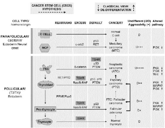
HYROID CANCER IS THE MOST frequent endocrine neoplasia and represents 1% of all cancers. Accord-ing to the American Cancer Society, the frequency of thyroid cancer in USA was 100 per million population in 2003, this incidence has been increasing at more than 5%/yr for a decade mostly due to increased diag-nosis of small tumours (1). The annual mortality from thyroid cancer in 2003 was 5–6 per million. The dis-crepancy between incidence and mortality reflects the good prognosis for most thyroid cancers. Thyroid neoplasms may appear as benign nodules and adeno-mas or malignant tumours that can be from follicular cell origin: differentiated or undifferentiated; from theparafollicular C cells: medullary thyroid carcinoma (MTC), or else. Differentiated carcinomas are hysto-logically divided into papillary thyroid carcinoma (PTC) or follicular thyroid carcinoma (FTC). Detailed descriptions of this classification can be consulted else-where, i.e.: http://www.meb.uni-bonn.de/cancer. gov/CDR0000062913.html. Most thyroid tumours are sporadic as a consequence of somatic mutations, although hereditary thyroid carcinoma resulting from germinal mutations also occurs. Currently there are 2 hypotheses to explain thyroid cancer onset that are summarised in figure 1. The classical view, depicted on the right, considers thyroid carcinoma as a complica-tion of a pre-existing follicular adenoma accumulating mutations, which drive the progression through a de-differentiation process. Differentiated thyrocytes
develop into differentiated thyroid cancer first, then undifferentiated and eventually to anaplastic thyroid carcinoma (ATC). Early thyroid tumour development correlates with mutation of signalling molecules encoded by five alternative genes: Ras, Ret, trk, gsp, and the TSH receptor; additional mutations in a genome caretaker gene such as p53 ensure genomic instability and would drive evolution towards ATC. Discrete de-differentiation steps difficult to explain are required and also it is rather infrequent that a benign adenoma evolves towards carcinoma; at present, it seems that most thyroid carcinomas are malignant from the onset. The original Ras and Ret mutations seen in FTC and PTC are hardly seen in ATC. These and other observations, particularly from Chernobyl irradiation studies, led to the formulation of an alter-native hypothesis based in the existence of cancer stem cells (CSCs) and represented by the horizontal arrows from left to right in figure 1. The CSCs would be derived from embryonic thyroid stem cells, thyroblasts or pro-thyrocytes intermediaries in the pathway of dif-ferentiation accumulating mutations that lead to car-cinogenesis. The CSCs hypothesis proposes that thy-roid cancer develops as a consequence of a block in a differentiation step from any of these intermediaries rather than the process of de-differentiation contem-plated by the classical view. The phenotype and inci-dence of a thyroid cancer type would remember those of the originating CSCs i.e.: the most undifferentiated and aggressive ATC would develop from the earliest and less abundant thyroblasts and would have the lower rate of incidence (2-4). The establishment of ES cell cultures able to differentiate into thyrocytes may clarify these views (5).
THYROTROPIN BINDING TO ITS RECEPTOR IS THE MAIN STIMULUS FOR THYROCYTE PROLIFERATION
Thyrotropin (TSH) secreted by the pituitary and bound to its receptor (TSHR) regulates thyroid growth and differentiation at late developmental stages but is not responsible for organogenesis or cell migration according to the phenotypes of the TSHR knockout mice [(6,7), reviewed in (8,9)]. TSH pro-motes growth of the thyrocyte directly binding to its receptor and also indirectly, stimulating secretion of autocrine growth factors and amyloid precursors (10-12) or the expression of growth factor receptors or of vascular endothelial growth factor, VEGF (13). Addi-tional growth factors, such as insulin/IGF-1 or serum factors (14,15) may be required for TSH induced pro-liferation of the thyrocyte. Thus, paracrine and autocrine factors secreted by follicular cells, the stro-mal apparatus and lymphocytes may be implicated in initiation and perpetuation of thyroid hyperplasia. TSH and its receptor are required not only for prolif-eration in the thyrocyte but also for the expression of differentiation markers such as thyroglobulin, thy-roperoxidase or the Na/I symporter (NIS) that is responsible for iodide uptake. These differentiation markers are required for the correct function of thyro-cytes that is the synthesis of thyroid hormones. TSHR molecules in the membrane are quite stable and sig-nalling in the thyrocyte will be controlled mainly through circulating TSH levels. TSH secretion is inhibited via negative feedback by thyroid hormones; in the absence of thyroid hormones there will be hyper-secretion of TSH and abnormal thyrocyte pro-liferation.
Figure 2.Known regulatory elements in the promoter of the TSHR: The promoter of TSHR is TATA-less and has multiple tran-scription start sites (arrows). GA-binding protein (GABP) dimers binding are sensitive to DNA methylation. The cAMP-response element (CRE-like) encompasses a constitutive enhancer that may bind the activators CREB or ATF2 and the repressor ICER. Binding of single strand binding protein (SSBP to the 5’ and 3’ flanking decanucleotide repeats modulates CRE-like site activi-ty. The CRE-like site overlaps with a thyroid hormone response element. Binding of the heterodimer TR/RXR (thyroid hormone receptor and retinoid X receptor) to this site represses transcription. The proximal promoter bears a binding site for the thyroid transcription factor 1, TTF1 and there is at least another binding site at the far 5’ end. The positions in the promoter relative to the transcription start site appear depicted in brackets underneath the sites. Inside of a transcription factor indicates modula-tion via phosphorylamodula-tion; ∆ indicates modulation via methylation.
•
[-881/-888] 1000
[-185/-179] [-139/-132] [-93/-85] [-89/-88]
THE RELEVANCE OF TSHR FOR THE ONSET/ EVOLUTION OF THYROID CANCER IS UNCLEAR
The importance of the TSHR signalling for the onset/evolution of thyroid cancer is supported by experiments in which regained expression of func-tional TSHR in a follicular thyroid cancer cell line (HTC) reduced angiogenesis and size of tumours of xenotransplanted HTC cells (16). Hyperactivated TSHR is commonly found in most adenomas, less common in differentiated carcinomas and the gene is silenced in undifferentiated cancers such as ATC (table 1, see also figure 1). From the point of view of the classical de-differentiation hypotheses, these data from case studies are difficult to interpret; two muta-tion events on the TSHR gene are highly improbable, but would be required in the evolution from ma to ATC: first hyperactivating mutations in adeno-mas and later silencing in ATC. It would be easier to imagine that the TSHR gene being a susceptible can-didate for mutation would mutate differently in dif-ferent precursor cells undergoing transformation to CSCs. According to the CSCs hypothesis, only one alteration would be required at the TSHR locus to render each transformed phenotype. TSHR implica-tion in the onset of thyroid cancer remains unclear but its functionality is important for thyroid function and treatment of thyroid cancers (see below). Addi-tionally, TSHR may also be relevant in diagnosis. For
example, based on the fact that most thyroid cancers still express the TSHR (17), its mRNA has been used as a highly sensitive and specific marker to detect thy-roid cancer cells in peripheral blood (18). Differenti-ated thyroid cancer has better prognosis because the cells express NIS and can be targeted by radioactive
131I. TSH administration to thyroid cancer patients
with functional TSHR ensures NIS expression, uptake of 131I and the removal of malignant cells left over
after ablation of the cancerous gland or nodule. Moreover, differentiated cancers produce thyroid hormones maintaining reduced pituitary TSH secre-tion. Undifferentiated thyroid carcinomas expressing TSHR but not NIS exist demonstrating that TSHR is required but not sufficient for NIS expression. These cancers are rather difficult to target because radioac-tive 131I will not be taken and insufficient thyroid
hor-mone production will result in elevated TSH levels inducing further proliferation even in cells with diminished TSHR numbers.
Hence, signalling from the TSHR may play a role in the onset, evolution, diagnostic and therapies of thyroid cancer.
THE TSHR GENE AND PROTEIN
The TSHR cloned in 1989 (19-22) belongs to the fa-mily of the G protein coupled receptors (GPCR) and
AA-codon S281N/T/G G431S V463M I486F I486M S505R/N V509A L512R L512Q V556F I568V V597F ∆613-621 T620I A623I M626I I630M F631V F631S F631I D633Y LOCATION Ectodomain TM1 TM2 ECL1 ECL1 TM3 (GL) TM3 (GL) TM3 TM3 TM4 ECL2 TM5 IC3 IC3 IC3 (S+GL) TM6 (S+GL) TM6 (6) TM6 (S+GL) TM6 TM6 TM6 PHENOTYPE
In vitro studies Hyperthyroidism hyperthyroidism adenoma/FTC Follicular adenoma Hyperthyroidism Congenital Hyperthyroidism,
Adenoma, follicular cancer PTC
Hyperthyroidism Multinodular goiter
HTN In vitro studies; non autoinmune hyperthyroidism Thytoxicosis and hyperthyroidism
Toxic thyroid nodules FTC
increased cell proliferation TSH independent Non autoimmune Hyperthyroidism
Hyperfunctioning thyroid nodules Hyperfunctioning thyroid nodules non autoinmune hyperthyroidism
Toxic FTC Toxic FTC
Comments
involved in G protein interaction increased cAMP (Gas) and I3P(Gaq) ECL1is involved in silencing unstimulated TSHR
Interactions TM3- TM5 are important for silencing the unstimulated receptor. Increased cAMP (Gas) and I3P(Gaq).
gain of function
Increased cAMP (Gas) and I3P(Gaq)
critical for G protein interaction
increased basal cAMP production Constitutive activation of Ga/cAMP
REF (131) (132, 133) (134) (123, 135) (135) (114, 136, 137)
(126) (138) (139) ; (140)
contains 7 transmembrane (TM) domains anchored to the basolateral plasma membrane of the thyrocytes and a number of other cells.The TSHR promoter contains functional binding sites for several transcription factors including GABP (23), TTF1 (24), TR/RXR (25), CREB and ICER (26), nevertheless there is little fluc-tuation in the TSHR mRNA levels and regulation of functional TSHR is mainly exerted at the postransla-tional level (27), specially by glycosylation and correct folding at the ER (28).
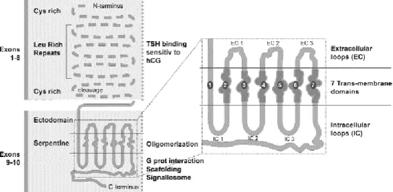
The mature TSHR is encoded by a single gene with 10 exons (29,30). The protein contains 2 sub-units: a large ectodomain also called A or αsubunit is encoded by exons 1–8 and binds TSH; a short trans-membrane and intracellular domain encoded by exons 9–10 called B or β subunit that will interact with G proteins to initiate signalling. Postranslational intramolecular cleavage of a 50 aa chain in the ectodomain close to the membrane and reduction of disulphide bridges releases the A and B subunits (29) followed by shedding of the A subunit from the mem-brane bound receptor (31,32). The presence of the TSHR ectodomain inhibits an otherwise constitutive-ly active β subunit (33) and the interactions between the ectodomain (α), the extracellular loops and trans-membrane domain TM6 (in the βsubunit) are critical for the maintenance of an inactive state (34). Figure 3 recapitulates the main features in the structure of TSHR. The white dotted line overlapping TM5 and
TM6 marks the position of the most frequently found mutations in adenomas. The ectodomain consists mainly of 9 leucine rich repeats (LRR) with intercon-necting loops important for receptor structure and activation (35). Precise delineation of the TSH bind-ing pocket of the receptor has been made through deletion-mutation analysis and a panel of antibodies (36,37). The β subunit contains the 7 TM domains joined by extracellular loops (ECL) and intracellular loops (ICL), and interacts selectively with G proteins when the TSHR is activated (34,38). Unstimulated TSHRs form oligomers that return to the monomer state with TSH (39,40). TSHR oligomerisation is an early posttranslational event detectable by FRET (flu-orescent resonance energy transfer) in the ER-Golgi (41). After TSH binding, a constitutively oligomeric TSHR dissociates into active monomers, which will be subsequently recruited to the lipid rafts to interact with G proteins (42,43). Systematic mutagenesis of the β-subunit has been used to identify critical residues and mechanisms of interactions as illustrated by the following examples. A repulsive separation between TM6 and TM3 in the context of a constitutively active TSHR mutant indicated the opening of the cytoplas-mic face of TSHR for G protein coupling (44); muta-tions in ECL2 and TM6 residues revealed dynamic interactions able to increase or decrease basal TSHR activity (45); deletions and substitutions at the N-ter-minus of ICL2 served to study TSHR coupling to
both Gαand Gqand to demonstrate that ICL2–ICL3
interactions are critical for selective Gqactivation (34).
Upon TSH binding, cytoplasmic proteins such as β-arrestins bind and desensitize GPCRs uncoupling them from G proteins and promoting their internal-ization. TSHR binds β-arrestin 2 but does not colo-calize with β-arrestins in endosomes (46). β-arrestins effects are pleiotropic and have been shown to act also in signal transduction and in transcription. β-arrestins scaffold some activated GPCRs with the Raf/MAPK/ ERK cascade inhibiting nuclear translocation of ERK (47) and some β-arrestins translocate to the nucleus and associate with transcription cofactors such as p300 and CBP at the promoters of genes targeted by the transcription factor CREB (cAMP-Response Element Binding Protein) (48). After desensitizing, TSHR is rapidly internalised by clathrin-coated pits (49,50). Most endocytosed receptors recycle back to the mem-brane, a vital process to maintain the levels. An adap-tor protein, hScrib, is crucial to maintain the correct number of TSHR molecules and to scaffold a correct signalling complex. hScrib interacts with TSHR at the cytoplasmic end of the basolateral membrane of the thyrocyte inhibiting basal TSHR internalization. Upon internalisation, hScrib interaction with the internalised TSHR promotes its recycling. To fulfil this function, hScrib interacts with the C-terminus of the TSHR and recruits several enzymatic activities including GTPase activating proteins and Guanine nucleotide exchange factor (GEF) in a complex required for receptor recycling (51). Finally we must mention here a phenomenon termed “specificity crossover” that consist in the binding and activation of TSHR not only by TSH but also by closely related hormones such as luteinizing (LH) and corionic gonadotropin (hCG) (52,53), a phenomenon that may become important in pathologies that curse with excessive hCG secretion. Indeed variants of hCG with increased TSHR affinity have been described in some patients (54).
THE CLASSICAL PATHWAY AND THE NETWORK THAT FRAMES TSH ACTION IN THE THYROCYTE
Thyrocyte growth occurs mainly through the TSHR mediated increase in cAMP. cAMP-dependent protein kinase (PKA) activation will follow as described below: 1. TSH stimulated TSHR dissociates the hete-rotrimeric G protein activating the Gαssubunit.
2. Gαs-dependent activation of adenilyl cyclase
(AC) follows, increasing cAMP production.
3. cAMP-dependent activates PKA by dissocia-tion of its regulatory subunits.
4. Activated PKA phosphorylates target pro-teins including membrane receptors, signalling mole-cules and transcription factors changing their activities to promote growth and differentiation. The variety of targets will further amplify and diversify the final out-come of this pathway. Perhaps the most classical target for PKA after translocation of its catalytic subunit to the nucleus is the transcription factor CREB, whose transcriptional activity will be promoted upon phos-phorylation by PKA.
Every intermediary in the pathway described may additionally interact with side molecules belonging to other pathways. This will build up a network responsi-ble for refining every response according to a contextu-al environment. Altered wiring of this network inter-connecting TSHR/PKA with other proliferation path-ways such as PI3K, BRaf/MAPK or Wnt plays a pivotal role in cancer, and common targets such as CREB or cyclin D1 may need the integrity of the network to be accurately regulated. We will describe next the classical pathway outlining possible important side branches for thyroid cancer, but since it is out of the scope of this review to analyse them in detail, excellent reviews on each pathway can be consulted elsewhere. Figure 4 out-lines the main effectors of TSHR and interactions with other signalling pathways.
STEP 1: The TSHR stimulated by TSH interacts with heterotrimeric G proteins
TSH induced dissociation of heterotrimeric G proteins leads to Gαand Gβγ activation. There are many sub-types of αand βγsubunits, each combination activat-ing a different set of pathways, and there are excellent reviews on the subject [i.e.: (55)].
Gαproteins
Photoaffinity labelling of the TSHR followed by inmunoprecipitation suggests that TSHR interacts with all four Gα subtypes (Gαs (L), Gαs (S), Gαq, Gα11,
Gi1-3, G0and G12) (56), however TSHR signalling in
the thyrocyte is mainly mediated by Gαsand Gαq
cou-pling to increased cAMP production and phospho-inositide turnover respectively. Gαs stimulation of AC
will increase cAMP, whose major effector is PKA. This path is further analysed below and activating muta-tions of the TSHR and Gαsthat increase AC activity
have been identified in hyperfunctioning benign follic-ular adenomas and less commonly in hypofunctioning adenomas and carcinomas of the thyroid. Gαq/11
stimulates phospholipase C (PLCβ) (57,58), which catalyses hydrolysis of phosphatidil inositols in the membrane yielding di-acyl-glycerol (DAG) and inosi-tol tri phosphate (IP3) as second messengers. DAG directly stimulates PKC. IP3 increase cytosolic Ca+2
levels (59), which act through a number of effectors including PKC. PKC stimulation is important in thy-roid cancer because it is the major effector of tumor promoters such as phorbol esters, and its activation leads to proliferation and de-differentiation in FRTL-5 and PC CL3 thyrocytes (60,61). TSHR mediated activation of the PLC-Ca+2 cascade has been
contro-versial because it requires very high TSH concentra-tions in human primary thyrocytes and in FRTL-5 cells (62-64), however, TSHR clearly increases Ca+2
mobi-lizations at least in certain contexts such as repeated stimulation or simultaneous activation of other GPCRs (65). Increases in PLC-PKC activities have been reported in thyroid carcinomas (66) but it could be due to activation of other receptors because TSHR is poorly expressed in neoplastic tissue (67); moreover,
there is a negative feed back from PKC to PLCβ in thyroid carcinomas (68), and constitutively active mutants of Gαqhave never been found in thyroid
neo-plasms (69), although the mutation induces thyroid hyperplasia in mice. Hence, the significance of the TSHR-Gαq-PLC-PKC in thyroid cancer still remains
obscure. Different G proteins compete for binding of the TSHR (70) mainly Gαsor Gαqin thyrocytes which
activate the PKA or PKC pathways respectively. Func-tional interference between the cAMP/PKA and PKC pathways has been described in normal thyrocytes (71,72) and in hyperfunctioning thyroid adenoma bearing Gαs mutants that induce the cAMP cascade
and suppress the PLC-Ca2+ signalling (73).
Trans-genic mice expressing Gαmutants that constitutively activate both AC and PLC suggest that these cascades may cooperate in vivo towards development of thyroid follicular malignancies (74).
Finally, TSHR may also couple to Gαi1-3, which
will inhibit AC and decrease cAMP levels providing a mechanism to desensitise the TSHR.
Gβγactivation
GPCR activated Gβγ dimers may induce the MAPK and PI3K signalling pathways, which are involved in processes important for tumorigenesis such as prolife-ration and cytoskeletal remodelling. The diversity of Gβγ subunits would allow at least 60 possible combi-nations, but the composition of the Gβγdimers in thy-rocytes has not been examined in that detail. More than 20 effectors have been reported for Gβγactivation including phospholipases (75), ACs (76), other GPCRs and PI3Ks (77). In the thyrocyte, Gβγdirectly activates PLCβ and PKC, which activates the PKB/Akt pathway. Our lab has shown that TSHR mediated Gβγactivation directly stimulates PI3K in rat thyrocytes leading to diminished NIS expression (Zaballos, MA et al. 2007 submitted).
In summary, TSHR coupling to G proteins pro-vides the opportunity for activation of the PKA, PKC and PI3K pathways, but mutations in G proteins hard-ly correlate with thyroid cancers, and hot thyroid nod-ules with constitutively active TSHR mutants very often bear reduced Gαprotein expression presumably as a mechanism of desensitizing the TSHR (78).
STEP 2: TSHR-dependent activation of Gααs
stimulates the AC increasing cAMP levels
cAMP has cell type specific effects and the outcome on proliferation is largely attributed to cross talk with the Ras-Raf-MAPK-ERK pathway. Besides PKA, a num-ber of cAMP effectors may cross talk with the MAPK pathway in the thyrocyte mainly GTP-exchange fac-tors (GEFs or Epac). Finally, other cAMP effecfac-tors in the thyrocyte include cAMP-gated membrane ion channels (79), and some phosphodiesterases (PDEs) such as PDE4. Steady state cAMP levels result from production by AC and degradation by PDEs. cAMP-PKA-dependent PDE4 activation feeds back negative-ly providing a mechanism to stop the signal (80). Given the enormous variety of cAMP effectors, our understanding of this very complex pathway is far from complete.
cAMP-dependent PKA activation by binding to its regulatory subunits is analysed in “Step 3” and here we briefly discuss other cAMP effectors. Although PKA activation in thyroid cells is necessary for cAMP mitogenic effects (15), it is not sufficient (81). In fact, cAMP inhibits growth in some human thyroid tumoral cell lines (82,83) perhaps involving negative feed back mechanisms such as over expression of PDE4, as described for autonomous hyperfunctioning thyroid nodules bearing constitutive activation of the cAMP pathway (84). In autonomous hyperplasic thyroid
ade-nomas with constitutive activation of the cAMP path-way by TSHR and Gαsmutants is not sufficient to
gen-erate toxic thyroid adenomas (85). Cooperation with other signalling pathways initiated by insulin/IGF-1, bFGF, EGF or serum factors (14,15) is required by TSH in thyrocytes to display full mitogenic activity. Other cAMP effectors include GEF or Epac, which activate the small GTPases Rap1, Rap2 and Ras, pro-viding another mechanism for diversification and tun-ing of the signal. The thyrocyte is highly enriched in Epacs (86-88). GTP bound Rap1 may activate Raf-1, B-Raf or C-Raf leading to activation of MAPK/ ERK1/2or p38 MAPKand mitogenesis; Rap1 is over expressed in thyroid follicular cancer (89), and muta-tions in its effector B-Raf are present in most PTC. Activated Ras signalling may lead to MAPK or to PI3K/PDK1activation, both pathways being involved in mitogenesis. Ras is required for cAMP dependent mitogenesis in several rat thyroid cell lines such as FRTL-5 and WRT (90,91) and in 60–70% follicular thyroid adenomas, and carcinomas components of the RET/Ras/B-RAF signalling pathway are mutated (92-96). The role of this pathway is further discussed under the epigraph “Networking the TSHR” and has been widely reviewed elsewhere.
STEP 3: cAMP-dependent activation of PKA
PKA is composed of 2 catalytic and 2 regulatory sub-units that bind cAMP. To date at least 3 different ca-talytic and 4 regulatory subunits have been found and depending on the combination the corresponding PKA molecules will be targeted to cytosolic substrates (PKA I) or to the membrane of certain organelles (PKA II) via a family of proteins called A kinase anchoring proteins or AKAPs (97). Selective activation of the cytosolic or membrane anchored forms of PKA in the thyrocyte has demonstrated specialised func-tions. PKA I stimulation increased iodide uptake in FRTL5 cells without affecting gene transcription while selective PKA II activation induced gene transcription and proliferation in FRTL5 cells (98).
II appears to be more clearly involved: silencing its regulatory subunit RIIβ impairs TSH nuclear effects, and loss of RIIβexpression in three human cancer cell lines suggests an essential role for PKA II in TSHR-mediated proliferation (98).
The transcription factor CREB is the classical nuclear target of PKA, although it is also targeted by other pathways, and PKA also targets other nuclear factors as we will see below.
STEP 4: PKA dependent activation of the transcription factor CREB
The ubiquitous transcription factor CREB binds to cAMP response elements and upon activation stimu-lates transcription at selected promoters (88). Tran-scription factors from the CREB/CREM family are required for cAMP-dependent proliferation in dog thy-roid primary cultures and are tightly regulated by TSH and other factors (26). CREB activity has been shown to be important for TSH dependent thyrocyte prolifer-ation in vitro and in vivo although it is not sufficient to mimic TSH-dependent DNA synthesis. In vitro, the importance of CREB was tested by stable transfection of FRTL-5 cells with either wild type CREB, which did not affect growth, or a dominant negative version dnCREB, which reduced up to 40% TSH-induced growth of the thyrocytes (100). In vivo, transgenic mice with targeted expression of dnCREB to their thy-roid glands exhibited severe growth retardation and primary hypothyroidism; dnCREB inhibited the expression of the genes for Pax8, TTF-1, and TTF-2, which are required for the expression of TSHR, thy-roglobulin and thyroperoxidase (101). Alterations of CREB family members can be observed in endocrine tumors (102), and levels of total CREB are markedly reduced in thyroid carcinomas (103), but their role in thyroid cancer remains highly controversial. Contradic-tory results have been reported in hyperfunctioning thyroid adenomas: some laboratories find a reduction of P-CREB (Ser 133) (104) while others do not find differences between nodular and extranodular tissue (103). Perhaps the interpretation of these results is too simplistic having in mind the multiple modifications that would affect CREB activity. It is generally believed that recruitment of the coactivators CREB-binding protein (CBP) and p300 after signal induced phospho-rylation of CREB at Ser 133 strongly enhances its tran-scriptional activity. However, a number of kinases may phosphorylate CREB in Ser 133 including PKA, PKB/Akt, PKC, Rsk1/2. CREB activation can also be promoted by Ser 133 independent mechanisms and not all the signals that induce Ser 133 phosphorylation
of CREB enhance its transcriptional activity. Further-more, CREB is subjected to other phosphorylations, de-phosphorylations (by PP2A and PP1), acetylation by CBP, ubiquitylation and SUMOylation and, depending on its modifications, CREB interacts with other proteins that may perturb its localisation and/or turnover leading to activity changes [reviewed in (105,106)]. Thus CREB acts as a platform targeted by multiple signalling pathways where interference or inte-gration takes place to render a gene expression profile extraordinarily tuned to incoming signals. CREB may use coactivators that are effectors of other signalling pathways (i.e. the Wnt effector β-catenin) and targets a number of genes involved in diverse aspects of prolifer-ation, for example cyclin D1, which is also targeted by the Wnt signalling pathway and others. This provides new opportunities for integration. A complete genome wide analysis of CREB target genes has recently been published (107). Furthermore, other transcription (co)factors involved in proliferation such as β-catenin and others are also activated by PKA.
TSHR ALTERATIONS RELATED TO THYROID CANCER
Excesses or defaults in TSHR activity may play a role in thyroid disease and cancer. Both can be achieved by a number of mechanisms including: improper epige-netic marking of the gene, incorrect transcriptional regulation or mutations in critical domains.
Altered levels of TSHR expression
Lack or low TSHR expression correlates with aberrant methylation of the promoter in human thyroid carci-nomas and in thyroid cancer cell lines (108). The silencing mechanism may also include the binding of the methylation-sensitive GABP transcription factor (23). TSHR function is required for NIS expression and iodide uptake. Low or absent TSHR expression and lack of NIS expression correlates with thyroid car-cinomas of the worse prognostic that cannot be elimi-nated with radioactive iodide (109). Excessive TSHR expression does not correlate with malignant thyroid cancer to our knowledge, although a number of con-stitutively activating mutations have been described in thyroid adenomas as we describe below.
Mutations also change the levels of functional TSHR
modifications or protein-protein interactions, including influences by and on other GPCRs, scaffolding, signal-ing mediators, desensitation, etc. TSHR activity is main-ly controlled through protein-protein interactions and constitutively active mutants correlate with thyroid ade-nomas and nodules, while inactivating mutations corre-late with diverse forms of hypothyroidism.
Inactivating mutations
More than 25 distinct loss-of-function mutations in the TSHR gene have been reported (41,110), including those occurring at the germ line and causing congenital hypothyroidism. So far none of these mutations appear to be related to the onset of thyroid cancer. Inactive TSHR fails to induce thyroid cell proliferation and is unlikely to induce nodule formation. Reduced TSHR expression in thyroid cancers may be secondary to ongo-ing de-differentiation, may happen in parallel or may cause disappearance of other differentiation markers.
The TSHR gene is highly susceptible to constitu-tively activating mutations
Structural studies discarded TSHR as a candidate onco-gene for thyroid tumor (111), however G protein cou-pling is a critical step and most gain of function muta-tions are located in exons 9 and 10 corresponding to TM domains (112,113), ICLs or ECLs (114,115). Not sur-prisingly, these sites are critical for G protein coupling (see figure 3). A list of TSHR mutants may be found at <http://www.uni-leipzig.de/innere/tshr> and we have updated it in table 1. Spontaneous activating mutations of the TSHR gene appear at the onset of autonomous functioning thyroid adenomas (116,117) and more rarely of thyroid carcinomas (118-124). Somatic tions cause autonomous nodules and germ-line muta-tions cause congenital hyperthyroidism and hereditary non-autoimmune toxic thyroid hyperplasia. Patients with activating TSHR mutations suffer hyperthyroidism; the disease activity directly correlates with the degree of TSHR activation measured as basal cAMP production (125). Likewise, thyroid nodules and goitre develop ear-lier in patients carrying TSHR variants with high consti-tutive receptor (126-130) [reviewed by (131)]. Further-more, in multinodular goiters different TSHR activating mutations have been found in separate hot nodules of the same gland suggesting a role in true nodule forma-tion (112). Constitutively activating mutaforma-tions lie also at the root of toxic thyroid adenomas and differentiated thyroid carcinomas (120). Meanwhile, Gαsmutations are
very infrequent in hyperthyroidism or toxic adenomas, suggesting that this subunit does not play an important role, a question intensely debated (132,133). Some
authors have compared the characteristics and location of the most frequently appearing germ-line and somatic mutations in the TSHR gene. Germ-line mutations are mostly transitions and affect residues [183, 505, 509, and 597] never involved in somatic mutations. Somatic mutations are usually transversions and affect residues 630 and 633 never affected by germ-line mutations. Finally, several residues located in a mutation cluster region [619–639] at TM6 are affected by both somatic and germinal mutations (134).
Thus, the question of whether or not TSHR mutations are involved in the onset of thyroid cancers remains controversial as explained in previous sections. Interpretations are complicated by the fact that biological effects of activating TSHR mutations vary with the ambi-ent iodide supply. In regions of iodine deficiency, higher incidence of toxic adenoma and toxic multi-nodular goitre has been reported, where 50–80% of these toxic adenomas are caused by TSHR mutations (135).
Postranslational modifications and protein interactions
NETWORKING THE TSH RECEPTOR SIGNALLING IN THYROID CANCER
Cancer arises from accumulation of mutations in impor-tant genes or oncogenes. Most known oncogenes are key-signalling molecules important for the integrity of the signalling networks that maintain cell homeostasis. Oncogenes relevant to thyroid carcinogenesis are nor-mally engaged in proliferation and/or survival pathways, the paradigms are RET/Ras/B-Raf, PTEN/Akt and E-cadherin/β-catenin representing the MAPK, PI3K and Wnt pathways respectively. These pathways are integrat-ed in the thyrocyte signalling network among them and with the cAMP/PKA pathway and altering their cross-talk may lead to carcinogenesis.
The MAPK pathway and its cross talk with TSHR/cAMP/PKA
The MAPK pathway in the thyrocyte conveys prolife-ration signals from tyrosine kinase receptors such as RET. Hyperactivating RET mutations in the kinase domain define MTCs and rearrangements (RET/ PTCs) are frequent at early onset of PTCs; RET mutants signal through B-Raf to activate the MAPK pathway in thyroid cells (160), and B-Raf mutations (V600E) are present in more than 40% of PTCs (161,162). The significance of the RET/Ras/B-Raf/ ERK pathway in thyroid carcinogenesis has been widely reviewed (163). Crosstalk between the Ras/ MAPK/ERK and the cAMP/PKA pathways has long been recognised and cAMP may either sup-press or induce the MAPK pathway depending on the cell type (164). RET/PTC2 is a particularly interesting rearrangement cross-linking the MAPK and PKA pathways because the kinase domain of RET is fused to the regulatory subunit of PKA II (165). In FRTL-5 rat thyrocytes, both TSH and cAMP induce ERK (166), and these cells require Ras for TSH-stimulated mitogenesis (90,91); in dog and human thyrocytes, indirect evidence suggest the same because a MEK inhibitor blocks TSH stimulat-ed DNA synthesis (167). cAMP may activate B-Raf and ERK through PKA-dependent or independent mechanisms that need further clarification at the thy-rocyte. Thus, in thyroid cells cAMP does not inhibit MAPK signalling but whether it activates it or not remains controversial due to differences among dis-tinct in vitro systems (15). The high sensitivity to B-Raf mutations exhibited by thyrocytes in cancer is intriguing and remarkable and perhaps is exacerbated by the lack of cAMP-dependent mechanisms to sup-press B-Raf/ERK activity.
The PI3K/AKT pathway and its crosstalk with TSHR/cAMP/PKA
PI3K/AKT is a pleiotropic kinase downstream of many growth factor receptors involved in cell survival, prolifer-ation and cancer. Indirect evidence of the role of PI3K in thyroid cancer comes from the use of an Akt inhibitor (KP372-1) that suppresses proliferation and induces apoptosis in thyroid cancer cells (168). Activating muta-tions of Akt or silencing its upstream suppressor PTEN (Phosphatase and TENsin homolog) is frequently asso-ciated with thyroid carcinoma (169). PTEN is a dual specificity phosphatase mutated or silenced in the major-ity of human advanced cancers. Also in thyroid cancers has been found loss, reduction, or inappropriate subcel-lular compartmentalization of PTEN (170,171). A rearrangement with the histone H4 gene has been found in irradiated thyroid cell lines, H4/PTEN (172). PTEN silencing has been suggested to be involved in the car-cinogenesis of highly malignant or late-stage thyroid cancers (170,173), and reintroduction of PTEN in thy-roid cancer cell lines causes G1 arrest in the differentiat-ed and apoptosis in the most undifferentiatdifferentiat-ed thyroid cancer cell lines (174). TSHR immunoprecipitates exhibit PI3K activity, which is greater after TSH treat-ment. Concomitantly, the kinase PDK1 is redistributed from the cytoplasm to the plasma membrane in a PI3K-and PKA-dependent manner after TSH treatment (175). The regulatory subunit of PI3K, p85 phosphorylated at Ser 83, binds the regulatory subunit of PKA, RIIβand mediate TSH-cAMP-PKA growth and survival signals (176). Moreover, the PI3K/Akt pathway has been sug-gested to be activated in thyroid tumors by RET onco-proteins that associate with RAI (ShcC/N-Shc) and recruit GAB 1 (Grb 2-associated binder 1) (177). Addi-tionally, PI3K is involved in IGF-1 or insulin mediated cooperation with TSH for DNA synthesis in thyrocytes. The interactions between the Ras/Raf/MEK/ERK and Ras/PI3K/PTEN/Akt pathways are crucial to regulate growth and may be altered in tumorigenesis.
The Wnt pathway
con-trol thyrocyte proliferation. PKA is known to potenti-ate Wnt signalling in some cells stabilizing β-catenin and inhibiting GSK3β (183,184). The CREB tran-scription factor might be targeted by both PKA and Wnt signalling, and recently its role in lithium-stimu-lated thyrocyte proliferation has been challenged (185). Although these authors conclude that CREB does not play a role in lithium-induced thyroid proli-feration, their experiments do not rule out a possible role of CREB in Wnt-dependent proliferation of thy-rocytes. Some components of the Wnt pathway (GSK3β) are also targeted by PI3K. Cyclin D1 is a transcriptional target for both CREB and β-catenin and is over expressed in thyroid papillary microcarci-noma with aberrant β-catenin (186).
In summary, mutations of individual compo-nents may cause fundamental functional changes well beyond the pathway they function in, due to the crosstalk between signalling pathways. One of the molecules most mutated in cancer in general and in ATC also is p53, another common target for many sig-nalling pathways including PI3K, cAMP, and ERK. p53 is inactivated in 50% of human cancers and 14% of malignant thyroid tumors; p53 mutations appear “late” in thyroid carcinogenesis and are associated with loss of differentiation and transformation to the anaplastic phenotype (132). p53 is a transcription fac-tor and plays multiple regulafac-tory functions in the cell cycle, DNA repair, and apoptosis. Reintroduction of wild type p53 in thyroid tumoral cell lines arrests growth and/or induces apoptosis (21,187). p53 pro-tein levels may be increased by cAMP, ERK, and also through the activation of PTEN, and recently a differ-ential proteomic approach has identified several tar-geted proteins for mutant p53 associated with thyroid cell transformation (188). The TSHR and p53 con-nection in thyrocytes still has to be clarified but it is remarkable that stable expression of a p53 mutant which does not bind DNA (V143A), induces loss of differentiation markers and TSH-independent growth in PC CL3 thyrocytes. In contrast, a p53 mutant (S392A), which does not interfere with DNA binding, causes only loss of TSH dependency for growth (188).
FUTURE PERSPECTIVES
Undifferentiated thyroid cancers are among the most rapid life-threatening and are characterised by abnormal CpG islands methylation patterns in critical promoters. Silencing of TSHR, PTEN and p53 appears to be relat-ed to the most aggressive behaviour of thyroid cancer,
and reintroduction of each of these genes in human thy-roid cancer cell lines either restores differentiation or arrest cell cycle or causes apoptosis. Hypomethylation of heterochromatin has been correlated with tumour pro-gression in thyroid cancer specimens (189), and hyper-methylation of tumor supressor promoters appears in undifferentiated thyroid cancer causing failure in clinical radioiodine treatment (171,173). Thus, aberrant DNA methylation patterns are some of the epigenetic marking processes more likely involved in the onset of thyroid cancer. Demethylating agents or inhibitors of the DNA methylase may be promising hopes in the treatment of the worse thyroid cancers, but their clinical value remains to be investigated and they have to be specifically target-ed. In general, the development of molecular-based therapies for thyroid carcinoma patients resistant to stan-dard radioiodine treatment such as histone deacetylase inhibitors combined with retinoids or the use of specific targeted RNAis for invasion/metastasis molecules repre-sent a critical issue and an exciting field in new drug development in thyroid cancer.
Large chromatin alterations related to thyroid cancer, such as the chromosomal rearrangements: RET/PTCs or Pax8/PPARγchimeras (190), coincide in their activation of the Ras/Raf/MEK/ERK path-way, which is also induced by TSHR signalling in the thyrocyte. Chromatin alterations related to cancer, especially epigenetic modifications, need to be further explored, especially lost of imprinting (LOI) and lost of heterozigosity (LOH) studies will reveal new clues to the onset of cancer.
Finally, an exciting aspect in thyroid cancer research is the definition and biological characterization of the precursor cells that give rise to thyroid carcinomas. Mouse embryonic stem cells might now be differentiat-ed into thyroid follicular cells in vitro (5), and this system will surely help to identify key events in thyroid carcino-genesis and understanding the role of TSHR.
ACKNOWLEDGEMENTS
We are grateful to grants URJC-CM-2006-BIO-0522 (to CG-J) and BFU 2004-03 169, and FIS PI 041216, PI 042374, RD06 0020 0060 (to PS) for supporting our work.
REFERENCES
2. Takano T. Fetal cell carcinogenesis of the thyroid: a hypothe-sis for better understanding of gene expression profile and genomic alternation in thyroid carcinoma. Endocr J 2004;51:509-15.
3. Takano T. Fetal cell carcinogenesis of the thyroid: Theory and practice. Semin Cancer Biol 2007;17(3):233-40.
4. Takano T, Amino N. Fetal cell carcinogenesis: a new hypoth-esis for better understanding of thyroid carcinoma. Thyroid 2005;15:432-8.
5. Arufe MC, Lu M, Kubo A, Keller G, Davies TF, Lin RY. Direct-ed differentiation of mouse embryonic stem cells into thyroid follicular cells. Endocrinology 2006;147:3007-15.
6. Marians RC, Ng L, Blair HC, Unger P, Graves PN, Davies TF. Defining thyrotropin-dependent and -independent steps of thyroid hormone synthesis by using thyrotropin receptor-null mice. Proc Natl Acad Sci U S A 2002;99:15776-81. 7. Postiglione MP, Parlato R, Rodriguez-Mallon A, Rosica A,
Mithbaokar P, Maresca M, et al. Role of the thyroid-stimulat-ing hormone receptor signalthyroid-stimulat-ing in development and differen-tiation of the thyroid gland. Proc Natl Acad Sci U S A 2002;99:15462-7.
8. Brown SR. Minireview: developmental regulation of thy-rotropin receptor gene expression in the fetal and newborn thyroid. Endocrinology 2004;145:4058-61.
9. Felice DM, Postiglione MP, Lauro DR. Minireview: thyrotropin receptor signaling in development and differentiation of the thyroid gland: insights from mouse models and human dis-eases. Endocrinology 2004;145:4062-7.
10. Takahashi S, Conti M, Wyk VJ. Thyrotropin potentiation of insulin-like growth factor-I dependent deoxribonucleic acid synthesis in FRTL-5 cells: mediation by an autocrine amplifi-cation factor(s). Endocrinology 1990;126:736-45.
11. Becks GP, Logan A, Phillips ID, Wang JF, Smith C, DeSousa D, et al. Increase of basic fibroblast growth factor (FGF) and FGF receptor messenger RNA during rat thyroid hyperplasia: temporal changes and cellular distribution. J Endocrinol 1994;142:325-38.
12. Pietrzik CU, Hoffmann J, Stober K, Chen CY, Bauer C, Otero DA, et al. From differentiation to proliferation: the secretory amyloid precursor protein as a local mediator of growth in thyroid epithelial cells. Proc Natl Acad Sci U S A 1998;95:1770-5.
13. Hoffmann S, Hofbauer LC, Scharrenbach V, Wunderlich A, Hassan I, Lingelbach S, et al. Thyrotropin (TSH)-induced pro-duction of vascular endothelial growth factor in thyroid can-cer cells in vitro: evaluation of TSH signal transduction and of angiogenesis-stimulating growth factors. J Clin Endocrinol Metab 2004;89:6139-45.
14. Medina DL, Suzuki K, Pietrarelli M, Okajima F, Kohn LD, Santisteban P. Role of insulin and serum on thyrotropin regulation of thyroid transcription factor-1 and pax-8 genes expression in FRTL-5 thyroid cells. Thyroid 2000 ;10:295-303.
15. Kimura T, Van Keymeulen A, Golstein J, Fusco A, Dumont JE, Roger PP. Regulation of thyroid cell proliferation by TSH and other factors: a critical evaluation of in vitro models. Endocr Rev 2001;22:631-56.
16. Hoffmann S, Maschuw K, Hassan I, Wunderlich A, Lingelbach S, Ramaswamy A, et al. Functional thyrotropin receptor attenuates malignant phenotype of follicular thyroid cancer cells. Endocrine 2006;30:129-38.
17. Ohta K, Endo T, Onaya T. The mRNA levels of thyrotropin receptor, thyroglobulin and thyroid peroxidase in neoplastic
human thyroid tissues. Biochem Biophys Res Commun
1991;174:1148-53.
18. Chia SY, Milas M, Reddy SK, Siperstein A, Skugor M, Brainard J, et al. TSH receptor mRNA measurement in blood as a marker for circulating thyroid cancer cells and its role in the preoperative diagnosis of thyroid cancer. J Clin Endocrinol Metab 2007;92(2):468-75.
19. Parmentier M, Libert F, Maenhaut C, Lefort A, Gerard C, Per-ret J, et al. Molecular cloning of the thyrotropin receptor. Sci-ence 1989;246:1620-2.
20. Libert F, Lefort A, Gerard C, Parmentier M, Perret J, Ludgate M, et al. Cloning, sequencing and expression of the human thyrotropin (TSH) receptor: evidence for binding
of autoantibodies. Biochem Biophys Res Commun
1989;165:1250-5.
21. Nagayama Y, Kaufman DK, Seto P, Rapoport B. Molecular cloning, sequence and functional expression of the cDNA for the human thyrotropin receptor. Biochem Biophys Res Commun 1989;165:1184-90.
22. Misrahi M, Loosfelt H, Atger M, Sar S, Guiochon-Mantel A, Milgrom E. Cloning, sequencing and expression of human
TSH receptor. Biochem Biophys Res Commun 1990;
166:394-403.
23. Yokomori N, Tawata M, Saito T, Shimura H, Onaya T. Regu-lation of the rat thyrotropin receptor gene by the methyRegu-lation- methylation-sensitive transcription factor GA-binding protein. Mol Endocrinol 1998;12:1241-9.
24. Ohe K, Ikuyama S, Takayanagi R, Kohn LD, Nawata H. Inter-feron-gamma suppresses thyrotropin receptor promoter activity by reducing thyroid transcription factor-1 (TTF-1) binding to its recognition site. Mol Endocrinol 1996; 10:826-36.
25. Chen TS, Lin DJ, Lin HK. Characterization of a thyroid hor-mone-mediated short-loop feedback control of TSH receptor gene in an anaplastic human thyroid cancer cell line. J Endocrinol 2002;175:459-65.
26. Uyttersprot N, Costagliola S, Dumont EJ, Miot F. Require-ment for cAMP-response eleRequire-ment (CRE) binding protein/CRE modulator transcription factors in thyrotropin-induced prolif-eration of dog thyroid cells in primary culture. Eur J Biochem 1999;259:370-8.
27. Seetharamaiah SG, Dallas SJ, Prabhakar SB. Glycosylated ectodomain of the human thyrotropin receptor induces anti-bodies capable of reacting with multiple blocking antibody epitopes. Autoimmunity 1999;29:21-31.
28. Siffroi PJ, Bourhis LC, Dadoune PJ. Collecting human sper-matozoa onto filters for FISH. Application to the study of extreme oligozoospermia. Acta Cytol 2002;46:1123-8. 29. Loosfelt H, Pichon C, Jolivet A, Misrahi M, Caillou B, Jamous
M, et al. Two-subunit structure of the human thyrotropin receptor. Proc Natl Acad Sci U S A 1992;89:3765-9. 30. Misrahi M, Ghinea N, Sar S, Saunier B, Jolivet A, Loosfelt H,
et al. Processing of the precursors of the human thyroid-stim-ulating hormone receptor in various eukaryotic cells (human thyrocytes, transfected L cells and baculovirus-infected insect cells). Eur J Biochem 1994;222:711-9.
31. Couet J, Sar S, Jolivet A, Hai MT, Milgrom E, Misrahi M. Shedding of human thyrotropin receptor ectodomain. Involvement of a matrix metalloprotease. J Biol Chem 1996;271:4545-52.
32. Tanaka K, Chazenbalk GD, McLachlan SM, Rapoport B. Sub-unit structure of thyrotropin receptors expressed on the cell surface. J Biol Chem 1999;274:33979-84.
33. Zhang M, Tong KP, Fremont V, Chen J, Narayan P, Puett D, et al. The extracellular domain suppresses constitutive activity of the transmembrane domain of the human TSH receptor: implications for hormone-receptor interaction and antagonist design. Endocrinology 2000;141:3514-7.
34. Neumann S, Krause G, Claus M, Paschke R. Structural deter-minants for g protein activation and selectivity in the second intracellular loop of the thyrotropin receptor. Endocrinology 2005;146:477-85.
35. Costagliola S, Khoo D, Vassart G. Production of bioactive amino-terminal domain of the thyrotropin receptor via inser-tion in the plasma membrane by a glycosylphosphatidylinos-itol anchor. FEBS Lett 1998;436:427-33.
36. Jeffreys J, Depraetere H, Sanders J, Oda Y, Evans M, Kiddie A, et al. Characterization of the thyrotropin binding pocket. Thyroid 2002;12:1051-61.
38. Vlaeminck-Guillem V, Ho SC, Rodien P, Vassart G, Costaglio-la S. Activation of the cAMP pathway by the TSH receptor involves switching of the ectodomain from a tethered inverse agonist to an agonist. Mol Endocrinol 2002;16:736-46. 39. Graves PN, Vlase H, Bobovnikova Y, Davies TF. Multimeric
complex formation by the thyrotropin receptor in solubilized thyroid membranes. Endocrinology 1996;137:3915-20. 40. Latif R, Graves P, Davies FT. Ligand-dependent inhibition of
oligomerization at the human thyrotropin receptor. J Biol Chem 2002;277:45059-67.
41. Calebiro D, de Filippis T, Lucchi S, Covino C, Panigone S, Beck-Peccoz P, et al. Intracellular entrapment of wild-type TSH receptor by oligomerization with mutants linked to dom-inant TSH resistance. Hum Mol Genet 2005;14:2991-3002. 42. Moffett S, Brown AD, Linder EM. Lipid-dependent targeting
of G proteins into rafts. J Biol Chem 2000;275:2191-8. 43. Davies T, Marians R, Latif R. The TSH receptor reveals itself.
J Clin Invest 2002;110:161-4.
44. Ringkananont U, Van Durme J, Montanelli L, Ugrasbul F, Yu YM, Weiss RE, et al. Repulsive separation of the cytoplasmic ends of transmembrane helices 3 and 6 is linked to receptor activation in a novel thyrotropin receptor mutant (M626I). Mol Endocrinol 2006;20:893-903.
45. Kleinau G, Brehm M, Wiedemann U, Labudde D, Leser U, Krause G. Implications for molecular mechanisms of glyco-protein hormone receptors using a new sequence-structure-function analysis resource. Mol Endocrinol 2007;21:574-80. 46. Frenzel R, Voigt C, Paschke R. The human thyrotropin recep-tor is predominantly internalized by beta-arrestin 2. Endocrinology 2006;147:3114-22.
47. Caunt CJ, Finch AR, Sedgley KR, Oakley L, Luttrell LM, McAr-dle CA. Arrestin-mediated ERK activation by gonadotropin-releasing hormone receptors: receptor-specific activation
mechanisms and compartmentalization. J Biol Chem
2006;281:2701-10.
48. Ma L, Pei G. β-arrestin signaling and regulation of transcrip-tion. J Cell Sci 2007;120:213-8.
49. Baratti-Elbaz C, Ghinea N, Lahuna O, Loosfelt H, Pichon C, Milgrom E. Internalization and recycling pathways of the thy-rotropin receptor. Mol Endocrinol 1999;13:1751-65. 50. Singh PS, McDonald D, Hope JT, Prabhakar BS. Upon
thy-rotropin binding the thythy-rotropin receptor is internalized and localized to endosome. Endocrinology 2004;145:1003-10. 51. Lahuna O, Quellari M, Achard C, Nola S, Meduri G, Navarro
C, et al. Thyrotropin receptor trafficking relies on the hScrib-βPIX-GIT1-ARF6 pathway. Embo J 2005;24:1364-74. 52. Graves NP, Davies FT. Absence of lutropin (LH) receptor
mRNA in the rat thyroid: further evidence for specificity cross-over at the thyroid-stimulating hormone receptor level. Mol Cell Endocrinol 1991;79:21-8.
53. Urizar E, Montanelli L, Loy T, Bonomi M, Swillens S, Gales C, et al. Glycoprotein hormone receptors: link between receptor homodimerization and negative cooperativity. Embo J 2005;24:1954-64.
54. Kraiem Z, Lahat N, Sadeh O, Blithe DL, Nisula BC. Desialylat-ed and deglycosylatDesialylat-ed human chorionic gonadotropin are superagonists of native human chorionic gonadotropin in human thyroid follicles. Thyroid 1997;7:783-8.
55. Cabrera-Vera TM, Vanhauwe J, Thomas TO, Medkova M, Preininger A, Mazzoni MR, et al. Insights into G protein struc-ture, function, and regulation. Endocr Rev 2003;24:765-81. 56. Laugwitz LK, Allgeier A, Offermanns S, Spicher K, Van Sande
J, Dumont JE, et al. The human thyrotropin receptor: a hepta-helical receptor capable of stimulating members of all four G protein families. Proc Natl Acad Sci U S A 1996;93:116-20. 57. Jhon DY, Lee HH, Park D, Lee CW, Lee KH, Yoo OJ, et al. Cloning, sequencing, purification, and Gq-dependent activa-tion of phospholipase C-β3. J Biol Chem 1993;268:6654-61. 58. Allgeier A, Offermanns S, Van Sande J, Spicher K, Schultz G, Dumont JE. The human thyrotropin receptor activates G-pro-teins Gs and Gq/11. J Biol Chem 1994;269:13733-5. 59. Newton AC. Regulation of protein kinase C. Curr Opin Cell
Biol 1997;9:161-7.
60. Gallo A, Benusiglio E, Bonapace IM, Feliciello A, Cassano S, Garbi C, et al. v-ras and protein kinase C dedifferentiate thy-roid cells by down-regulating nuclear cAMP-dependent pro-tein kinase A. Genes Dev 1992;6:1621-30.
61. Portella G, Vitagliano D, Li Z, Sferratore F, Santoro M, Vec-chio G, et al. TPA induces a block of differentiation and increases the susceptibility to neoplastic transformation of a rat thyroid epithelial cell line. Oncol Res 1998;10:441-7. 62. Wang JF, Hill DJ, Becks GP. Role of 3’, 5’ cyclic adenosine
monophosphate and protein kinase C in the regulation of insulin-like growth factor-binding protein secretion by thy-roid-stimulating hormone in isolated ovine thyroid cells. J Endocrinol 1994;141:231-42.
63. D’Arcangelo D, Silletta MG, Di Francesco AL, Bonfitto N, Di Cerbo A, Falasca M, et al. Physiological concentrations of thy-rotropin increase cytosolic calcium levels in primary cultures
of human thyroid cells. J Clin Endocrinol Metab
1995;80:1136-43.
64. Yanagita Y, Okajima F, Sho K, Nagamachi Y, Kondo Y. An adenosine derivative cooperates with TSH and Graves’ IgG to induce Ca2+mobilization in single human thyroid cells. Mol
Cell Endocrinol 1996;118:47-56.
65. Metcalfe RA, Findlay C, Robertson WR, Weetman AP, Mac Neil S. Differential effect of thyroid-stimulating hormone (TSH) on intracellular free calcium and cAMP in cells
trans-fected with the human TSH receptor. J Endocrinol
1998;157:415-24.
66. Kobayashi K, Shaver JK, Liang W, Siperstein AE, Duh QY, Clark OH. Increased phospholipase C activity in neoplastic thyroid membrane. Thyroid 1993;3:25-9.
67. Brabant G, Maenhaut C, Kohrle J, Scheumann G, Dralle H, Hoang-Vu C, et al. Human thyrotropin receptor gene: expres-sion in thyroid tumors and correlation to markers of thyroid differentiation and dedifferentiation. Mol Cell Endocrinol 1991;82:R7-12.
68. Broecker M, Mayr GW, Derwah MI. Suppression of thy-rotropin receptor-G protein-phospholipase C coupling by activation of protein kinase C in thyroid carcinoma cells. Endocrinology 1997;138:3787-96.
69. Ringel MD, Saji M, Schwindinger WF, Segev D, Zeiger MA, Levine MA. Absence of activating mutations of the genes encoding the αsubunits of G11 and Gq in thyroid neoplasia. J Clin Endocrinol Metab 1998;83:554-9.
70. Cleator JH, Ravenell R, Kurtz DT, Hildebrandt JD. A dominant negative Gαs mutant that prevents thyroid-stimulating hor-mone receptor activation of cAMP production and inositol 1,4,5-trisphosphate turnover: competition by different G pro-teins for activation by a common receptor. J Biol Chem 2004;279:36601-7.
71. Sho KM, Okajima F, Abdul Majid M, Kondo Y. Reciprocal modulation of thyrotropin actions by P1-purinergic agonists in FRTL-5 thyroid cells. Inhibition of cAMP pathway and stim-ulation of phospholipase C-Ca2+ pathway. J Biol Chem
1991;266:12180-4.
72. Laglia G, Zeiger MA, Leipricht A, Caturegli P, Levine MA, Kohn LD, et al. Increased cyclic adenosine 3’,5’-monophos-phate inhibits G protein-coupled activation of phospholi-pase C in rat FRTL-5 thyroid cells. Endocrinology 1996; 137:3170-6.
73. Kamiya Y, Murakami M, Yanagita Y, Koitabashi H, Naga-machi Y, Hosoi Y, et al. Primary culture of cells from hyper-functioning thyroid adenoma with an activating mutation of Gαs. Mol Cell Endocrinol 1998;138:137-42.
74. Ledent C, Denef JF, Cottecchia S, Lefkowitz R, Dumont J, Vas-sart G, et al. Costimulation of adenylyl cyclase and phospho-lipase C by a mutant α-1B-adrenergic receptor transgene pro-motes malignant transformation of thyroid follicular cells. Endocrinology 1997;138:369-78.
77. Vanhaesebroeck B, Leevers SJ, Panayotou G, Waterfield MD. Phosphoinositide 3-kinases: a conserved family of signal transducers. Trends Biochem Sci 1997;22:267-72. 78. Holzapfel HP, Bergner B, Wonerow P, Paschke R. Expression
of Gαs proteins and TSH receptor signalling in hyperfunc-tioning thyroid nodules with TSH receptor mutations. Eur J Endocrinol 2002;147:109-16.
79. Kaupp UB, Seifert R. Cyclic nucleotide-gated ion channels. Physiol Rev 2002;82:769-824.
80. MacKenzie SJ, Baillie GS, McPhee I, MacKenzie C, Seamons R, McSorley T, et al. Long PDE4 cAMP specific phosphodi-esterases are activated by protein kinase A-mediated phos-phorylation of a single serine residue in Upstream Conserved Region 1 (UCR1). Br J Pharmacol 2002;136:421-33. 81. Dremier S, Vandeput F, Zwartkruis FJ, Bos JL, Dumont JE,
Maenhaut C. Activation of the small G protein Rap1 in dog thyroid cells by both cAMP-dependent and -independent
pathways. Biochem Biophys Res Commun 2000
;267:7-11.
82. Derwahl M, Kuemmel M, Goretzki P, Schatz H, Broecker M. Expression of the human TSH receptor in a human thyroid carcinoma cell line that lacks an endogenous TSH receptor: growth inhibition by cAMP. Biochem Biophys Res Com-mun 1993;191:1131-8.
83. Ohta K, Pang XP, Berg L, Hershman JM. Growth inhibition of new human thyroid carcinoma cell lines by activation of adenylate cyclase through the β-adrenergic receptor. J Clin Endocrinol Metab 1997;82:2633-8.
84. Persani L, Lania A, Alberti L, Romoli R, Mantovani G, Filetti S, et al. Induction of specific phosphodiesterase isoforms by constitutive activation of the cAMP pathway in autonomous
thyroid adenomas. J Clin Endocrinol Metab 2000;
85:2872-8.
85. Derwahl M, Manole D, Sobke A, Broecker M. Pathogenesis of toxic thyroid adenomas and nodules: relevance of activating mutations in the TSH-receptor and Gαs gene, the possible role of iodine deficiency and secondary and TSH-indepen-dent molecular mechanisms. Exp Clin Endocrinol Dia-betes 1998;106(suppl 4):S6-9.
86. Kawasaki H, Springett GM, Mochizuki N, Toki S, Nakaya M, Matsuda M, et al. A family of cAMP-binding proteins that directly activate Rap1. Science 1998;282:2275-9.
87. Kawasaki H, Springett GM, Toki S, Canales JJ, Harlan P, Blu-menstiel JP, et al. A Rap guanine nucleotide exchange factor enriched highly in the basal ganglia. Proc Natl Acad Sci U S A 1998;95:13278-83.
88. Richards JS. New signaling pathways for hormones and cyclic adenosine 3’,5’-monophosphate action in endocrine cells. Mol Endocrinol 2001;15:209-18.
89. Puskas LG, Juhasz F, Zarva A, Hackler L Jr, Farid NR. Gene profiling identifies genes specific for well-differentiated epithelial thyroid tumors. Cell Mol Biol (Noisy-le-grand) 2005;51:177-86.
90. Miller MJ, Rioux L, Prendergast GV, Cannon S, White MA, Meinkoth JL. Differential effects of protein kinase A on Ras effector pathways. Mol Cell Biol 1998;18:3718-26. 91. Ciullo I, Diez-Roux G, Di Domenico M, Migliaccio A,
Avvedi-mento EV. cAMP signaling selectively influences Ras effec-tors pathways. Oncogene 2001;20:1186-92.
92. Fukushima T, Suzuki S, Mashiko M, Ohtake T, Endo Y, Take-bayashi Y, et al. BRAF mutations in papillary carcinomas of the thyroid. Oncogene 2003;22:6455-7.
93. Kimura ET, Nikiforova MN, Zhu Z, Knauf JA, Nikiforov YE, Fagin JA. High prevalence of BRAF mutations in thyroid can-cer: genetic evidence for constitutive activation of the RET/PTC-RAS-BRAF signaling pathway in papillary thyroid carcinoma. Cancer Res 2003;63:1454-7.
94. Soares P, Maximo V, Sobrinho-Simões M. Molecular pathol-ogy of papillary, follicular and Hurthle cell carcinomas of the thyroid. Arkh Patol 2003;65:45-7.
95. Xu X, Quiros RM, Gattuso P, Ain KB, Prinz RA. High preva-lence of BRAF gene mutation in papillary thyroid carcinomas and thyroid tumor cell lines. Cancer Res 2003;63:4561-7.
96. Williams SF, Smallridge RC. Targeting the ERK pathway: novel therapeutics for thyroid cancer. Curr Drug Targets Immune Endocr Metabol Disord 2004;4:199-220. 97. Wong W, Scott JD. AKAP signalling complexes: focal points
in space and time. Nat Rev Mol Cell Biol 2004;5:959-70. 98. Calebiro D, de Filippis T, Lucchi S, Martinez F, Porazzi P,
Triv-ellato R, et al. Selective modulation of protein kinase A I and II reveals distinct roles in thyroid cell gene expression and growth. Mol Endocrinol 2006;20:3196-211.
99. Kirschner LS, Carney JA, Pack SD, Taymans SE, Giatzakis C, Cho YS, et al. Mutations of the gene encoding the protein kinase A type I-alpha regulatory subunit in patients with the Carney complex. Nat Genet 2000;26:89-92.
100.Woloshin PI, Walton KM, Rehfuss RP, Goodman RH, Cone RD. 3’,5’-cyclic adenosine monophosphate-regulated enhancer binding (CREB) activity is required for normal growth and differentiated phenotype in the FRTL5 thyroid fol-licular cell line. Mol Endocrinol 1992;6:1725-33.
101.Nguyen LQ, Kopp P, Martinson F, Stanfield K, Roth SI, Jame-son JL. A dominant negative CREB (cAMP response element-binding protein) isoform inhibits thyrocyte growth, thyroid-specific gene expression, differentiation, and function. Mol Endocrinol 2000;14:1448-61.
102.Rosenberg D, Groussin L, Jullian E, Perlemoine K, Bertagna X, Bertherat J. Role of the PKA-regulated transcription factor CREB in development and tumorigenesis of endocrine tis-sues. Ann N Y Acad Sci 2002;968:65-74.
103.Luciani P, Buci L, Conforti B, Tonacchera M, Agretti P, Elisei R, et al. Expression of cAMP response element-binding pro-tein and sodium iodide symporter in benign non-functioning
and malignant thyroid tumours. Eur J Endocrinol
2003;148:579-86.
104.Brunetti A, Chiefari E, Filetti S, Russo D. The 3’,5’-cyclic adenosine monophosphate response element binding pro-tein (CREB) is functionally reduced in human toxic thyroid adenomas. Endocrinology 2000;141:722-30.
105.Johannessen M, Delghandi MP, Moens U. What turns CREB on? Cell Signal 2004;16:1211-27.
106.Johannessen M, Moens U. Multisite phosphorylation of the cAMP response element-binding protein (CREB) by a diversi-ty of protein kinases. Front Biosci 2007;12:1814-32. 107.Zhang X, Odom DT, Koo SH, Conkright MD, Canettieri G, Best
J, et al. Genome-wide analysis of cAMP-response element binding protein occupancy, phosphorylation, and target gene activation in human tissues. Proc Natl Acad Sci U S A 2005;102:4459-64.
108.Xing M, Usadel H, Cohen Y, Tokumaru Y, Guo Z, Westra WB, et al. Methylation of the thyroid-stimulating hormone recep-tor gene in epithelial thyroid tumors: a marker of malignancy and a cause of gene silencing. Cancer Res 2003;63:2316-21. 109.Mirebeau-Prunier D, Guyetant S, Rodien P, Franc B, Baris O, Rohmer V, et al. Decreased expression of thyrotropin recep-tor gene suggests a high-risk subgroup for oncocytic adeno-ma. Eur J Endocrinol 2004;150:269-76.
110.Fricke-Otto S, Pfarr N, Muhlenberg R, Pohlenz J. Mild con-genital primary hypothyroidism in a Turkish family caused by a homozygous missense thyrotropin receptor (TSHR) gene
mutation (A593 V). Exp Clin Endocrinol Diabetes
2005;113:582-5.
111.Matsuo K, Friedman E, Gejman PV, Fagin JA. The thyrotropin receptor (TSH-R) is not an oncogene for thyroid tumors: structural studies of the TSH-R and the alpha-subunit of Gs in
human thyroid neoplasms. J Clin Endocrinol Metab
1993;76:1446-51.
112.Krohn K, Paschke R. Somatic mutations in thyroid nodular disease. Mol Genet Metab 2002;75:202-8.
113.Rodien P, Ho SC, Vlaeminck V, Vassart G, Costagliola S. Acti-vating mutations of TSH receptor. Ann Endocrinol (Paris) 2003;64:12-6.