ABSTRACT
Iodine is a trace element that is essential for the synthesis of thyroid hormone. Both chronic iodine deficiency and iodine excess have been associated with hypertrophy and hyperplasia of follicular cells, attributed to excessive secretion of TSH. This may be associated to thyroid cancer risk, particularly in women. Exper-imental studies have documented thyroid cancer induction by elevation of endogenous TSH, although in a small number of animals. Iodine deficiency asso-ciated with carcinogenic agents and chemical mutagens will result in a higher inci-dence of thyroid malignancy. Inadequate low iodine intake will result in increased TSH stimulation, increased thyroid cell responsiveness to TSH, increased thyroid cell EGF-induced proliferation, decreased TGFβ1 production and increased angio-genesis, all phenomena related to promotion of tumor growth. Epidemiological studies associating iodine intake and thyroid cancer led to controversial and con-flicting results. There is no doubt that introduction of universal iodine prophylax-is in population previously in chronic iodine-deficiency leads to a changing pat-tern of more prevalent papillary thyroid cancer and declining of follicular thyroid cancer. Also anaplastic thyroid cancer is practically not seen after years of iodine supplementation. Iodine excess has also been indicated as a possible nutritional factor in the prevalence of differentiated thyroid cancer in Iceland, Hawaii and, more recently, in China. In conclusion: available evidence from animal experi-ments, epidemiological studies and iodine prophylaxis has demonstrated a shift towards a rise in papillary carcinoma, but no clear relationship between overall thyroid cancer incidence and iodine intake. (Arq Bras Endocrinol Metab 2007;51/5:701-712)
Keywords:Iodine; Thyroid cancer; Iodine deficiency; Iodine excess; Carcino-genic agents; Environmental effect
RESUMO
Relevância da Ingestão de Iodo como Fator Predisponente ao Câncer de Tireóide.
O iodo é essencial para a síntese de hormônios tireóideos e tanto a deficiência crônica deste halogeno como o excesso nutricional de iodo levam a hiperplasia e hipertrofia dos elementos foliculares (por excesso de TSH). Esse fenômeno pode se associar a maior risco de câncer de tireóide, especialmente no sexo feminino. Estudos experimentais documentam indução de câncer de tireóide após prolongado excesso circulante de TSH, o qual induz aumento da pro-liferação celular medida por fator de crescimento epidermal (EGF), decréscimo de síntese de fator de transformação do crescimento (TGFβ1) e aumento da angiogenese. Estudos epidemiológicos entre nutrição de iodo e câncer de tireóide são conflitantes. É, todavia, aceito que a correção de prévia deficiência de iodo com aporte nutricional adequado deste halogeno leva à maior pre-valência de carcinoma papilífero (e decréscimo de carcinoma folicular). Em alguns países, o excesso de iodo foi apontado como causa aparente de maior prevalência de câncer de tireóide. Em conclusão: não existe uma relação causa-efeito entre iodo nutricional e prevalência de câncer de tireóide, e outros fatores intervenientes ambientais devem ser considerados. (Arq Bras Endocrinol Metab 2007;51/5:701-712)
Descritores:Iodo nutricional; Câncer de tireóide; Deficiência de iodo; Excesso de iodo; Agentes carcinogênicos; Efeito ambiental
revisão
MEYERKNOBEL
GERALDOMEDEIROS-NETO
Thyroid Unit, Division of
Endocrinology and Metabolism, Hospital das Clínicas (MK), and Department of Internal Medicine University of São Paulo Medical School (GM-N), São Paulo, SP.
T
HYROID CARCINOMA IS THE MOST commonly encountered endocrine malignancy (1). Its inci-dence among all ages rose steadily in different countries in the last 10 years, whereas its mortality rates dropped (2). The declining mortality rates are largely due to early diagnosis and effective therapy applied at an early tumor stage when it is most amenable to surgery and 131I therapy. Those tumors have a marked variability in aggressiveness from highly differentiated papillary carcinomas with a good prognosis, if found and treated early, to undifferentiated anaplastic cancers occurring mainly in older people and with a poor prognosis (3).Iodine is a trace element that is essential in the synthesis of thyroid hormones. Both chronic iodine deficiency and chronically high iodine intake have been associated with the development of goiter (i.e., hypertrophy and hyperplasia of the thyroid cells) and attributed to excessive secretion of TSH by the pitu-itary. In turn, goiter has been associated with thyroid cancer risk, particularly in women (4).
A clear relationship between iodine supply to the population and thyroid cancer incidence has not been proven and is still debated (3). In general, chron-ic iodine defchron-iciency and residence in an endemchron-ic goiter area are associated with an increased risk of follicular histological type of cancer, whereas chronically high iodine intake may increase the risk of the more com-mon papillary histological type of thyroid cancer.
This outline summarizes available data from animal experiments, epidemiological studies and from the introduction of iodine prophylaxis on the possible association between iodine deficiency and the devel-opment of differentiated thyroid carcinoma.
EXPERIMENTAL INDUCTION OF THYROID TUMORS
Thyroid neoplasia may be induced by exposure of experimental animals to a variety of treatment regi-mens, exogenous chemicals or physical agents.
Tumor induction by elevation of TSH
It has been recognized for some time that neoplasms induced in experimental animals by a number of treat-ments result in thyroid gland dysfunction, more fre-quently hypothyroidism.
Among the thyroid cancer-causing factors induc-ing a hypothyroid state are iodine deficiency (5,6) and subtotal thyroidectomy (7). In addition, thyroid tumors can result from the transplantation of TSH-secreting pituitary tumors (7-9). The one factor common to each
of these conditions is that they all lead to increased pro-duction of TSH and prolonged stimulation of the thyroid gland by continuously elevated serum TSH. In the first two conditions, elevated TSH results from chronic stim-ulation of the pituitary in response to a deficiency in the circulating levels of thyroid hormone. It is worth mention note that nothing has been administered to these animals. Therefore the tumors developed in the absence of factors that are normally present in the animals (i.e., iodine and thyroid gland mass). It should rightfully be pointed out, however, that the animals were under chronic stress due to deficiency of thyroid hormone. In the third case, excess TSH comes from the transplanted pituitary tumor. Thus, irrespective of the cause, it appears that prolonged stimu-lation of the thyroid pituitary that results in release of ele-vated levels of TSH by the thyrotrophs may lead to thy-roid gland neoplasia (10).
Tumor induction by low iodine intake
The hypothesis of an association between iodine intake and the risk for thyroid cancer is based on the results of studies in experimental animals, which showed that animals on an iodine-restricted diet were more likely to develop cancer (6,11). On this basis, overstimula-tion by thyroid-stimulating hormone (TSH) was con-sidered to play a major role in tumorigenesis. At vari-ance, recent studies have shown, however, that the serum levels of TSH are in fact lower in individuals with mild-moderate iodine deficiency (12).
In golden hamsters submitted to long-term low-iodine diet, it has been experimentally shown hyperplasia, then nodular goiter and, eventually, in a small percentage of animals, carcinomas. The sponta-neous malignancies reported in the iodine-deficient animals were follicular adenocarcinomas; 18% of females, but no thyroid lesions were found in males after a 70-week iodine-deficient diet. (11).
The risk that a benign thyroid tumor may develop after iodine deficiency or excess is 2- to 3-fold higher than in controls. It was suggested that under the experimental conditions both long-term iodine deficiency and excess are insufficient to stimulate carcinogenesis, whereas benign tumors, detectable in approximately 90% of the animals after 110 weeks of treatment, were clearly more frequently found as compared to the control group with normal iodine supply. It seems that previous studies contradict these data (5,14).
Iodine-deficient Fisher rats were shown to develop chromosomal abnormalities (aneuploidy) of some heterogeneous thyroid cell population. These cells may have acquired neoplastic characteristics, for when such a cell population is transplanted, a neoplas-tic tumor is produced (15).
Studies with combination of tumor-inducing factors
Several investigations have indicated that combined treatment regimens are associated with thyroid car-cinogenetic responses in excess of that produced by either single treatment alone.
A few direct acting exogenous chemical com-pounds have been identified that produce thyroid tumors that do not appear to influence the thyroid-pituitary status, as these compounds did not increase thyroid weights unrelated to tumor development or cause a persistent elevation of serum TSH levels (16). Two of them are nitrosomethylurea (NMU) and N-bis(2-hydroxypropyl)-nitrosamine (DHPN), classified as genotoxic, since involves direct carcinogenic effects at the DNA level in follicular cells.
Non-genotoxic agents, as iodine, may alter the negative feedback system of the thyroid-pituitary hypothalamus axis, leading to TSH increase and its prolonged stimulation of follicular epithelium (17) as already mentioned.
The thyroid combined treatment studies are consistent with the concepts of initiation-promotion. The genotoxic agent might permanently alter the thy-roid cell so that its accentuated growth under a goitro-genic stimulus would result in neoplasms (6).
Rats previously treated with NMU and then placed on an iodine-deficient diet, had increased inci-dence of thyroid follicular cell carcinomas (90%) in comparison to 10% of rats exposed to iodine deficiency alone. On the other hand, about 20% of the rats devel-oped follicular carcinomas when treated with the same carcinogen, but in normal conditions for iodine nutri-tion. These observations indicated that the iodine-defi-cient diet is a potent promoter of thyroid tumors
initi-ated by NMU and also a carcinogen by itself (18). Moreover, the results suggested that the non-genotox-ic component, i.e., elevation of TSH, plays a key role in the development of thyroid lesions (19).
In another study, F344 rats were given the chemical mutagen DHPN and supplemented with var-ious amounts of potassium iodide in drinking water to generate conditions that extended to chronic iodine deficiency to severe iodine excess. In DHPN-treated rats, both conditions significantly increased thyroid follicular tumorigenesis. In DHPN-untreated rats, iodine deficiency produced diffuse thyroid hyperplasia, together with a decrease in serum thyroxine and an increase in serum TSH. On the other hand, iodine excess produced colloid goiter, together with normal serum T4 and slightly decreased TSH. Under influ-ence of DHPN there was a rapid rise in tumor inci-dence with increasingly low levels of dietary iodine. Interestingly, in this study there was a slight rise in tumor incidence at high iodide levels. These effects were directly proportional to the severity of iodine deficiency or extent of iodine excess and suggest that each condition has a different thyroid tumor promo-tion mechanism (20) (figure 1).
In rats, iodine deficiency is much more effective as a tumor promoter as compared to its carcinogenic effect, suggesting that a similar relationship may exist in human populations. However, it should be empha-sized that data from animal studies in relation to thy-roid tumors may not be extended to human thythy-roid.
Figure 1.In this experimental study conducted by Kanno et al. (20), tumor-promoting effects were observed clearly in iodine deficiency, when using a chemical carcinogen. Data includes adenomas and carcinomas of the thyroid. Note, however, that animals fed excessive nutritional iodine had an increase in tumor prevalence.
80
60
40
20
0
0.1 1 10 100 1000
Iodine intake, µg/day
In the former, thyroid tumors can be reproduced pre-cisely and studied exactly, but the relevance of these tumors to human lesions is doubtful since they show a pattern of morphology and behavior that is strictly dif-ferent from human tumors.
CELL BIOLOGY OF THYROID CARCINOGENESIS
Thyroid carcinogenesis is a multi-stage phenomenon and is considered to involve basically an oncogene mutation, response to growth factors and the clonali-ty of the neoplastic tissue architecture (21). All condi-tions that favor prolonged stimulation of cell prolifer-ation are apt to facilitate pre-malignant transforma-tion. The increased number of cell division gives to protooncogenes an enhanced probability to be activat-ed and to oncosuppressor genes to be damagactivat-ed (22).
The characteristic feature of malignancy is an increased growth rate of the cell, and the development of a tumor can be thought of as a series of steps, each marked by the growth of a clone with higher growth rate than the rest of the lesion. Moreover, the impor-tance of post-mutagen growth in the thyroid is well shown by two observations: a) there is a great increase in incidence of rodent thyroid tumors if TSH is ele-vated by goitrogen treatment or by a low-iodide diet after mutagen administration, as DHPN or NMU, and b) thyroid carcinogenesis is abolished if suppressive thyroxine treatment or hypophysectomy is used imme-diately after mutagen exposure preventing any TSH-induced growth (23,24).
Unlike many other highly specialized cells, thy-roid cells are not irreversibly terminally differentiated. When they proliferate in response to certain growth signals they temporarily lose the ability to concentrate iodide and to synthesize thyroglobulin. Certain unreg-ulated growth signals associated with malignant trans-formation may contribute to the loss of differentiated thyroid function. The thyroid cell turns over, on aver-age, no more than five times through adulthood (25). Turnover increases in early infancy and adolescence (25,26). Because of the homogeneity of the growth potential of follicular cells (27), a fraction of these fol-licular cells in adults would likely turnover much more than five times. It is assumed that cycles of cell division may be more frequent in benign or malignant tumors (27,28), although evidence from human tumors does not always support this notion (29).
An understanding of the factors involved in nor-mal human thyroid cell growth, differentiation, and sig-naling is an essential prelude to understanding aberrations
related to thyroid cell transformation. Unfortunately, the picture is incomplete due to the variability among species of factors that sustain thyroid proliferation and the path-ways used (30). Growth factors also play a key role, along with TSH, in the complex regulation of thyroid cell pro-liferation, but there are few data yet to explain how these trophic factors interact with the cell cycle to stimulate or inhibit cell division in the thyroid.
A variety of mechanisms cooperate to deter-mine thyroid cell stimulation to proliferate in condi-tions of chronic iodine deficiency (31).
Increased TSH stimulation
Thyrotropin, through activation of its receptor, has been shown to stimulate more than one signal trans-duction pathway in the regulation of both growth and differentiated function. Each pathway may be related to specific cellular events. The main effector of TSH on proliferation and differentiation in a variety of species, humans and rats included, is the cAMP signal transduc-tion pathway, that is, the cascade involving activatransduc-tion of adenylate cyclase resulting in cAMP generation (33).
Evidence concerning the mitogenic role of TSH for thyroid cells in vivo has been further consol-idated over recent years. Studies from various labora-tories using tritiated thymidine labeling, metaphase arrest techniques for mitotic index, or immunohisto-chemical decoration of statin (a non-proliferation-spe-cific nuclear antigen identifying quiescent Go-phase cells) show that TSH stimulates, in a dose- and time-dependent way, the recruitment of noncycling Gocells into the cycling compartment, as well as entry into S-phase and the entry of G2cells into mitosis (32).
Recent work has indicated that the normal rodent (and human) thyroid may have an intrinsic het-erogeneity in the follicular cell population regarding the capacity for proliferative response to TSH. One hypothesis suggests that there are a few subsets of stemlike follicular cells with a high growth potential compared to the majority of the population and that this trait is stable and heritable (34). According to this model, the clones of cells with extensive proliferative potential are the origin of the adenomas that develop under conditions of chronic TSH stimulation (35).
select preexisting thyrocyte clones with the greatest proliferative potential and thus with the greater risk of neoplastic transformation. An alternative model for explaining the self-limited nature of hyperthyroidism and the development of hormone-responsive tumors in the chronically stimulated rat thyroid proposes that clones of cells escape from the desensitization mecha-nism through mutational events and natural selection, leading ultimately to tumor formation (19). Both models agree that thyroid carcinogenesis involves rare subsets of cells responsive to continued TSH stimula-tion but differ concerning the origin of follicular cell heterogeneity. Additional support for the intrinsic subset concept and/or the controlling influence of TSH on the development of selected clones of thyroid follicular cells comes from other studies on cell prolif-eration (33) and from those using transplantation methodology (38).
The impaired synthesis of thyroid hormones, due to the iodine shortage, will result in (subclinical) hypothy-roidism and increased TSH secretion. TSH induces human thyroid cell growth at a higher concentration than is necessary for induction of differentiated function (39).
Increased thyroid cell responsiveness to TSH Iodine deficient thyroid cells will have an increased content of the two major intracellular messengers for the TSH signaling pathways, cAMP and Ca++ (39). Under this condition, the thyroid cell is sensitized to TSH stimulation, which elicit responses that are inap-propriately high for a given stimulation.
There is evidence that iodine is a major media-tor of thyroid aumedia-toregulation, involving numerous inhibitory actions (41). One of these is a decrease in cAMP formation in response to TSH, resulting in an inhibition of all cAMP-mediated stimulatory effects of TSH on the thyroid. Excess iodide therefore exerts a negative control on different thyroid parameters, inhibiting iodide uptake and organification, protein and RNA biosynthesis, hormone secretion, as well as paracrine mitogenic activity on endothelial cells and fibroblasts (42).
Most of these actions appear to be mediated by an intracellular organified iodine intermediate of unknown identity. Various derivatives of arachidonic acid have been proposed as this putative regulator(s), including the iodinated eicosanoid, delta-iodolactone (42).
The iodolactone growth inhibitory mechanism, which is decreased in iodine-deficient thyroid cells, is believed to occur via inhibition of the specific EGF-dependent production of IP3, a signal transducer that
modulates thyroid cell proliferation (43). However, this compound had no effect on TSH-mediated cAMP formation in porcine follicles (43). Considered to be a more likely candidate is 2-iodohexadecanal (44), a major iodolipid formed in horse thyroid when incu-bated with iodide (45), but which is also detectable in the thyroid of other species, including humans and rats.
Increased thyroid cell EGF-induced proliferation Growth factors also play a key role, along with TSH, in the complex regulation of thyroid cell proliferation, but there are few data yet to explain how these troph-ic factors interact with the cell cycle to stimulate or inhibit cell division in the thyroid.
These autocrine/paracrine regulators of thyroid growth, with potent mitogenic activity for thyroid cells demonstrable in vitro, include epidermal growth factor (EGF) and basic fibroblast growth factor (bFGF). EGF is synthesized within the thyroid gland and induces proliferation in thyroid cells from a wide range of species at the expense of dedifferentiation and loss of specialized thyroid-specific function (46). Over expression of EGF and GF-receptor in RNAS were detected in dyshormonogenetic goiters that were under constant and prolonged endogenous TSH stim-ulation (47). Basic FGF (fibroblast growth factor) is present in human thyroid tissue (48) and there are stores of FGF in the basement membrane of follicles in normal adult rat thyroid (49). Moreover, bFGF expression increases during thyroid hyperplasia in rodents (50).
EGF has been shown to stimulate the growth and invasion of differentiated human thyroid cancer cells in culture and in nude mice (51). In some tissue culture systems TSH does not stimulate thyroid cell growth unless other growth factors as EGF are present (52). EGF binding in thyroid neoplasms is also some-what higher than in normal thyroid tissue from the same patients (53).
Decreased TGFβproduction
a striking correlation being observed between the dual presence of TGFβ1 expression and arginine substitution at codon 61 of the H-ras oncogene (56). Lazzereschi et al. (57) demonstrated a significant reduction of TGFβ
receptor type II mRNA and protein expression in thy-roid carcinomas but found no reduction in thythy-roid ade-nomas. These studies suggest that escape from the growth inhibitory effect of TGFβ1 may play an impor-tant role in the progression of human thyroid cancer.
Iodine increases the thyroid production of TGFβ1 (58), a process that is related to iodine organ-ification. Iodine deficiency, therefore, may result in a decrease of thyroid cell growth negative regulation.
Increased angiogenesis
Iodine deficiency causes thyroid enlargement (goiter), which is characterized also by formation of new vessels. The factors regulating angiogenesis in thyroid are not yet well established. Many of the factors elaborated by the thyroid, e.g., IGF-I (59) and FGF (60), also act in vivo on endothelial cells to induce neovascularization (59) and to enhance adhesion molecule synthesis to support thyroid growth. Data from animal thyroid tissue support the hypothesis that capillary growth during thyroid enlargement occurs, at least in part, as a result of a parenchymal-stromal (epithelial-mesenchymal) paracrine interaction mediated by specific endotheliotropic (angio-genic) factors released by follicular epithelial cells (62). FGF is also a potent thyroid cell mitogen (63). In agree-ment, serum vascular endothelial growth factor (VEGF) is significantly elevated in patients with metastatic differ-entiated thyroid cancer but not in those with poorly dif-ferentiated thyroid cancer metastases (64). Furthermore, in hypothyroid subjects, serum VEGF levels correlate with TSH values (65).
Thus, the proposed mechanisms implicating growth factors associating low iodine intake to thyroid carcinogenesis can be summarized as follows:
•Increased TSH stimulation: low T4 synthesis associ-ated to higher TSH secretion will promote follicular cell proliferation
•Thyroid cell responsiveness to TSH: is increased in iodine deficient thyroid cell (increased Ca++ and cAMP pathways)
•Increased thyroid cells EGF-induced proliferation: decreased intracellular organified iodine intermediate (iodolactone or 2-iodohexadecanal) will result in EGF-induced cell proliferation
•Decreased TGFβ production: thyroid cell prolifera-tion is inhibited by TGFβ. Iodine deficiency will result in a decrease of growth negative regulation
•Increased angiogenesis: may promote tumor growth.
EPIDEMIOLOGICAL STUDIES, RELATIONSHIP BETWEEN IODINE INTAKE AND THYROID CANCER
HISTOTYPES
Thyroid carcinoma is an infrequent tumor with geo-graphic variation in its prevalence. Papillary carcinoma accounts for 85–90% of differentiated thyroid cancers in iodide-sufficient countries (66), while the remaining epithelial thyroid cell tumors are predominantly follicu-lar carcinomas. Anaplastic carcinoma, whose incidence has been decreasing, often arises from preexisting well-differentiated (usually follicular) carcinoma (67).
Epidemiologists search for clues to causes of disease and to factors that increase an individual’s risk of disease (risk factors) by examining descriptive data or designing analytic studies. Descriptive data consist of morbidity, mortality or incidence rates of diseases in population groups. Incidence rates (newly diagnosed cases in a population over a given time period) reveal patterns of disease by age, race, sex, ethnic group and geographic localization. Those rates and their changes over time identify high-risk groups and provide indi-rect evidence for causes of disease. Analytical epidemi-ology consists of case control, often termed retrospec-tive and cohort or prospecretrospec-tive studies. These studies permit greater control of confounding factors and an opportunity to link exposure and response information in individuals. Thus, evidence for causes of disease is more direct.
Numerous studies have been carried out in the populations of distinct geographical areas known to have different iodine intake and in the same area, before and after changes in population iodine intake, in order to evaluate the discrepancies in the prevalence of thyroid cancer. Most of them, however, are consid-ered to be biased by either ethnic differences in the studies populations and also by environmental factors, other than iodine deficiency, that may also influence thyroid tumorigenesis, like exposure to radiation, goitrogens or diet. The distinct procedures used by the investigators for both surveying cancer and evalu-ating iodine deficiency may also have affected the results obtained.
The epidemiologic approach to investigating whether goiter leads to thyroid cancer in humans is based in the following procedures:
1.examine descriptive data
2.compare the thyroid cancer rates between endemic goiter areas and goiter-free areas
4.evaluate whether goitrous individuals have a greater risk of thyroid cancer or whether thyroid cancer cases have a more frequent history of hyperplasia and nodules than controls.
Clinically evident thyroid nodules and benign goiters occur frequently in iodine-deficient areas. A population-based study carried out in Sicily in two adjacent areas with different iodine intake but similar ethnic and life-style characteristics, first surveyed cold thyroid nodules and then using fine needle aspiration and cytology which percentage of cancer occurred among the cold nodules in two populations (68). This study indicated that the risk of thyroid cancer is increased (relative risk = 1.4) in the population living in the iodine deficient area.
The relationship of thyroid cancer to endemic goiter is widely accepted (69,70). In agreement, it was found a geographical correlation between endemic areas for goiter and thyroid cancer. A higher incidence of thyroid cancer was found in England, Wales and Switzerland in areas with endemic goiter (70,71). However, no association was found in the United States between goitrous and non-goitrous areas (72). Another study performed in Nordic countries as Fin-land, IceFin-land, Norway and Sweden over periods rang-ing from 1 to 14 years found no association between the presence of endemic goiter and the risk of follicu-lar carcinoma. Further, there was a marked negative association between endemic goiter and the risk of papillary carcinoma (73).
The search for a similar relationship between the occurrence of malignant thyroid tumors and iodine intake has been controversial and with conflicting results. The overall incidence of thyroid carcinoma is generally considered without influence from the iodine intake in a given population (3), although a study from two regions in Italy has recently demon-strated a two-fold increase in thyroid cancer incidence in an iodine-deficient area compared to an iodine-suf-ficient one (68). Contrary to this, some of the highest incidence rates of thyroid cancer are found in Iceland (74) and Hawaii (75) (table 1), both known to have long-standing high iodine intake, although exposure to volcanic activity (where the natural radiation is higher and radiation is known to increase the develop-ment of thyroid carcinomas) has been suggested as an explanation for the high incidence in these islands.
Very recently, another study compared the thy-roid cancer incidence by morphological subtypes in the eastern (with mild iodine deficiency) and western (with moderate iodine deficiency) Denmark, in the period of 1973– 1997. No regional difference was found in the overall incidence of follicular or papillary thyroid cancer (76).
Overall the above data indicate that:
•nutritional iodine intake and incidence of cancer remains a controversial issue
•there is a weak evidence that low iodine intake would increase the temporal incidence of thyroid malignancy in a given population
Table 1.Iodine-rich areas and incidence of thyroid cancer.
Country* (year) Thyroid cancer after Incidence rate Reference surgery x 105inhabitants (%)
Iceland (1977) 186 6.0 74
Hawaii (1988) 1472 8.1 75
3.1 * Both countries have a very high iodine intake through seafood.
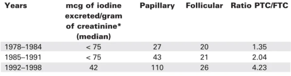
Table 2.Changing incidence of thyroid cancer in Tasmania during transition from iodine sufficiency to iodine deficiency: histologic types.
Years mcg of iodine Papillary Follicular Ratio PTC/FTC excreted/gram
of creatinine* (median)
1978–1984 < 75 27 20 1.35
1985–1991 < 75 43 21 2.04
1992–1998 42 110 26 4.23
•high iodine intake, however, is also associated with an increased incidence of thyroid cancer (other envi-ronmental factors may be present).
Differences in iodine intake also affect the dis-tribution of morphological subtypes of thyroid cancer, although the findings are also equivocal.
It has been hypothesized that iodine deficiency causes a high incidence of follicular tumors and that iodine supplementation shifts the distribution towards papillary tumors, as observed in Switzerland (70), Ger-many (77) and in Austria (78) (figure 1).
A study in Tasmania during a period when iodine intake changed from sufficient to deficient (1978–1998) showed a similar trend, although this followed a period of iodine supplementation, which complicates the interpretation (79) (table 2).
In a Swedish study, papillary thyroid cancer was commoner in iodine-rich areas, and follicular cancer was more common in iodine-depleted areas. After an iodine supplementation program, the incidence of papillary cancer rose and that of follicular cancer fell, but these trends were the same in both areas, and thus unrelated to the supplementation program (80). These inconsis-tencies in the results of these studies might be explained by different causation patterns for the different histolog-ical subtypes. In line with this argument, a case control study in Sweden of 484 cases of thyroid cancers during 1980–1992 (80) showed that the length of residence in an iodine-deficient area where goiter had been endemic was most strongly associated with an increased risk of thyroid follicular cancer (RR = 1.3–1.5). Moreover, for papillary cancer, an increased risk (RR) to 2.5 was observed only for women exposed to iodine-deficiency during puberty (81). Overall there remains a general view that no convincing evidence has yet emerged to link environmental thyroid cancer with areas of iodine defi-ciency (82-84). In a recent study on the effect of iodine intake on thyroid diseases in China, three representative communities, respectively with deficient iodine intake (A), normal iodine intake (B) and excessive iodine intake (C), were surveyed for thyroid diseases in a period of five years. No cases of thyroid cancer were identified in areas A and B but, at baseline, 10 patients with thyroid cancer were identified in the area C (excessive iodine intake). Moreover 13 new cases of thyroid cancer were diag-nosed in this area C but none were found in the two other regions (A, B). Although this study has some lim-itations, it points to the fact that excessive iodine intake may be linked to a higher prevalence of thyroid cancer as compared to populations with low or adequate Iodine nutrition (85). More recently, Camargo et al. (86) stud-ied urinary excretion of iodine, thyroid function tests,
thyroid antibodies and sonographic characteristics of 829 inhabitants of a suburban area of great São Paulo city. This population had been on excessive iodine intake (59.5% excreted more than 300 µg Iodine/L of urine) for six years (1998–2003). Although there was an increase in prevalence of autoimmune thyroiditis (17.9%), 18 nodules (2.18%) were considered suspicious for malignancy (none have yet been submitted to surgery). In order to reach a conclusion about excessive intake and thyroid cancer it would be necessary to fol-low-up subjects with nodules form this population for at least two more years and to obtain histological evidence (after surgery) for suspicious nodules.
There is also some further evidence that has come from studies in areas with previous iodine defi-ciency before and after iodine prophylaxis, that the incidence rates of the type of thyroid carcinoma may be changing. In one such study from an area in Argentina (82), the ratio of papillary to follicular car-cinomas rose from 1.7:1 to 3.1:1 (table 3).
Another study demonstrated that the histo-pathological types of thyroid cancer are different in iodi-ne deficient areas and iodiiodi-ne sufficient oiodi-nes (83).
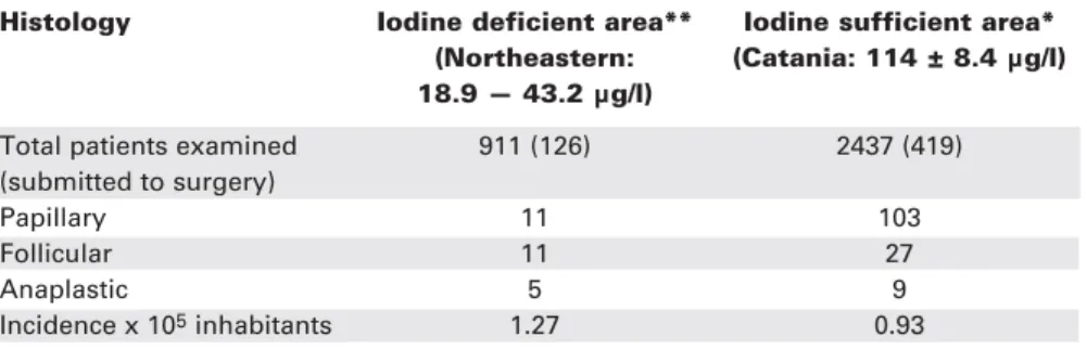
A striking difference in the relative prevalence in the thyroid cancer histotypes was also observed in the two adjacent areas of Sicily (68) with different iodine intake (table 4) indicating that even moderate iodine deficiency is associated with an increased frequency of more aggressive histopathological types of thyroid can-cer. However, it should be emphasized that the long-standing program of supplementation of food items with iodine in Sweden has not affected thyroid cancer trends in iodine-deficient or iodine-rich areas (80).
An alternative explanation for the observed increase in the number of papillary cancers could be the introduction and wider use of more modern and precise examination equipment through the years, as the reversal of iodine deficiency coincides with the dis-semination of better techniques in the health system, including thyroid scintigraphy, ultrasound and fine-needle biopsy. Furthermore, in areas without iodine deficiency, and hence less goiter, patients are more likely to notice a slight change in their thyroid gland and visit their doctor for examination (3). Autopsy studies support the possibility of a pool of slowly growing, undiagnosed papillary thyroid tumors (84).
The importance of accurate histological typing in cancer epidemiology cannot be overemphasized, and great caution is needed in comparison of different types of thyroid tumors in different populations if not evi-denced by the same pathologist. The reproducibility of the WHO classification has been questioned and a con-siderable observer variation has been reported, partic-ularly as regards to follicular and mixed papillary-fol-licular cancers (87). Differences in the detection and inclusion of occult thyroid cancers in the studies could also play a role in the observed cancer incidences. Final-ly, it should be noted that a concomitant selenium
defi-ciency in iodine-deficient areas has not been taken into account, since selenium (Se) deficiency in itself has recently been associated with an increased incidence of thyroid carcinoma, based on converging data from epi-demiological and clinical studies (88). Other studies, however, failed to find links between Se and thyroid car-cinogenesis (89). To our knowledge, up to now there are no reliable experimental data that supports the plau-sibility of Se as a risk factor in thyroid cancer.
All of the above reservations are probably rea-sons why comparison of thyroid carcinoma epidemiol-ogy in different populations and geographic areas are so difficult to perform and interpret, and why no spe-cific solutions to the questions concerning environ-mental influence of iodine intake have been provided, despite a large number of studies over many years.
Thus, regarding iodine prophylaxis and varia-tions on thyroid cancer incidence, the data indicate that: • After iodine prophylaxis it has been demonstrated a clear relationship between increased iodine nutrition and elevation of the PTC/FTC ratio
• This has occurred even in modest increases in iodine urinary excretion
• A decrease in prevalence of anaplastic thyroid cancer was also observed in most areas.
Table 4.Histologic pattern of thyroid tumors in two areas of Sicily, Italy, with low and adequate iodine intake.
Histology Iodine deficient area** Iodine sufficient area* (Northeastern: (Catania: 114 ± 8.4 µg/l) 18.9 — 43.2 µg/l)
Total patients examined 911 (126) 2437 (419)
(submitted to surgery)
Papillary 11 103
Follicular 11 27
Anaplastic 5 9
Incidence x 105inhabitants 1.27 0.93
Adapted from ref. 68.
Table 3.Frequency of thyroid cancer by tumor type in Salta, Argentina, before and after iodine supplementation.
Period of study of pathological specimens (years after iodine supplementation)
10 11–26
Total number of cases (n) 59 85
Papillary carcinoma (n) 26 46
Follicular carcinoma (n) 15 15
Undifferentiated carcinoma (n) 9 13
Others (n) 9 11
Ratio PTC/FTC 1.7 3.1
CONCLUSION
Available evidence from animal experiments, epidemi-ological studies and from the introduction of iodine prophylaxis has demonstrated a relationship between iodine intake and the types of thyroid carcinoma, while no clear evidence exists for a relationship between the overall cancer incidence and iodine intake. All the studies are in general hampered by difficulty in com-paring populations since many factors have to be con-sidered other than the iodine intake, such as ethnicity, other dietary factors (e.g., selenium), histological examination and radiation. Knowledge of all these fac-tors has an influence also on the diagnostic work-up and management of patients in each population.
REFERENCES
1. Parkin DM, Muir CS, Whelan SL, et al. Cancer incidence in five continents. IARC Sci Publ 1992;(120):984-7.
2. Whelan SL, Parkin DM, Masuyer E. Patterns of cancer in five continents. IARC Sci Publ 1990;102:152-3.
3. Feldt-Rasmussen U. Iodine and cancer. Thyroid
2001;11:483-6.
4. Horn-Ross PL, Morris JS, Lee M, West DW, Whittemore AS, McDougall IR, et al. Iodine and thyroid cancer risk among women in a multiethnic population: the Bay Area Thyroid Cancer Study. Cancer Epidemiol Biomarkers Prev 2001;10:979-85.
5. Axelrad AA, Leblond CP. Induction of thyroid tumors in rats by low-iodine diet. Cancer 1955;8:339-67.
6. Schaller RT, Stevenson JK. Development of carcinoma of the thyroid in iodine-deficient mice. Cancer 1966;19:1063-80. 7. Dent JN, Godsden EL, Furth J. Further studies on induction
and growth of thyrotropic pituitary tumors in mice. Cancer Res 1956;16:171-4.
8. Haran-Guera N, Pullar P, Furth J. Induction of thyrotropin-dependent thyroid tumors by thyrotropes. J Endocrinol 1960;66:694-701.
9. Sinha D, Pascal R, Furth J. Transplantable thyroid carcinoma induced by thyrotropin. Arch Pathol 1965;79:192-8. 10. Williams ED. TSH and thyroid cancer. Horm Metab Res
Suppl 1990;23:72-5.
11. Fortner JG, George PA, Sternberg SS. Induced and sponta-neous thyroid cancer in the Syrian (golden) hamster.
Endocrinology 1960;66:364-76.
12. Laurberg P, Nohr SB, Pedersen KM, Hreidarsson AB, Ander-sen S, Bulow PederAnder-sen I, et al. Thyroid disorders in mild iodine deficiency. Thyroid 2000;10:951-63.
13. Boltze C, Brabant G, Dralle H, Gerlach R, Roessner A, Hoang-Vu C. Radiation-induced thyroid carcinogenesis as a function of time and dietary iodine supply: an in vivo model of tumorigenesis in the rat. Endocrinology 2002 ;143(7):2584-92.
14. Correa P, Welsh RA. The effect of excessive iodine intake on the thyroid gland of the rat. Arch Pathol 1960;70:247-51. 15. Al-Saadi A. Precursor cytogenetic changes of transplantable
thyroid carcinoma in iodine deficient goiters. Cancer Res 1968;28:739-45.
16. Hiasa Y, Kitahori Y, Konishi N, Ohshima M. Chemical car-cinogenesis in the thyroid gland. Toxicol Lett 1992;64/65:389-95.
17. Hill RN, Erdreich LS, Paynter OE, Roberts PA, Rosenthal SL, Wilkinson CF. Thyroid follicular cell carcinogenesis: a review.
Fundam Appl Toxicol 1989;12:629-97.
18. Ohshima M, Ward JM. Dietary iodine deficiency as a tumor promoter and carcinogen in male F344/NCr rats. Cancer Res 1986;46:877-83.
19. Thomas GA, Williams ED. Evidence for and possible mecha-nisms of non-genotoxic carcinogenesis in the rodent thyroid.
Mutat Res 1991;248:357-70.
20. Kanno J, Onodera H, Furuta K, Maekawa A, Kasuga T, Hayashi Y. Tumor-promoting effects of both iodine deficiency and iodine excess in rat thyroid. Toxicol Pathol 1992;20:226-35. 21. Williams ED. Mechanisms and pathogenesis of thyroid
can-cer in animals and man. Mutat Res 1995;333:123-9. 22. Cohen SM, Ellwein LB. Cell proliferation in carcinogenesis.
Science 1990;249:1007-11.
23. Jemec B. Studies of the goitrogenic and tumorigenic effect of two goitrogens in combination with hypophysectomy or thy-roid hormone treatment. Cancer 1980;45:2138-48.
24. Nadler NJ, Mandavia M, Goldberg M. The effect of hypophy-sectomy on the experimental production of thyroid neo-plasms. Cancer Res 1970;30:1909-11.
25. Coclet J, Foureau F, Ketelbant P, Galand P, Dumont JE. All population kinetics in dog and human adult thyroid. Clin Endocrinol (Oxf) 1989;31:655-65.
26. Conde E, Martin-Lacave I, Utrilla JC, Moreno A, Gonzales-Campora R, Galera-Davidson H. Mitotic activity of the endocrine cells in rat thyroid glands during postnatal life.
Endocrinology 1992;131:436-40.
27. Studer H, Peter HJ, Gerber H. Natural heterogeneity of thy-roid cells: the basis for understanding thythy-roid function-and nodular goiter growth. Endocr Rev 1989;10:125-35. 28. Knobel M, Bisi H, Peres CA, Medeiros-Neto G. Studies on
functional and morphological aspects in human multinodular simple goiter tissues. Endocr Pathol 1993;4:205-14. 29. Dawson TP, Wyllie FS, Wynford-Thomas D. In vitro
respon-siveness to serum growth factors is inversely related to in vivo malignancy in human thyroid epithelial cells. Br J Can-cer 1991;63:827-900.
30. Farid NR, Shi Y, Zou M. Molecular basis of thyroid cancer.
Endocr Rev 1994;15:202-32.
31. World Health Organization, United Nations Children’s Found and International Council for Control of Iodine Deficiency Disor-ders; Elimination of iodine deficiency disorder (IDD) in Central and Eastern Europe, the Commonwealth of Independent States and the Baltic States. Proceedings of a conference held in Munich, Germany, 3-6 September 1997. WHO/Euro/NUT/98.1. 32. Dumont JE, Lamy F, Roger P, Maenhaut C. Physiological and
pathological regulation of thyroid cell proliferation and dif-ferentiation by thyrotropin and other factors. Physiol Rev 1992;72:667-97.
33. Bayer I, Mitmaker B, Gordon PH, Wang E. Modulation of nuclear statin expression in rat thyroid follicle cell following administration of thyroid stimulating hormone. J Cell Phys-iol 1992;150:276-82.
34. Smeds S, Peter HJ, Jortso E, Gerber H, Studer H. Naturally occurring clones of cells with high intrinsic proliferation potential within the follicular epithelium of mouse thyroids.
Cancer Res 1987;47:1646-51.
35. Groch KM, Clifton KH. The plateau phase rat goiter contains a sub-population of TSH-responsive follicular cells capable of proliferation following transplantation. Acta Endocrinol 1992;126:85-96.
36. Christov K. Cell proliferation kinetics and DNA content during thyroid carcinogenesis. Cell Tissue Kinet 1985;18:119-31. 37. Wynford-Thomas D, Stringer BMJ, Williams ED.
Desensitiza-tion of rat thyroid to the growth-stimulating acDesensitiza-tion of TSH during prolonged goitrogen administration. Persistence of refractoriness following withdrawal of stimulation. Acta Endocrinol 1982;101:562-9.
38. Domann FE, Mitchen JM, Clifton KH. Restoration of thyroid function after total thyroidectomy and quantitative thyroid transplantation. Endocrinology 1990;127:2673-8.
40. Corvilain B, Laurent E, Lecomte M, Vansande J, Dumont JE. Role of the adenosine 3’,5’-monophosphate and the phos-phatidylinositol-Ca2+cascade in mediating the effects of thy-rotropin and iodide on hormone synthesis and secretion in
human thyroid slices. J Clin Endocrinol Metab
1994;79:152-9.
41. Wolff J. Excess iodide inhibits the thyroid by multiple mech-anisms. Adv Exp Med Biol 1989;261:211-44.
42. Gartner R, Dugrillon A, Bechtner G. Evidence that thyroid growth autoregulation is mediated by an iodolactone. Acta Med Austriaca 1990;17(suppl 1):124-6.
43. Dugrillon A, Gartner R. Delta-Iodolactones decrease epider-mal growth factor-induced proliferation and inositol-11,4,5-triphosphate generation in porcine thyroid follicles – possible mechanism of growth inhibition by iodide. Eur J Endocrinol 1995;132:735-43.
44. Boeynaems JM, van Sande J, Dumont JE. Which iodolipids are involved in thyroid in thyroid autoregulation: iodolac-tones or iodoaldehydes? Eur J Endocrinol 1995;132:733-4. 45. Pereira A, Braekman JC, Dumont JE, Boeynaems JM. Identi-fication of a major iodolipid from the horse thyroid gland as 2-iodohexadecanal. J Biol Chem 1990;265:17018-25. 46. Nilsson M. Actions of epidermal growth factor and its
recep-tor in the thyroid. Trends Endocrinol Metab 1995 ;6:175-82.
47. Pedrinola F, Rubio I, Santos CL, Medeiros-Neto G. Over expression of epidermal growth factor and epidermal growth factor-receptor mRNAS in dyshormonogenetic goiters. Thy-roid 2001;11:15-20.
48. Taylor AH, Millatt LJ, Whitley GS, Johnstone AP, Nussey SS. The effect of basic fibroblast growth factor on the growth and function of human thyrocytes. J Endocrinol 1993;136:339-44. 49. Logan A, Black AG, Gonzales AM, et al. Basic fibroblast growth factor: an autocrine mitogen of rat thyroid follicular cells. Endocrinology 1992;130:2363-72.
50. Becks GP, Logan A, Phillips ID, Wang JF, Smith C, DeSousa D, et al. Increase of basic fibroblast growth factor (FGF) and FGF receptor messenger RNA during rat thyroid hyperplasia: temporal changes and cellular distribution. J Endocrinol 1994;142:325-38.
51. Hoelting T, Siperstein AE, Clark OH, Duh QY. Epidermal growth factor enhances proliferation, migration and invasion of follicular and papillary thyroid cancer in vitro and in vivo.
J Clin Endocrinol Metab 1994;79:401-8.
52. Westermark K, Karlsson FA, Westermark B. Epidermal growth factor modulates thyroid growth and function in cul-ture. Endocrinology 1983;112:1680-6.
53. Duh QY, Gum ET, Gerend PL, Raper SE, Clark OH. Epidermal growth factor receptors in normal and neoplastic thyroid tis-sue. Surgery 1985;98:1000-7.
54. Roger PP. Thyrotropin-dependent transforming growth factor beta expression in thyroid gland. Eur J Endocrinol 1996;134:269-71.
55. Hoelting T, Zielke A, Siperstein AE, Clark OH, Duh QY. Trans-forming growth factor-beta 1 is a negative regulator for dif-ferentiated thyroid cancer: studies of growth, migration, inva-sion, and adhesion of cultured follicular and papillary thyroid cancer cell lines. J Clin Endocrinol Metab 1994;79:806-13. 56. Jasani B, Wyllie FS, Wright PA, Lemoine NR, Williams ED, Wynford-Thomas D. Immunocytochemically detectable TGFβ associated with malignancy in thyroid epithelial neoplasia.
Growth Factors 1990;2:149-55.
57. Lazzereschi D, Ranieri A, Mincione G, Taccogna S, Nardi F, Colletta G. Human malignant thyroid tumors displayed reduced levels of transforming growth factor βreceptor type II messenger RNA and protein. Cancer Res 1997;57:2071-6. 58. Yuasa R, Eggo MC, Meinkoth J, Dillmann WH, Burrow GN. Iodide induces transforming growth factor beta 1 (TGFβ1) mRNA in sheep thyroid cells. Thyroid 1992;141-5.
59. Minuto F, Barreca A, Del Monte P, Cariola G, Torre GC, Gior-dano G. Immunoreactive insulin-like growth factor I (IGF-I) and IGF-I-binding protein content in human thyroid tissue. J Clin Endocrinol Metab 1989;68:621-6.
60. Thompson SD, Franklyn JA, Watkinson JC, Verhaeg JM, Sheppard MC, Eggo MC. Fibroblast growth factors 1 and 2 and fibroblast growth factor receptor 1 are elevated in thyroid hyperplasia. J Clin Endocrinol Metab 1998;83:1336-41. 61. Eggo MC, Hopkins JM, Franklyn JA, Johnson GD, Sanders
DS, Sheppard MC. Expression of fibroblast growth factors in thyroid cancer. J Clin Endocrinol Metab 1995;80:1006-11. 62. Goodman AL, Rone JD. Thyroid angiogenesis: endothe-liotropic chemoattractant activity from rat thyroid cells in cul-ture. Endocrinology 1987;121:2131-40.
63. Cocks HC, Thompson S, Turner FE, Logan A, Franklyn JA, Watkinson JC, et al. Role and regulation of the fibroblast growth factor axis in human thyroid follicular cells. Am J Physiol Endocrinol Metab 2003;285:E460-9.
64. Tuttle RM, Fleisher M, Francis GL, Robbins RJ. Serum vascular endothelial growth factor levels are elevated in metastatic dif-ferentiated thyroid cancer but not Increased by short-term TSH stimulation. J Clin Endocrinol Metab 2002;87:1737-42. 65. Iitaka M, Miura S, Yamanaka K, Kawasaki S, Kitahama S,
Kawakami Y, et al. Increased serum vascular endothelial growth factor levels and intrathyroidal vascular area in patients with Graves’ disease and Hashimoto’s thyroiditis. J Clin Endocrinol Metab 1998;83:3908-12.
66. Hay ID. Papillary thyroid carcinoma. Clin Endocrinol Metab North Am 1990;19:545-76.
67. Venkatesh YS, Ordonez NG, Schultz PN, Hickey RC, Goepfert H, Samaan NA. Anaplastic carcinoma of the thyroid: a clini-copathologic study of 121 cases. Cancer 1990;66:321-30. 68. Belfiore A, La Rosa GL, Padova G, Sava L, Ippolito O, Vigneri
R. The frequency of cold thyroid nodules and thyroid malig-nancies in patients from an iodine-deficient area. Cancer 1987;60:3096-102.
69. Mellemgaard A, From G, Jorgensen T, Johansen C, Olsen JH, Perrild H. Cancer risk in individuals with benign thyroid dis-orders. Thyroid 1998;8:751-4.
70. Levi F, Franceschi S, La Vecchia C, Negri E, Gulie C, Duruz G, et al. Previous thyroid disease and risk of thyroid cancer in Switzerland. Eur J Cancer 1991;27:85-8.
71. dos Santos Silva I, Swerdlow AJ. Thyroid cancer epidemiolo-gy in England and Wales: time trends and geographical dis-tribution. Br J Cancer 1993;67:330-40.
72. Pendergrast WJ, Milmore BK, Marcus SC. Thyroid cancer and thyrotoxicosis in the United States: their relation to endemic goiter. J Chronic Dis 1961;13:22-38.
73. Franssila K, Saxén E, Teppo L, Bjarnason O, Tulinius H, Nor-mann T, et al. Incidence of different morphological types of thyroid cancer in the Nordic countries. Acta Pathol Micro-biol Scand [A] 1981;89:49-55.
74. Williams ED, Doniach I, Bjarnason O, Michie W. Thyroid can-cer in an iodide rich area: a histopathological study. Cancer 1977;39:215-22.
75. Goodman MT, Yoshizawa CN, Kolonel LN. Descriptive
epi-demiology of thyroid cancer in Hawaii. Cancer
1988;61:1272-81.
76. Sehestedt T, Knudsen N, Perrild H, Johansen C. Iodine intake and incidence of thyroid cancer in Denmark. Clin Endocrinol 2006;65:229-33.
77. Farahati J, Geling M, Mader U, Mortl M, Luster M, Muller JG, et al. Changing trends of incidence and prognosis of thyroid carcinoma in lower Franconia, Germany, from 1981–1995.
Thyroid 2004;14:141-7.
78. Gomez Segovia I, Gallowitsch HJ, Kresnik E, Kumnig G, Igerc I, Matschnig S, et al. Descriptive epidemiology of thyroid car-cinoma in Carinthia, Austria: 1984–2001. Histopathologic fea-tures and tumor classification of 734 cases under elevated general iodination of table salt since 1990: population based age-stratified analysis on thyroid carcinoma incidence. Thy-roid 2004;14:277-86.
80. Pettersson B, Coleman MP, Ron E, Adami HO. Iodine supple-mentation in Sweden and regional trends in thyroid cancer inci-dence by histopathologic type. Int J Cancer 1996;65:13-9. 81. Galanti M, Sparen P, Karlsson A, Grimelius L, Ekbom A. Is
residence in areas of endemic goiter a risk factor for thyroid cancer? Int J Cancer 1995;61:615-21.
82. Harach HR, Williams ED. Thyroid cancer and thyroiditis in the goitrous region of Salta, Argentina, before and after iodine prophylaxis. Clin Endocrinol 1995;43:701-6.
83. Lind P, Langsteger W, Molnar M, Gallowitsch HJ, Mikosch P, Gomez I. Epidemiology of thyroid diseases in iodine suffi-ciency. Thyroid 1998;8:1179-83.
84. Franceschi S. Iodine intake and thyroid carcinoma – a potential risk factor. Exp Clin Endocrinol Diabetes 1998;106:38-44. 85. Teng W, Shan Z, Teng X, Guan H, Li Y, Teng D, et al. Effect of
iodide intake on thyroid diseases in China. N Engl J Med 2006;354:2783-93.
86. Camargo RYA, Tomimori EK, Neves SC, Knobel M, Medeiros-Neto G. Prevalence of chronic autoimmune thyroiditis in the urban area neighboring a petrochemical complex and a con-trol area in São Paulo, Brazil. Clinics 2006;61:307-12.
87. Saxén E, Franssila K, Bjarnason O, Normann T, Ringertz N. Observer variation in histologic classification of thyroid
clas-sification. Acta Pathol Microbiol Scand [A]
1978;86A:483-6.
88. Duntas LH. The role of selenium in thyroid autoimmunity and cancer. Thyroid 2006;16:455-60.
89. Rayman MP. Selenium in cancer prevention: a review of the evidence and mechanism of action. Proc Nutr Soc 2005;64:527-42.
Address for correspondence:
Meyer Knobel
Thyroid Unit, Division of Endocrinology and Metabolism Hospital das Clínicas, University of São Paulo Medical School Av. Dr. Enéas Carvalho de Aguiar 155, 8 A, bl 3, PAMB 05403-900 São Paulo, SP
Fax: (11) 3069-7970