Ar
ti
cl
e
0103 - 5053 $6.00+0.00
*e-mail: [email protected]
A Novel and Simple Synthetic Route for a Piperazine Derivative
Maria A. S. da Silva,a Solange de O. Pinheiro,a Thiago dos S. Francisco,a Francisco O.
N. da Silva,a Alzir A. Batista,b Javier Ellena,c Idalina M. M. Carvalho,a Jackson R. de
Sousa,a Francisco A. Dias-Filho,a Elisane Longhinottid and Izaura C. N. Diógenes*,a
aDepartamento de Química Orgânica e Inorgânica, Universidade Federal do Ceará, CP 6021,
60455-970 Fortaleza-CE, Brazil
bDepartamento de Química, Universidade Federal de São Carlos, 13565-905 São Carlos- SP, Brasil
cInstituto de Física, Universidade de São Paulo, 13656-905 São Carlos-São Paulo, Brasil
dDepartamento de Química Analítica e Físico-Química, Universidade Federal do Ceará, CP 6035,
60455-970 Fortaleza-CE, Brasil
Um novo derivado da piperazina, 5-oxopiperazinio-3-sulfonato monohidratado, foi produzido
a partir de uma rota sintética simples como resultado da adição do íon bisulito, HSO3−, ao anel
e do ataque nucleofílico de moléculas de água a moléculas de pirazina. O material isolado foi caracterizado por RMN, espectrometria de massa, infravermelho e difração de raios-X.
A new derivative of piperazine, 5-oxopiperazinium-3-sulfonate monohydrate, was produced
from a simple synthetic route as a result of the nucleophilic addition to HSO3−bisulphite ion and
of the nucleophilic attack of water molecules on pyrazine molecules. The isolated material was characterized by means of NMR, mass spectrometry, infrared, and X-ray diffraction.
Keywords: piperazine, nucleophilic addition, mass spectrometry, NMR
Introduction
Synthetic methods and strategies have been extensively investigated to enable access to piperazine derivatives, particularly the oxo species, due to the importance of this class of compounds in a wide range of biological
activities.1-11 Also some of these species have been recently
probed to be versatile as a probe for crystal structure. For instance, the propensity to form macromolecular arrays in the solid state enables the formation of planar or non-planar
type structures.12,13
We report herein the synthesis of a novel piperazine derivative obtained from the direct reaction of pyrazine
and SO2 in aqueous solution.
Results and Discussion
5-Oxopiperazinium-3-sulfonate monohydrate was
prepared by the direct reaction of pyrazine with SO2 gas
in water. The isolated material crystallizes as pale yellow monoclinic prisms in the space group P1. Figure 1 and
Table 1 present, respectively, the ORTEP14 view of the
compound and selected bond lengths and angles. In addition, an illustration of the hydrogen bonds involved in the packing of the water molecule in the crystal is also
presented in Figure 1. Bond lengths (Å)and angles (°) of
the intermolecular hydrogen bonds are presented in Table 1. The elemental analysis data are consistent with the chemical formulation C4H8N2O4S•H
2O.
The distances observed between the carbon atoms are far shorter than those reported for piperazine (1.614 Å), morpholine (1.599 Å), thiomorpholine (1.588 Å), and thioxane (1.575 Å). However, the C(1)-C(2) and C(3)-C(4) bond lengths are higher than those observed for benzene
ring (1.40 Å).16 This result suggests a non-aromatic ring as
evidenced by the ORTEP view illustrated in Figure 1. In addition, the conformation of the ring is that of a distorted chair as suggested by the C(4)-N(1)-C(1) and C(3)-N(2)-C(2) angles. This is probably due to the strain induced by the
is reinforced by the different bond lengths N(1)-C(4) (1.342 Å) and N(1)-C(1) (1.4478 Å) which relect different
withdrawing capability of the SO3 and CO fragments.
The infrared spectrum of the isolated compound presents signals typically assigned to substituted piperazine. Two absorptions characteristic of the piperazine ring, assigned to the CN stretching vibrational
modes,17,18 are observed at 1130 and 1168 cm−1. A very
sharp and intense band is observed at 1037 cm−1 and is
assigned to the ring CH2 rocking motions. According to
Spell,17 this is one of the most useful band for detecting the
presence of di-substituted piperazines. The band observed
at 1680 cm−1 is assigned17,18 to the carbonyl stretching
frequency thus indicating the presence of this group in
the molecule. Two sharp absorptions assigned,17,18 to SO
stretching modes of the SO3 fragment are observed at
1005 and 957 cm−1.
1H and 13C NMR data of the
5-oxopiperazinium-3-sulfonate monohydrate are reported in the experimental
section. HMQC spectrum, illustrated in Figure 2, was acquired to undoubtedly assign the protons.
The singlet at 4.06 ppm in the 1H NMR spectrum is
assigned to the H6a and H6b protons based on the correlation
with the C6 carbon in the HMQC spectrum. The doublet of
doublet at 3.82 ppm is assigned to the geminal (2J 12 Hz),
and vicinal (3J = 5 Hz) coupling of the H
2a proton with the
H2b and H3 protons, respectively. According to the HMQC
spectrum, the signals at 3.82 and 4.01 ppm are correlated to the same carbon atom. This assignment is reinforced by the
Table 1. Selected bond lengths (Å) and angles (°)
N(1)-C(4) 1.342(2) C(4)-N(1)-C(1) 125.46(12)
N(1)-C(1) 1.4478(19) C(3)-N(2)-C(2) 112.24(11) N(2)-C(3) 1.4869(18) N(1)-C(1)-C(2) 111.33(12)
N(2)-C(2) 1.4882(18) N(2)-C(3)-C(4) 115.07(12) C(1)-C(2) 1.511(2) N(2)-C(2)-C(1) 110.07(12) C(3)-C(4) 1.507(2) N(1)-C(4)-C(3) 118.89(13)
S-C(1) 1.8157(15) C(2)-C(1)-S 111.77(10) O(4)-C(4) 1.342(2) O(4)-C(4)-C(3) 118.06(13)
Figure 1. (A) ORTEP14 view showing the atoms labelling and the 50% probability ellipsoids and (B) illustration of the hydrogen bonds involved in the packing of the solvated water molecule of the 5-oxopiperazinium-3-sulfonate monohydrate.
data obtained from COSY spectrum in which a correlation
between the H2a and H2b protons and between these protons
and the H3 proton is observed. The signal at 4.77 ppm is
assigned to the H3 proton. Although the COSY spectrum
indicates a correlation between this proton and the H2a and
H2b protons, it is not possible to assign the multiplicity due
to the solvent signal. In fact, for piperazine compounds, the exchange between the protons of the amine fragment and deuterium atoms is frequently observed resulting in a
single signal in the water region (4.8 ppm).19
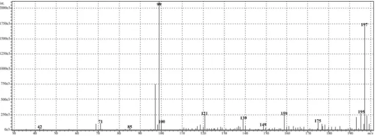
The mass spectrum of the 5-oxopiperazinium-3-sulfonate monohydrate, illustrated in Figure 3, presents
two metastable ions at m/z 197 and 99.
Figure 3. Mass spectrum of the 5-oxopiperazinium-3-sulfonate monohydrate in water/methanol solution.
N N H
SO3– O
H H
+
N N O
H H
+
m/z = 99 N N H
SO3– O
H
m/z = 197 H2O
H2O
N H N O
+
m/z = 97
Figure 4. Suggested mechanism for the formation of the major ions from the fragmentation of the 5-oxopiperazinium-3-sulfonate monohydrate in water/methanol solution.
The fact that the peak at m/z 99 is more intense than that
at m/z 197 is consistent with the current observation that
in heteroatom-containing molecules, the amino fragment presents lower abundance. This effect is indeed observed
for some diketopiperazine species.20,21 Figure 4 presents
a suggestion of a mechanism for the formation of these major ions.
Attempts were made in order to apply the same synthetic approach starting with pyridine, pyrazinamide, and imidazole. However, none of these molecules was reduced as pyrazine, meaning not only that two nitrogen atoms in the ring are required, but also that these atoms
process to occur. In addition, the procedure was carried out in dried methanol instead of water. In such condition, no reaction was observed even after 24 h under vigorous
stirring and SO2 low indicating that water molecules play a
fundamental role in the mechanism. Based on these results and as conclusion, the mechanism presented in Figure 5 is suggested.
The reaction was carried out in acidic medium
(1.5 < pH < 4.0) saturated with SO2 gas. In such condition,
it is well known that the most stable form of SO2 molecules
is the HSO3−bisulphite ion.22 Therefore, a nucleophilic
addition is suggested to occur with the attack initiated
by HSO3− to one of the C=N bond of the ring (I). Then, a
positively charged intermediate is formed and experiences successive attacks of water molecules, which act as nucleophiles like in a hydrolysis reaction. The hydrogen
ions thus formed are pulled out of the ring by the HSO3− ion
acting as a Bronsted base. Upon the elimination of water molecule, an enol (compound II) is formed and suffers tautomerism, furnishing compound III. The ORTEP view
illustrated in Figure 1 is, indeed, the zwitterionic structure of the inal product (III), which is the most stable form in acidic medium.
Experimental
The water used throughout was puriied by a Milli-Q system (Millipore Co.).
Pyrazine, pyridine, pyrazinamide, and imidazole,
from Aldrich, were used as received. Pure SO2 (purity >
99.9%) delivered in a bottle as liqueied gas, was purchased from White Martins Praxair Inc.. All other chemicals and solvents were of analytical grade.
Elemental analyses were performed by on a FISIONS CHNS, mod. EA 1108 micro analyzer at the Microanalytical Laboratory at Universidade Federal de São Carlos in São Carlos, SP. LCMS (liquid chromatography mass spectrometry) analyses were conducted using isocratic elution (water/methanol, 90:10 v/v) with
a Shimadzu C18 column (250×2.0 mm, 4.6 µm).
N N H H N N H H
SO3H
H O H H H + N N H H
SO3H O H H N N H H
SO3H H O H H N N H H
SO3H H HO O H H N N H H
SO3H H HO O H H H N N H H
SO3H O
H
N N H
SO3H O
H H
HSO3
-HSO3
-(II) (III) (I)
N N H
SO3 -O
H H
Table 2. Crystal data and structure reinement parameters
Empirical formula C4H10N2O5S
Formula weight 198.03
Temperature 293(2) K
Wavelength 0.71073 Å
Crystal system Monoclinic
Space group P-1
Unit cell dimensions a = 6.5211(2) Å α= 100.405(2)° b = 6.6655(2) Å β= 103.914(2)°
c = 9.7323(3) Å γ = 102.886(2)°
Volume 387.82(2) Å3
Z 2
Density (calculated) 1.697 Mg m−3 Absorption coeficient 0.405 mm-1
F(000) 208
Crystal size 0.24×0.18×0.15 mm3 Theta range for data
collection
3.24 to 27.54°
Index ranges –8 ≤ h ≤ 8, –8 ≤ k ≤ 8, –12 ≤ l ≤ 12
Relections collected 3408
Independent relections 1770 (R(int) = 0.0130)
Completeness to theta = 27.54°
98.4%
Absorption correction23 Semi-empirical from equivalents
Max. and min. transmission 0.904 and 0.880 Reinement method Full-matrix
least-squares on F2
aComputing24-26 COLLECT, HKL Denzo and Scalepack SHELXS-97, SHELXL-97
Data / restraints / parameters 1770 / 0 / 149 Goodness-of-it on F2 1.092 Final R indices [I>2σ(I)] R1 = 0.0321,
wR2 = 0.0830
R indices (all data) R1 = 0.0339, wR2 = 0.0840 Largest diff. peak and hole 0.264 and –0.383 e.Å-3
a Data collection, data processing, structure solution and structure reinement respectively.
The experiments were carried out on a Shimadzu
LCMS-2010 equipment and the flow rate was set at
0.2 mL min-1. The measurements were performed in
positive mode by scanning between m/z 30 and 300
using an APCI interface and SIM technique. The APCI
parameters were set asfollows: probe voltage (kV), 3.50;
probe temperature, 250 °C; blocktemperature: 200 °C;
CDL temperature: 230 °C; Q-array voltage: 0and 20 V;
gas flow: 2.5 L min-1. The electronic spectrum was
acquired with a Hitachi model U-2000 spectrophotometer. The transmission infrared spectrum of the compound dispersed in KBr was obtained by using a Perkin–Elmer
instrument model Spectrum 1000. 1H and 13C NMR
normal and two-dimension COSY 1H-1H and HMQC
1H-13C spectra were recorded on Bruker AVANCE 500
spectrometer.
The general synthetic procedure was followed using pyrazine (150 mg, 0.83 mmol) in water (2 mL), at room
temperature, in a Schlenk lask. A low of SO2 was bubbled
for 30 s at each 30 min of reaction. According to SO2
equilibrium,22 in the acidic condition (1.5 < pH < 7.0)
in which the reaction was carried out, the HSO3− form is
favored. Just after the beginning of SO2 addition, a color
change is observed. After 1 h of reaction, pale yellow crystals start to be produced. The mixture was kept under
stirring and SO2 addition for 3 h when it seems that the
precipitate was no longer formed thus suggesting the complete consumption of the starting material. Calc. for
C4H8N2O4S.H2O: C, 24.24; H, 5.09; N, 14.13; S, 16.18%.
Found: C, 24.12; H, 5.01; N, 14.09; S, 15.93%. Yield: 98%.
Pale yellow crystals, mp > 250 0C. λ
max (H2O): 238 nm.
1H NMR (500 MHz, D
2O) d 3.82 (dd, H2b), 4.01
(dd, H2a), 4.06 (s, H3a e H3b), 4.77 (s, H1). 13C{1H} NMR
(125 MHz, D2O) d 163.16 (s, C4), 61.45 (s, C1), 44.57
(s, C3), 40.98 (s, C2). Internal reference: DSS (sodium
4,4-dimethyl-4-silapentane-1-sulfonate). Crystallographic data and refinement parameters are reported in Table 2.
Supplementary Information
Crystallographic data for C4H10N2O5S (excluding
structure factors) for the structures in this paper have been deposited with the Cambridge Crystallographic Data Centre as supplementary publication no CCDC 746269. Copies of the data can be obtained, free of charge via www.ccdc. cam.ac.uk/conts/retrieving.html (or from the Cambridge Crystallographic Data Centre, CCDC, 12 Union Road, Cambridge CB2 1EZ, UK; fax: +44 1223 336033; or e-mail: [email protected]).
Acknowledgments
References
1. Brown, D. A.; Mishra, M.; Zhang, S.; Biswas, S.; Parrington, I.; Antonio, T.; Reith, M. E. A.; Dutta, A. K.; Bioorg. Med. Chem. 2009, 17, 3923.
2. Pichowicz, M.; Simpkins, N. S.: Blake, A. J.; Wilson, C.; Tetrahedron 2008, 64, 3713.
3. Martini, E.; Ghelardini, C.; Dei, S.; Guandalini, L.; Manetti, D.; Melchiorre, M.; Norcini, M.; Scapecchi, S.; Teodoria, E.; Romanelli, M. N.; Bioorg. Med. Chem. 2008, 16, 1431. 4. Tullberg, M.; Grøtli, M.; Luthman, K.; J. Org. Chem. 2007, 72,
195.
5. Jam, F.; Tullberg, M.; Luthman, K.; Grøtli, M.; Tetrahedron 2007, 63, 9881.
6. Tullberg, M.; Luthman, K.; Grøtli, M. J. Comb. Chem. 2006, 8, 915.
7. Wang, H.; Usui, T.; Osada, H.; Ganesan, A.; J. Med. Chem. 2000, 43, 1577.
8. Wang, H. Ganesan, A.; Org. Lett. 1999, 1, 1647.
9. Fresno, M.; Alsina, J.; Royo, M.; Barany, G.; Albericio, F.; Tetrahedron Lett. 1998, 39, 2639.
10. Kennedy, A. L.; Fryer, A. M.; Josey, J. A.; Org. Lett. 2002, 4, 1167.
11. Wang, T.; Kadow, J. F.; Zhang, Z.; Yin, Z.; Gao, Q.; Wu, D.; Parker, D. D.-G.; Yang Z.; Zadjura L.; Robinson, B. A.; Gong, Y.-F.; Blair, W. S.; Shi, P.-Y.; Yamanaka, G.; Lin, P.-F.; Meanwell, N. A.; Bioorg. Med. Chem. Lett. 2009, 19, 5140. 12. Weatherhead, K. R.; Selby, H. D.; Miller III, W. B.; Mash, E.
A.; J. Org. Chem. 2005, 70, 8693.
13. Du, Y.; Creighton, C. J.; Tounge, B. A.; Reitz, A. B.; Org. Lett. 2004, 6, 309.
14. Farrugia, L. J. ORTEP3 for Windows. J. Appl. Crystallogr. 1997, 30, 565.
15. Nuzhdin, K. B.; Nesterov, S. V.; Tyurin, D. A.; Feldman, V. I.; Wei, L.; Lund, A.; J. Phys. Chem. A 2005, 109, 6166. 16. Fox, M. A.; Whitesell, J. K.; Organische Chemie 1994,
Spektrum.
17. Spell, H. L.; Anal. Chem. 1969, 41, 902.
18. Hendra, P. J.; Powell, D. B.; Spectrochim. Acta 1962, 18, 305. 19. Ermatchkov, V.; Kamps, A. P.-S.; Maurer, G.; J. Chem.
Thermodyn. 2003, 35, 1277.
20. Eriksen, S.; Fagerson, I. S.; J. Agric. Food Chem. 1976, 24, 1242.
21. Biemann, K.; Seibl, J.; Gapp, F.; J. Am. Chem. Soc. 1961, 83, 3795.
22. Brandt, C.; Eldik, R.-Van.; Chem. Rev. 1995, 95, 119 23. Blessing, R. H.; Acta Crystallogr., Sect. D: Biol. Crystallogr.
1995, A51, 33.
24. Enraf-Nonius, Collet, Nonius BV, Delft: The Netherlands, 1997. 25. Otwinowski, Z.; Minor, W.; Macromolecular Crystallography,
Pt A, Academic Press: New York, 1997, vol. 276.
26. Sheldrick, G. M.; Acta Crystallogr., Sect A: Found. Cristallogr. 2008, A64, 112.
Submitted: November 3, 2009
Published online: June 8, 2010