0103 - 5053 $6.00+0.00
Article
*e-mail: [email protected]
Novel Zwitterionic Oxorhenium(V) Complexes: Synthesis, Characterization and
Crystal Structure of [ReOX
2(Hdhp)(PPh
3)] (X = Cl, Br; H
2dhp = 2,3-Dihydroxypyridine)
Elizeu J. de Souza,a André G. de A. Fernandes,a Victor M. Deflon,*,a Karl E. Bessler,a Sebastião S. Lemos,a Alzir A. Batista,b Javier Ellena,c Eduardo E. Castellano,c
Ulrich Abramd and Adelheid Hagenbachd
a
Instituto de Química, Universidade de Brasília, 70919-970 Brasília – DF, Brazil
b
Departamento de Química, Universidade Federal de São Carlos, 13565-905 São Carlos – SP, Brazil
c
Instituto de Física de São Carlos, Universidade de São Paulo, 13560-970 São Carlos – SP, Brazil
d
Institut für Chemie, Freie Universität Berlin, D-14195 Berlin, Germany
Dois novos complexos zwitteriônicos de oxorrênio(V), [ReOCl2(Hdhp)(PPh3)] (1) e
[ReOBr2(Hdhp)(PPh3)] (2) (H2dhp = 2,3-dihidroxipiridina), foram sintetizados e
caracterizados por espectroscopia de absorção no infravermelho, ressonância magnética nuclear de 1H e 31P, análise elementar e determinação da estrutura cristalina e molecular
por difração de raios X em monocristais. Os complexos apresentam geometria de coordenação octaédrica bastante distorcida, com os dois ligantes haletos arranjados em posições cis equatoriais, o ligante trifenilfosfina em posição trans a um dos haletos e o
ligante Hdhp– coordenado de forma bidentada através de seus átomos de oxigênio, sendo
um em posição trans ao ligante oxo e o outro em posição trans com relação ao outro haleto.
Este ligante tem seu átomo de nitrogênio protonado. Os compostos 1 e 2 apresentam
empacotamento cristalino bastante diferente, influenciado em ambos os casos por ligações de hidrogênio intermoleculares dos tipos N–H⋅⋅⋅X (X = Cl, Br) e N–H⋅⋅⋅O.
Two novel zwitterionic oxorhenium(V) complexes, [ReOCl2(Hdhp)(PPh3)] (1) and [ReOBr2(Hdhp)(PPh3)] (2) (H2dhp = 2,3-dihydroxypyridine), were synthesized and characterized
by infrared spectroscopy, 1H and 31P nuclear magnetic resonance, elemental analysis and crystal
and molecular structure determination by X-ray diffraction on single crystals. Both complexes show distorted octahedral coordination geometry, with the halide ligands arranged in equatorial
cis positions, the triphenylphosphine ligand in a trans position to one of the halides and the
Hdhp– ligand coordinated in a bidentate form through its oxygen atoms, one in trans position to
the oxo-ligand and the other in trans position to the second halide. The nitrogen atom of this
ligand is protonated. Compounds 1 and 2 show quite different crystal packing, both influenced
by hydrogen bonds of the types N–H⋅⋅⋅X (X = Cl, Br) and N–H⋅⋅⋅O.
Keywords: oxorhenium(V), zwitterionic complexes, 2,3-dihydroxypyridine
Introduction
The ability of 2,3-dihydroxypyridine (H2dhp) to act as a chelating ligand is well known. Previous X-ray diffraction studies showed a bidentate coordination via the oxygen atoms, as Hdhp–, in complexes with the trivalent ions Al3+, Cr3+ and Fe3+. The pyridine nitrogen atoms in these examples remain protonated, resulting in zwitterionic structures.1,2 To the best of our knowledge no rhenium complex was
reported so far involving the Hdhp– anion as a ligand. In a recent work we described new zwitterionic rhenium complexes containing 2-hydroxypyridine as ligand.3
This work describes the synthesis, characterization and the X-ray crystal structure of novel rhenium complexes with Hdhp– ligands. The zwitterionic [ReOX
Experimental
Reagents and apparatus
The rhenium starting complexes [ReOX3(PPh3)2] (X = Cl, Br) were prepared as previously described.4 2,3-Dihydroxypyridine was used as purchased (Aldrich) without further purification. Solvents of analytical grade were degassed by purging with argon for about 15 minutes prior to use. Melting points were measured on a Melt-Temp II apparatus. Microanalytical data for C, H and N were obtained on a CHNS analyzer, FISONS model EA 1108. IR spectra were recorded on a BOMEM MICHELSON FT BM 102 spectrophotometer, using CsI pellets, within the 4000-200 cm-1 range. NMR spectra were recorded at room temperature, in CD2Cl2 solutions, on a Varian Mercury Plus spectrometer, 7.05 T, operating at 300.07 MHz for 1H and 121.47 MHz for 31P. The 1H spectra were internally referenced to TMS and 31P{1H} spectra were externally referenced to H3PO4 (85%, δ 0). The data collections for crystal structure determinations were performed on a NONIUS KAPPA CCD diffractometer, equipped with Mo-Kα radiation (71.073 pm) and graphite monochromator, applying standard procedures.
Preparation of [ReOCl
2(Hdhp)(PPh3)] (1)
H2dhp (33.3 mg, 0.30 mmol) was added to a suspension of [ReOCl3(PPh3)2] (249.9 mg, 0.30 mmol) in degassed dichloromethane (25 mL). The mixture was stirred and heated to reflux for 3 h. After cooling, the green precipitate was filtered off and washed with dichloromethane followed by hexane. Yield: 92% (176 mg, 0.27 mmol). Melting point 178-181 °C (dec.). Anal. Calc. for C23H19NO3Cl2PRe (645.50 g mol-1): C, 42.80; H, 2.97; N, 2.17%. Found: C, 43.09; H, 3.16; N, 2.59%. IR (CsI) νmax/cm-1: 3266 ν(N-H), 1627 ν(C=O), 1599 and 1542 ν(C=C + C=N), 1186 ν(C-O), 1482 and 1435
ν(C=C, PPh3), 1096 ν(P-C), 692 γ(ring C6H5, PPh3), 978
ν(Re=O), 335 ν(Re-Cl). 1H NMR: δ 6.166 (t, J 7,5 Hz, 1H, Hdhp–), 6.599 (d, J 7.5 Hz, 1H, Hdhp–), 6.765 (m, 1H, Hdhp–), 7.35 – 7.73 (m, 15H, PPh
3), and 10.7 (br, 1H, NH). 31P{1H} NMR (ppm, CD
2Cl2): -21.06 (s, Re-PPh3).
Preparation of [ReOBr2(Hdhp)(PPh3)] (2)
The reaction was conducted as described for 1, but using
[ReOBr3(PPh3)2] (298.8 mg, 0.30 mmol). After the reflux time, the partially precipitated product was separated by filtration and washed with dichloromethane and hexane. Addition of more hexane (10 mL) and storing overnight at -15 °C resulted in more product. Overall yield: 80% (176.6 mg, 0.24 mmol). Melting point 169-172 °C (dec.). Anal. Calc. for C23H19NO3Br2PRe (734.40 g mol-1): C, 37.62; H, 2.61; N, 1.91%. Found: C, 38.67; H, 2.74; N, 1.90%.
IR (CsI) νmax/cm-1: 3224 ν(N-H), 1627 ν(C=O), 1605 and 1546 ν(C=C + C=N), 1197 ν(C-O), 1482 and 1435
ν(C=C, PPh3), 1097 ν(P-C), 693 γ(ring C6H5, PPh3), 978
ν(Re=O), 273 ν(Re-Br). 1H NMR: δ, 6.219 (t, J 7.5 Hz, 1H, Hdhp–), 6.651 (d, J 7.5 Hz, 1H, Hdhp–), 6.900 (m, 1H, Hdhp–), 7.36 – 7.90 (m, 15H, PPh
3), and 11.1 (br, 1H, NH). 31P{1H} NMR: d -20.60 (s, Re-PPh
3).
Crystal structure determinations
Suitable single crystals of 1 and 2 were obtained in the
form of green needles by crystallization from dichloro-methane. The cell constants were calculated from 33906 reflections for 1 and 77905 reflections for 2. Direct methods5
were used for the solution of the structures. Except for the hydrogen atoms, which were calculated at idealized positions, all other atoms were refined with anisotropic displacement parameters.6 Complex 1 crystallizes in the trigonal crystal system, space group R–3, with one complex molecule in the asymmetric unit. Complex 2 crystallizes
in the triclinic system, space group P1, with two complexes– in the asymmetric unit. This compound presented radiation damage which prevented the acquisition of an ideally complete data set. Additional information on the crystal structure analyses is given in Table 1.
Results and Discussion
Synthesis of the complexes
Complexes 1 and 2 are formed in good yields by
reactions starting from [ReOX3(PPh3)2] (X = Cl, Br) and H2dhp, with elimination of HX and PPh3, after 3 h of stirring under reflux.
[ReOX3(PPh3)2] + H2dhp →
[ReOX2(Hdhp)(PPh3)] + HX + PPh3 (1)
form by crystallization from their dichloromethane solutions. The air stable compounds are moderately soluble in dichloromethane, but only slightly soluble in chloroform.
Spectroscopic characterization
The IR spectra of 1 and 2 are very similar in the range
between 4000 and 400 cm-1, which excludes the absorption bands related to ν(Re-X) (X = Cl in 1 and Br in 2), indicating
that they have an analogous coordination sphere. The
ν(Re=O) bands appear at 978 cm-1 for both complexes. The
O,O-coordination mode of the Hdhp– ligand is indicated by shifts of the ν(C=O) (1627 cm-1 for 1 and 2) and ν(C-O) bands (1186 cm-1 for 1 and 1197 cm-1 for 2) with respect to the values found in uncoordinated H2dhp (1663 cm-1 and 1188 cm-1). The ν(C=C + C=N) bands are found at 1599 cm-1 and 1542 cm-1 in 1 and at 1605 and 1546 in 2, respectively. The ν(N-H) bands at 3266 cm-1 for 1 and at 3224 cm-1 for 2 are consistent with the zwitterionic structure of the complexes. The presence of the triphenylphosphine ligands in the complexes is evidenced by the presence of
characteristic bands at 1482 cm-1 and 1435 cm-1 (ν(C=C)), and 1096 cm-1 and 1097 cm-1 (ν(P-C)) for 1 and 2, respectively. The ν(Re-Cl) band appears at 335 cm-1 for 1, while a band at 273 cm-1 was tentatively assigned for ν (Re-Br) for 2.
The 1H-NMR spectra of 1 and 2 (for details see the Experimental Section) are consistent with the structures determined in the solid state. The 31P{1H}-NMR spectra show predictable singlet peaks for the phosphorus donor atoms at -21.06 ppm and -20.60 ppm for 1 and 2,
respectively.
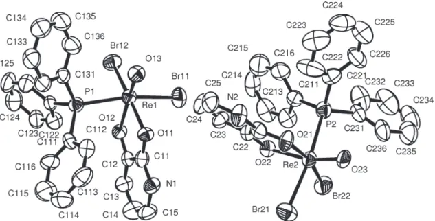
Crystal structures of 1 and 2
Complexes 1 and 2 possess a similar arrangement of
the ligands around the hexacoordinate rhenium(V) centers. The coordination spheres of the rhenium atoms are distorted octahedra, each one formed by one oxo ligand, two halides (Cl– for 1 and Br– for 2), a triphenylphosphine and two oxygen atoms of the Hdhp- ligand, which coordinates as a
O,O-chelate. The pyridine nitrogen atom of the chelating
ligands is protonated. The halides have a cis arrangement Table 1. Crystal data and structure refinement for [ReOCl2(Hdph)(PPh3)] (1) and [ReOBr2(Hdph)(PPh3)] (2)
Complex 1 2
Empirical formula C23H19Cl2NO3PRe C23H19Br2NO3PRe
Formula weight / g mol-1 645.46 734.38
Temperature / o C 20(2) 20(2)
Crystal System Trigonal Triclinic
Space group R3– P1–
a / pm 3652.3(1) 993.90(9)
b / pm 3652.3(1) 1521.7(2)
c / pm 962.60(3) 1772.3(2)
α / o 90 110.714(4)
β / o 90 98.726(7)
γ / o 120 98.346(7)
Volume / nm3 11.1201(7) 2.4202(4)
Chemical units per cell, Z 18 4
Absorption coefficient / mm-1 5.222 8.413
F(000) 5616 1392
Crystal size / mm3 0.18 × 0.02 × 0.02 0.145 × 0.020 × 0.016
Crystal description green needle green needle
θ range for data collection / o 2.95 - 24.99. 2.71 – 21.83.
Index ranges (h, k, l) -43→h→43, -42→k→43, -11→l→9 -10→h→10, -15→k→15, -18→l→18
Reflections collected 33123 9707
Independent reflections / Rint 4344 / 0.1003 5675 / 0.0515
Reflections observed [I>2σ(I)] 3194 4296
Absorption correction multi-scan12 Gaussian13
Max. and min. transmission 0.754 and 0.731 0.903 and 0.704
Refinement method full-matrix least-squares on F2 full-matrix least-squares on F2
Hydrogen treatment riding model riding model
Final R-factors [I>2σ(I)] R1 = 0.0408; wR2 = 0.0871 R1 = 0.0382; wR2 = 0.0699 Final R-factors (all data) R1 = 0.0685; wR2 = 0.0973 R1 = 0.0594; wR2 = 0.0763
“Goodness-of-fit, S, on F2 1.051 1.022
Final peak and hole in the last difference map 1.725 and -0.793 e Å-3 1.070 and -0.958 e Å-3
to each other. The triphenylphosphine ligand is in trans
position to one of the halides. One of the oxygen donor atoms from Hdhp– is located trans to the oxo ligand, while the other is trans to the second halide. The molecular
structure of 1 is shown in Figure 1. The molecular structures
of the two independent molecules of 2 are virtually identical
and essentially the same as 1, and are therefore not shown
here. Selected bond lengths and angles for both compounds are given in Table 2. The molecular labeling scheme of 1
has also been adopted for the bromo complex.
Two crystal structures of complexes with ReO3Cl2P coordination spheres, in similar modes as observed in 1, have
been reported previously, namely [ReOCl2(acac)(PPh3)]7 (3) and [ReOCl2(benzac)(PPh3)]8 (4) (acac– = acetylacetonate, benzac– = benzoylacetonate).
As in 1, in 4 the chlorine atoms are disposed in cis
positions, while the structure of 3 presents these ligands
in trans positions. No crystallographic data were found
in the literature for a complex with a ReO3Br2P coordination sphere, as observed in 2.
As observed in 4, the Re-Cl bond in 2 is longer for the
chloro ligand trans to PPh3. This results from the trans
-labilizing effect of the phosphine ligand. The Re-O bonds
trans to the oxo ligands are shorter than those trans to the
chloro ligands in 1 and in 4. This phenomenon is also
observed in other rhenium complexes with similar coordination spheres and is assigned to delocalization of
π-electron density from the oxo bonds to the trans-situated
Re–O bonds.9-11 The Re–O and Re–P bond lengths in 2 are similar to the values observed in 1. The distances
between the rhenium atoms and the oxo ligands correspond to values normally found for Re=O double bonds in oxorhenium(V) complexes.9
The bond lengths in the Hdhp- ligands in 1 and 2 do not differ significantly from the values found for the uncoordinated H2dhp, which is also protonated at the nitrogen atom.1 The following resonance structures (a, b and c) should best represent the structure of the Hdhp– ligands in 1 and 2.
Table 2. Selected bonds (pm) and angles (º) for [ReOCl2(Hdhp)(PPh3)] (1) and [ReOBr2(Hdhp)(PPh3)] (2)
1 2*
Re–O(3) 167.0(5) 169.2(5) 169.1(5)
Re–O(1) 213.5(5) 212.5(5) 212.5(5)
Re–O(2) 200.5(5) 203.9(5) 198.8(5)
Re–X(1) 241.7(2) 253.30(10) 255.19(12)
Re–X(2) 232.0(2) 247.13(11) 247.41(10)
Re–P 245.1(2) 246.1(2) 247.1(3)
O(1)–C(1) 128.4(8) 127.2(10) 126.8(10)
O(2)–C(2) 133.9(9) 135.7(10) 136.4(9)
N(1)–C(1) 132.8(9) 134.5(11) 134.1(9)
N(1)–C(5) 135.5(10) 137.4(11) 136.1(11)
C(2)–C(3) 135.3(10) 134.6(11) 135.3(10)
C(3)–C(4) 140.5(11) 139.1(13) 141.1(10)
C(4)–C(5) 132.9(11) 135.2(11) 131.7(11)
O(1)–Re–X(1) 86.16(15) 85.23(14) 84.37(17)
O(1)–Re–X(2) 169.21(14) 167.42(14) 166.83(16)
O(2)–Re–X(1) 87.10(15) 85.62(15) 88.00(16)
O(2)–Re–X(2) 94.50(14) 91.48(16) 92.66(15)
O(3)–Re–X(1) 99.54(19) 100.48(19) 100.1(2)
O(3)–Re–X(2) 103.2(2) 103.0(2) 103.73(18)
O(1)–Re–P 94.64(15) 93.63(16) 97.32(18)
O(2)–Re–P 82.07(14) 81.49(15) 83.42(17)
O(3)–Re–P 91.85(19) 92.5(2) 89.1(2)
Cl(1)–Re–P 168.61(7) 166.97(6) 170.64(7)
Cl(2)–Re–P 89.57(8) 89.61(6) 89.83(6)
Cl(2)–Re–X(1) 87.77(8) 88.87(4) 86.83(4)
* The molecular labeling scheme was adopted from 1 given in Figure 1; values correspond to two complex molecules in the asymmetric unit of 2.
Figure 1. Ellipsoid representation14 of 1, with the thermal ellipsoids
The C(1)-O(1) bond is expectedly shorter than C(2)-O(2), showing a larger double bond character. This results from the contribution of the resonance structure a. The
N-C(1) and N-C(5) distances, however, are similar and have considerable double bond character, showing that the zwitterionic resonance structures b and c also give
significant contributions to the resonance hybrid.
Crystal packings in 1 and 2 are quite different, being
apparently influenced by the type of intermolecular H-bonds involved in each structure. The trigonal structure of 1 contains channels formed by groups of six molecules,
as shown in Figure 2, which are arranged in a cyclic form by N–H⋅⋅⋅Cl hydrogen bonds. No solvent molecules were found in these voids. In the triclinic structure of 2,
N–H⋅⋅⋅Br and N–H⋅⋅⋅O H-bonds give rise to a network of molecules, as seen in Figure 3.
Conclusions
The zwitterionic complexes 1 and 2 can be prepared
in good yields and purities following a relatively simple procedure. A distorted octahedral ReO3X2P coordination (X = Cl in 1 and Br in 2) is observed for them. The crystal
structures of the compounds show different kinds of complex molecular packing, influenced by intermolecular hydrogen bonds.
Acknowledgments
This work was supported by CNPq, FAPESP and FINEP.
Supplementary Information
Crystallographic data have been deposited with the Cambridge Crystallographic Data Centre as supplementary material (deposition numbers CCDC 610190 and 610191 for 1 and 2, respectively). Copies
of the data can be obtained, free of charge, via www.ccdc.cam.ac.uk/conts/retrieving.html (or from the Cambridge Crystallographic Data Centre, CCDC, 12 Union Road, Cambridge CB2 1EZ, UK ; fax: +44 1223 336033 ; or e-mail: [email protected]).
The ellipsoid representation of 2 with the thermal
ellipsoids representing 50% probability is available free of charge at http://jbcs.sbq.org.br, as PDF file.
References
1. Deflon, V. M.; Bessler, K. E.; Kretschmar, M.; Abram, U.; Z.
Anorg. Allg. Chem.2000, 626, 1545.
2. Scarrow, R. C.; Riley, P. E.; Abu-Dari, K; White, D. L.; Raymond, K. N.; Inorg. Chem.1985, 24, 954.
3. de Souza, E. J.; Deflon, V. M.; Fernandes, A. G. de A.; Lemos, S. S.; Hagenbach, A.; Abram, U.; Inorg. Chim. Acta2006, 359,
1513.
4. Johnson, N. P.; Lock, C. J. L.; Wilkinson, G; Inorg. Synth.1967,
9, 145.
Figure 2. View of the unit cell of 1 in the direction [001].14 The molecules
are linked by H-bonds of the type N-H⋅⋅⋅Cl, forming channels in the crys-tal structure, which are parallel to the cryscrys-tallographic c axis. N(1)-H(1)
= 86.0 pm, H(1)⋅⋅⋅Cl(1)(x-y, x-1, -z+1) = 232.6 pm, N(1)⋅⋅⋅Cl(1)(x-y, x-1, -z+1) = 317.6
pm, N(1)-H(1)⋅⋅⋅Cl(1)(x-y, x-1, -z+1) = 169.76 º.
Figure 3. Perspective view14 of the unit cell of 2, showing the
intercon-nection of the complex molecules by H-bonds of the type N-H⋅⋅⋅Br and N-H⋅⋅⋅O. N(1)-H(1) = 86.0 pm, H(1)⋅⋅⋅Br(21)(-x, -y, -z+1) = 244.6 pm,
N(1)⋅⋅⋅Br(21)(-x, -y, -z+1) = 330.3 pm, N(1)-H(1)⋅⋅⋅Br(21)(-x, -y, -z+1) = 173.90 º;
N(2)-H(2) = 86.0 pm, H(2)⋅⋅⋅O(12)( x-1, y, z) = 227.5 pm, N(1)⋅⋅⋅O(12)( x-1, y, z)
5. Sheldrick, G. M; SHELXS-97A Program for Automatic Solution
of Crystal Structures, University of Göttingen: Germany, 1997,
Release 97-2.
6. Sheldrick, G. M; SHELXL-97A Program for Crystal Structure
Refinement, University of Göttingen: Germany, 1997, Release
97-2.
7. Lock, C. J. L.; Wan, C.; Can. J. Chem.1975, 53, 1548.
8. Sawusch, S; Schilde, U; Starke, I; Lehmann, A.; Uhlemann,
E.; Inorg. Chim. Acta1998, 268, 109.
9. Abram, U. In Comprehensive Coordination Chemistry II,
Elsevier: Oxford, 2004, vol. V, p. 271 and refs. cited therein. 10. Bolano, S.; Bravo, J.; Carballo, R.; Freijanes, E.; Garcia-Fontan,
S.; Rodriguez-Seoane, P.; Polyhedron2003, 22, 1711.
11. Cavell, R. G.; Hills, R. W.; Luo, H; McDonald, R.; Inorg. Chem.
1999, 38, 897.
12. Blessing, R. H.; Acta Crystallogr. 1995, A51, 33.
13. Coppens, R.; Leiserowitz, L.; Rabinovich, D.; Acta Crystallogr.
1965, 18, 1035.
14. Spek, A. L.; PLATONA Multipurpose Crystallographic Tool,
University of Utrecht: Netherlands, 2004.
Received: June 12, 2006 Published on the web: November 14, 2006
0103 - 5053 $6.00+0.00
Supplementary Information
*e-mail: [email protected]
Novel Zwitterionic Oxorhenium(V) Complexes: Synthesis, Characterization and
Crystal Structure of [ReOX
2(Hdhp)(PPh
3)] (X = Cl, Br; H
2dhp = 2,3-Dihydroxypyridine)
Elizeu J. de Souza,a André G. de A. Fernandes,a Victor M. Deflon,*,a Karl E. Bessler,a Sebastião S. Lemos,a Alzir A. Batista,b Javier Ellena,c Eduardo E. Castellano,c
Ulrich Abramd and Adelheid Hagenbachd
a
Instituto de Química, Universidade de Brasília, 70919-970 Brasília – DF, Brazil
b
Departamento de Química, Universidade Federal de São Carlos, 13565-905 São Carlos – SP, Brazil
c
Instituto de Física de São Carlos, Universidade de São Paulo, 13560-970 São Carlos – SP, Brazil
d
Institut für Chemie, Freie Universität Berlin, D-14195 Berlin, Germany
![Table 1. Crystal data and structure refinement for [ReOCl 2 (Hdph)(PPh 3 )] (1) and [ReOBr 2 (Hdph)(PPh 3 )] (2)](https://thumb-eu.123doks.com/thumbv2/123dok_br/18990902.460609/3.892.71.793.132.638/table-crystal-structure-refinement-reocl-hdph-reobr-hdph.webp)
![Figure 2. View of the unit cell of 1 in the direction [001]. 14 The molecules are linked by H-bonds of the type N-H⋅⋅⋅Cl, forming channels in the crys-tal structure, which are parallel to the cryscrys-tallographic c axis](https://thumb-eu.123doks.com/thumbv2/123dok_br/18990902.460609/5.892.116.363.103.199/figure-direction-molecules-channels-structure-parallel-cryscrys-tallographic.webp)