Article
Organotin(IV) Derivatives of 2-Acetylpyridine-N(4)-Phenylthiosemicarbazone, HAP4P,
and 2-Hydroxyacetophenone-N(4)-Phenylthiosemicarbazone, H
2DAP4P. Crystal and
Molecular Structure of [SnMe
2(DAP4P)] and [SnBu
2(DAP4P)]
Gerimário F. de Sousaa*, Regina H. P. Franciscob, M. Teresa do P. Gambardellab, Regina H. de A. Santosb and Anuar Abrasc
a
Instituto de Química, Universidade de Brasília, 70919-970 Brasília - DF, Brazil b
Instituto de Química de São Carlos, Universidade de São Paulo, 13560-970 São Carlos - SP, Brazil c
Departamento de Física, Universidade Federal de Minas Gerais, 30123-970 Belo Horizonte - MG, Brazil
As reações de 2-acetilpiridina-N(4)-feniltiosemicarbazona, HAP4P, e 2-hydroxiacetofenona-N(4)-feniltiosemicarbazona, H2DAP4P, com R4-mSnXm (m = 2, 3; R = Me, nBu, Ph e X = Cl, Br)
levaram à formação de complexos organoestânicos hexa- e penta-coordenados, que foram estudados por análise elementar, espectroscopias no IV, RMN de 1H e Mössbauer. As estruturas moleculares dos complexos [SnMe2(DAP4P)] e [SnnBu
2(DAP4P)] foram determinadas por análises de difração de raios X. Nos compostos [SnClMe2(AP4P)] e [SnBrMe2(AP4P)], o ligante desprotonado AP4P -está N,N,S-ligado aos átomos de Sn(IV) que exibem cordenação octaédrica fortemente distorcida. As estruturas dos complexos [SnMe2(DAP4P)] e [SnnBu
2(DAP4P)] revelaram que o ânion DAP4P 2-age como um ligante O,N,S-tridentado. Nestes casos os átomos de Sn(IV) adotam coordenação com geometria bipiramidal trigonal fortemente distorcida, com o átomo de N e os dois átomos de C no plano equatorial, enquanto que os átomos de O e S ocupam as posições axiais.
The reactions of 2-acetylpyridine-N(4)-phenylthiosemicarbazone, HAP4P, and 2-hydroxyacetophenone-N(4)-phenylthiosemicarbazone, H2DAP4P, with R4-mSnXm (m = 2, 3; R = Me, nBu, Ph and X = Cl, Br) led to the formation of hexa- and penta-coordinated organotin(IV)
complexes, which were studied by microanalysis, IR, 1H-NMR and Mössbauer spectroscopies. The molecular structures of [SnMe2(DAP4P)] and [SnnBu
2(DAP4P)] were determined by single-crystal X-ray diffraction studies. In the compounds [SnClMe2(AP4P)] and [SnBrMe2(AP4P)], the deprotonated ligand AP4P- is N,N,S-bonded to the Sn(IV) atoms, which exhibit strongly distorted octahedral coordination. The structures of [SnMe2(DAP4P)] and [SnnBu
2(DAP4P)] revealed that the DAP4P2- anion acts as a O,N,S-tridentate ligand. In these cases, the Sn(IV) atoms adopt a strongly distorted trigonal bipyramidal configuration where the azomethine N and the two C atoms are on the equatorial plane while the O and the S atoms occupy the axial positions.
Keywords: thiosemicarbazone complexes, organotin(VI) complexes, crystal structure analyses
Introduction
N,N,S- and O,N,S-tridentate thiosemicarbazones derived from 2-formyl- and 2-acetylpyridine form two important classes of compounds possessing biological activity1-3. In
this context, a number of complexes of first-row transition elements have been extensively studied by X-ray structural analysis and a number of other spectroscopic techniques2,4.
However organotin(IV) complexes of thiosemicarbazones have received less attention.
The chelating behaviour of N,N,S-tridentate thiosemicarbazones has been investigated and three different
complexation modes have been identified. In the most common one, the compound acts as a mononegative N,N,S-tridentate ligand and coordinates to the metal through both the nitrogen and the thiolate sulphur atoms. This coordination mode was observed in [SnX3(FPT)] (X = Cl, Br, I and HFPT = 2-formylpyridinethiosemicarbazone)5. A
second coordination mode was reported for a series of octahedral 1:1 adducts of the general formula [SnCl2R2(HFPT)]6, where the thiosemicarbazone molecule
acts as a neutral N(azomethine),S-bidentate ligand. Finally, a third coordination mode was found in [SnClMe2(FPT)]2.
a mononegative N,S-bidentate ligand, while the pyridine nitrogen atom remains uncoordinated.
A number of organotin(IV) complexes containing O,N,S-and O,N,O-tridentate ligO,N,S-ands O,N,S-and exhibiting biological or pharmacological activity have been studied, but structural features which may affect the antitumor activity and cytotoxicity of organotin(IV) are still uncertain7.
These observations have increased our interest in the structural properties of these kinds of ligands and have motivated us to study the reactions of 2-acetylpyridine-N(4)-phenylthiosemicarbazone, HP4P, and 2-hydroxy-acetophenone-N(4)-phenylthiosemicarbazone, H2DAP4P, with organotin(IV) compounds. The structures of these ligands are shown below.
[SnCl2Me(AP4P)] (1). Anal. Calc. for C15H17Cl2N4SSn:
C, 37.95; H, 3.61; N, 11.80. Found: C, 38.81; H, 3.56; N, 12.81%.
[SnClPh2(AP4P)] (2). Anal. Calc. for C26H23ClN4SSn: C,
54.06; H, 4.02; N, 9.70. Found: C, 52.48; H, 4.22; N, 11.10%.
[SnClMe2(AP4P)] (3). Anal. Calc. for C16H19ClN4SSn:
C, 42.37; H, 4.22; N, 12.35. Found: C, 42.31; H, 4.19; N, 12.25%.
[SnBrMe2(AP4P)] (4). Anal. Calc. for C16H19BrN4SSn:
C, 38.59; H, 3.85; N, 11.25. Found: C, 37.07; H, 3.55; N, 11.12%.
[SnClnBu
2(AP4P)] (5). Anal. Calc. for C22H31ClN4SSn:
C, 49.14; H, 5.81; N, 10.42. Found: C, 48.82; H, 5.75; N, 10.36%.
[SnCl2Me(HDAP4P)] (6). Anal. Calc. for C16H17Cl2N3SSn:
C, 39.29; H, 3.48; N, 8.59. Found: C, 41.89; H, 4.00; N, 8.69%.
[SnPh2(DAP4P)] (7). Anal. Calc. for C27H23N3OSSn:
C, 58.30; H, 4.17; N, 7.55. Found: C, 59.56; H, 4.40; N, 7.75%. [SnMe2(DAP4P)] (8). Anal. Calc. for C17H19N3OSSn:
C, 47.25; H, 4.43; N, 9.72. Found: C, 47.13; H, 4.29; N, 9.71%. [SnnBu
2(DAP4P)] (9). Anal. Calc. for C23H31N3OSSn:
C, 53.51; H, 6.05; N, 8.14. Found: C, 53.38; H, 5.82; N, 8.13%. Infrared spectra were recorded on a Nicolet 5ZDX-FT spectrophotometer in the 4000-400 cm-1 range using KBr
pellets. Due to the poor solubility of some of the complexes, it was possible to obtain 1H NMR spectra only for 3, 4 and 8, in CDCl3, using a 250 MHz Bruker spectrometer.
Chemical shifts are relative to internal tetramethylsilane.
119Sn Mössbauer spectra were measured using a Model
AM-1 Mössbauer efect spectrometer, moving a AM-15-mCi BaSnO3 source at room temperature. The isomer shift values are given with respect to this source. The samples were measured at liquid nitrogen temperature and all spectra were computer fitted assuming Lorentzian line shapes.
Crystal structure determinations
Single-crystal X-ray diffraction data were collected on an Enraf-Nonius CAD-4 automatic diffractometer, with a graphite monochromated KαMo radiation (λ = 0.71073 Å), obtained in a fine focus sealed tube9.
Experimental parameters and crystallographic data for both complexes are shown in Table 1. In both cases, the data reductions were carried out with the XCAD-4 software10.
The structures were solved using the heavy-atom method11.
The SHEL97 software12 was used for refinement by
full-matrix least-squares calculations. N
C N CH3
C N H
S H OH
N N C
N CH3
C N H
S H
H2DAP4P
HAP4P
Experimental
Syntheses
HAP4P and H2DAP4P were prepared from a 1:1 molar ratio of N(4)-phenylthiosemicabazide and the appropriate ketone (2-acetylpyridine or 2-acetylphenol) in boiling EtOH (15 mL) for 30 min. After cooling, the compounds were obtained as pale-yellow needles (HAP4P: mp 171-174 °C; H2DAP4P: mp 120-122 ºC) which were filtered and dried under vacuum over CaCl2. The organotin(IV) complexes were obtained by the following procedure: 0.20 mmol of the appropriate ligand were dissolved by refluxing in dry MeOH for 5 min. To this solution were added 0.21 mmol of one organotin(IV) species in 5 mL of MeOH, and the resulting mixture was refluxed for 1 h and filtered to give a clear solution. Cooling the solution and slowly evaporating the solvent led to crystalline products with yields on the order of 70%. Single crystals suitable for X-ray diffraction were isolated only for complexes 2, 3, 7 and 8. The structures of 2 and 3 were solved and
described previously8. The microanalyses were
Results and Discussion
Crystal structures of [SnMe2(DAP4P)](8) and [Sn nBu2(DAP4P)] (9).
The structure determinations of compounds 8 and 9
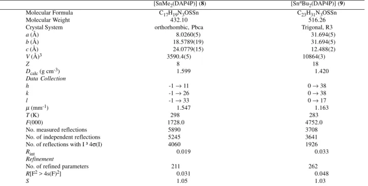
revealed similar molecular structures with the occurrence of pentacordinated Sn(IV) complexes, with a trigonal bipyramidal (TBP) geometry. In the coordination polyhedron of the two compounds, two carbons (from alkyl groups) and the N(1) atom (from the thiosemicarbazone) occupy the equatorial plane. Atoms S and O occupy axial positions. Figures 1 and 2 show the molecules along with the labeled atoms.
In both complexes, the largest bond angle involving the Sn(IV) atom was O–Sn–S: 151.32(6) and 156.96(15)°, respectively, indicating strong deviations from the ideal value of 180°. Dihedral angles of 88 and 87° were observed between the planes through Sn, C(16), C(17), N(1) and Sn, O, S (8) and Sn, C(16), C(20), N(1) and Sn, O, S (9),
respectively. Selected bond distances and angles of the two complexes are shown in Table 2.
The structures of the complexes [SnPh2(Hdaptsc)]Cl13
(A), H2daptsc = 2,6-diacetylpyridinebis(thiosemicarbazone),
[SnMe2(Hdapf)]2[SnCl4Me2]14 (B), H
2dapf =
2,6-diacetyl-pyridinebis(2-furoylhydrazone) and [SnMe2(Hdapt)]
Br.H2O15 (C), H
2dapt = 2,6-diacetylpyridinebis
(2-tenoyl-hidrazone) showed that one proton from the azomethine group of the ligand molecules has been eliminated upon coordination. The proton on the other azomethine group remained bonded
Table 1. Crystallographic data and experimental parameters.
[SnMe2(DAP4P)] (8) [SnnBu
2(DAP4P)](9)
Molecular Formula C17H19N3OSSn C23H31N3OSSn
Molecular Weight 432.10 516.26
Crystal System orthorhombic, Pbca Trigonal, R3
a (Å) 0000008.0260(5) 000031.694(5)
b (Å) 00000018.5789(19) 000031.694(5)
c (Å) 00000024.0779(15) 000012.488(2)
V (Å)3 3590.4(5) 10864(3)....
Z 8. 18..
Dcalc (g cm-3) 0001.599 0001.420
Data Collection
h -1 → 11 0 → 38
k -1 → 26 0 → 38
l -1 → 33 0 → 17
µ (mm-1) .00.1.547 0001.163
T (K) 298.00 283....
F(000) 1728.000 4752.000
No. measured reflections 5890.00. 3708....0.
No. of independent reflections 5245.00. 3641....0.
No. of reflections with I ³ 4σ(I) 4060.00. 1926....0.
Rint 0000.019 0000.033
Refinement
No. of refined parameters 211.... 262...0
R[F2 > 4s(F)2] 0000.031 0000.048
S 001.05 001.03
Table 2. Selected bond distances (Å) and angles (º) for [SnMe2(DAP4P)]
(8) and [SnnBu
2(DAP4P)] (9).
8 9
Sn–O 002.095(2) 002.120(4)
Sn–N(1) 002.233(2) 002.194(6)
Sn–S 002.5296(8) 002.547(2)
S–C(1) 001.734(3) 001.754(8)
N(1)–N(2) 001.390(3) 001.382(8) C(1)–N(2) 001.291(3) 001.292(8) C(1)–N(3) 001.375(3) 001.365(9) N(3)–C(10) 001.417(4) 001.396(9) N(1)–C(2) 001.307(3) 001.311(8)
O–C(9) 001.336(3) 001.338(8)
Sn–C(16) 002.117(3) 002.129(7) Sn–C(17) 002.123(3) 002.165(8)a
O–Sn–S 151.32(6) 156.69(15)
O–Sn–N(1) 079.50(7) 080.7(2) O– Sn–C(16) 088.80(11) 094.1(2) O–Sn–C(17) 098.55(12) 085.5(3)b
S – Sn – N(1) 076.71(6) 076.24(15) S–Sn–C(16) 095.54(11) 097.6(2) S–Sn–C(17) 102.82(11) 100.2(3)c
N(1)–Sn–C(16) 132.48(11) 100.2(3) N(1)–Sn–C(17) 104.81(11) 112.6(3)d
O–C(9)–C(4) 122.7(2) 121.5(6)
C(16)–Sn–C(17) 122.50(13) 134.4(4)e a Sn–C(20), b O-Sn-C(20), c S-Sn-C(20), d S-Sn-C(20), e C(16)-Sn-C(20)
(thiolate form) 2.592(1), S(1)–C(13) 1.731(5), Sn–S(2) (thione form) 2.703(1), and S(2)–C(23) 1.694(6) Å; in complex B14, the distances are: Sn(2)–O(1) (enolate
form) 2.227(4), O(1)–C(4) 1.276(8), Sn(2)–O(2) (keto form) 2.474(4) and O(2)–C(14) 1.242(7) Å; for C15 the
values are: Sn–O(1) (keto) 2.493(4), O(1)–C(3) 1.241(7), Sn–O(2) (enolate) 2.209(4) and O(2)–C(13) 1.286(7) Å.
In the cases, of compounds 8 and 9, part of the
thiosemicarbazone derivative molecule is planar. The observed dihedral angle between the phenyl rings was 51° for complex 8 and 57° for complex 9. The packing
mode is determined by the nature of the alkyl ligands. The presence of butyl groups in 9, larger and with larger
displacement parameters, prevents the orthorhombic symmetry once the average distances between the thio-semicarbazones are not very different. In both structures there are intermolecular interactions involving N(3)– HΛO. The geometric features of these interactions are respectively: N(3)–H 0.85 Å, N(3)–HΛO (1/2 + x, 1/2 -y, 1 - z) 3.033 Å, N(3)–HΛO 179.1º for compound 8 and
N(3)–H 0.86 Å, N(3)–HΛO 174.6º for compound 9.
In complexes 3 and 4, whose structures are nearly
identical, except for the presence of a chloride ligand in 3
and a bromide ligand in 4, the ligand molecule bears only
one substituent on the pyridine ring8. The observed
distances were: Sn–S 2.4728(8), S–C(10) 1.750(3) Å for the chloride complex and Sn–S 2.4743(12), S–C(10)
Figure 1. Perspective view of [SnMe2(DAP4P] (8) showing the atom numbering scheme. Displacement ellipsoids are drawn at the 50% probability level.
Figure 2. Perspective view of [SnnBu
1.739(5) Å for the bromide complex, indicating that the S atom is coordinated in the thiolate form.
1H NMR spectroscopy
The 1H NMR spectrum of complex 8 showed two
singlets at δ 2.7 and 0.7 [2J(119Sn–CH
3) 73 Hz], due to
two magnetically non-equivalent methyl groups assigned to N=C–CH3 and Sn–CH3, respectively. According to the literature, the use of the Lockhart-Manders16 equation
shown below and of the observed coupling constant of 73 Hz yields a C–Sn–C angle of 123º , in excellent agreement with the angle of 122.50(13)º observed in the solid state. This data suggests that the basic structural features of the solid-state phase remain in solution.
θ = 0.0161{2J(119Sn–CH 3)}2
- 1.32{2J(119Sn–CH
3)] + 133.4
A similar result has been reported for a trigonal bipyramidal (TBP) complex with an analogous coordination geometry, [SnMe2(L)]17 (H
2L =
salicyl-aldeydethiosemicarbazone). In this case, the 2J(119Sn–
CH3) was 70 Hz.
Infrared spectroscopy
Table 3 shows the assignment of the main IR absorption bands for the ligands and their complexes. In the 3463-1168 cm-1 region the HAP4P and H
2DAP4P
ligands show bands attributed to hydrogen-bonded O–H and N–H overlapping with the C–H stretching absorptions of phenyl and pyridyl rings. As a result of the monodeprotonation of the HAP4P ligand and double deprotonation of the H2DAP4P ligand (except in complex
6), the spectra of compounds 1-5 and 7-9 lack bands
located at 3241 and 3168 cm-1, attributed to ν(NN–H),
and retain the absorption bands found in the free ligands at 3301 and 3305 cm-1, attributed to ν(PhN–H) vibrations,
which are shifted to lower frequencies. Thus, the bands
in the 3426-3237 cm-1 range are assigned to the ν(NN–H) stretching frequencies.
The ν(C=N) absorptions at 1523 and 1521 cm-1 for
HAP4P and H2DAP4P, respectively, are shifted to higher frequencies by ca. 15-30 cm-1 in the spectra of the complexes,
indicating coordination of the azomethine nitrogen N(2) to the metal ion18. The same trend is exhibited by the bands at
1360 cm-1 for HAP4P and at 1366 cm-1 for H
2DAP4P.
The bands at 1189 and 782 cm-1 in HAP4P and at 1197
and 757 cm-1 in H
2DAP4P, which have a significant
contribution from ν(C=S) stretching vibrations, are shifted to lower frequencies in the spectra of the complexes, suggesting coordination through the sulfur atom18,19. The
far IR bands observed in the 509-504 cm-1 range were
tentatively assigned to the ν(Sn–O) mode.
Mössbauer spectroscopy
119Sn Mössbauer spectroscopy was performed on all
nine complexes, giving the results shown in Table 4, which include parameters from the literature for comparison.
Complexes 1-6 exhibit octahedral coordination while
complexes 7-9 adopt a strongly distorted trigonal
bipyramidal (TBP) configuration where the anions AP4P
-and HDAP4P- act as N,N,S- and O,N,S-tridentate ligands,
respectively. Their isomer shifts (δ) are lower than those of the parent acids15 [SnCl
3Me] (1.20 mm s-1), [SnCl2Ph2]
(1.32 mm s-1), [SnCl
2Me2](1.49 mm s-1), [SnBr2Me2]
(1.59 mm s-1) and [SnCl
2nBu2](1.75 mm s-1).
Isomer shifts always decrease upon adduct formation, as a result of rehybridization to higher coordination for the Sn(IV) atoms in the complexes in which s orbital participation is less than 25%. The reduction in the isomer shift in complex 6
(0.90 mm s-1), compared to 1 (0.98 mm s-1), is due to the
structural differences between AP4P- and HDAP4P-. The
former ligand has one ligating pyridine N atom, whereas the latter one has a phenol O atom. Therefore, AP4P- is a much
less electronegative ligand than HDAP4P-, which accounts
for the lower value of the isomer shift in complex 6.
Table 3. Main IR bands (cm-1) for the ligands HAP4P and H2DAP4P and their complexes
Compound ν(N–H) ν(C=N, C=C) ν(C–S) + ν(C–N) ν(C=S) ν(Sn–O)
HAP4P 3301, 3241 1598, 1523, 1468, 1444 1360 1189, 782
[SnCl2Me(AP4P)] (1) 3280 1597, 1544, 1497, 1453 1428 1163, 761
-[SnClPh2(AP4P)] (2) 3237, 3178 1596, 1535, 1498, 1457 1433 1160, 762
-[SnClMe2(AP4P)] (3) 3276 1596, 1532, 1497, 1458 1435 1160, 766
-[SnBrMe2(AP4P)] (4) 3243 1596, 1548, 1499, 1459 1433 1152, 761
-[SnBrnBu
2(AP4P)] (5) 3426, 3335 1601, 1520, 1493, 1480 1401 1159, 767
-H2DAP4P* 3305, 3168 1593, 1521, 1444 1366 1197, 757
-[SnCl2Me(HDAP4P)] (6) 3407, 3388 1599, 1574, 1501 1434 1178, 757 505
[SnPh2(DAP4P)] (7) 3325 1599, 1543, 1494 1436 1183, 745 504
[SnMe2(DAP4P)] (8) 3241 1595, 1570, 1537, 1494 1438 1190, 756 509
[SnnBu
2(DAP4P)] (9) 3245 1596, 1567, 1540, 1494 1434 1191, 744 509
Table 4. 119Sn Mössbauer spectroscpy data for Sn(IV) complexes.
Complex C.N. δ (mm s-1) ∆ (mm s-1) θ (C–Sn–C)
(exp.)
[SnCl2Me(AP4P)] (1) 6 0.98(1) 1.96(1)
-[SnClPh2(AP4P)] (2) 6 1.26(1) 2.92(1)
-[SnClMe2(AP4P)] (3) 6 1.38(1) 3.35(1) 145º
[SnBrMe2(AP4P)] (4) 6 1.42(1) 3.40(1) 144º
[SnBrnBu
2(AP4P)] (5) 6 1.54(1) 3.50(1)
-[SnCl2Me(HDAP4P)] (6) 6 0.90(1) 2.02(1)
-[SnPh2(DAP4P)] (7) 5 1.09(1) 2.03(1)
-[SnMe2(DAP4P)] (8) 5 1.18(1) 2.41(1) 122º
[SnnBu
2(DAP4P)] (9) 5 1.31(1) 2.62(1) 134º
[ClSnCl2(FPT)]a (10) 6 0.58 0.00
-[MeSnCl3(BtSOMe)]b (11) 6 0.98(1) 2.07(1)
-[Ph2SnCl(of)]c (12) 6 1.10 2.61
-[Ph2Sn(L1)]d (13) 5 1.30 2.30 127º
[Me2Sn(L1)]d (14) 5 1.27 2.43 127º
[Bu2Sn(L2)]e (15) 5 1.28 2.85
-Abreviations: C.N. = coordination number, HAP4P = 2-acetylpiridine-N(4)-phenylthiosemicarbazone, H2DAP4P = 2-hydroxyacetophenone-N(4)-phenylthiosemicarbazone, BtSOMe = (2-methylsulphinyl)benzothiazole, Hof = 3-hydroxyflavone, H2L1 = salicilaldehydethiosemicarbazone, H2L2 = salicilaldehyde-2-furanthiosemicarbazone, aRef. 5, bRef. 23, cRef. 22, dRef. 18, eRef. 24.
In complexes 2 to 5, the ligand remains unchanged, but
the phenyl groups in complex 2 (1.26 mm s-1) were replaced
by methyl groups in 3 (1.38 mm s-1), the chloride in complex 3 was replaced by bromide in 4 (1.42 mm s-1) and the methyl
groups in complex 4 were replaced by n-butyl groups in 5
(1.54 mm s-1). Again, a similar effect is observed as before,
and the isomer shifts decrease on going from complex 5 to 2. Similar results were observed for complexes 7 (1.09 mm
s-1), 8 (1.18 mm s-1) and 9 (1.31 mm s-1).
A somewhat analogous trend can be seen in literature examples, as shown in Table 4. Although the ligands are not the same in all cases, they are related, and one can see that lower δ values correspond to the complexes containing electronegative groups (Cl ou Ph), whereas alkyl groups (Me, nBu), of low electronegativity, lead to higher δ values. Quadrupole splitting (∆) values, presented in Table 4, are not sufficient in themselves to characterize a Sn(IV) complex as either tetra-, penta-, hexa-, or hepta-coordinated20. However, ∆ has been useful in distinguishing
between cis-, trans-[SnX4R2]and [SnX3R2]configurations
in complexes with octahedral and trigonal bipyramidal geometries, respectively. Thus, a complex with a trans
-[SnX4R2] configuration usually has ∆ values of ca. 4 mm
s-1, while a value of ca. 2 mm s-1 would be expected for a
complex with a cis-SnX4R2 configuration. Sham and
Bancroft21, also using point charge calculations, have
shown that the quadrupole splittings (∆) for trans-[SnX4R2]
compounds decrease smoothly from the value (4 mm s-1)
for a regular octahedral geometry (θ = 180º) as the structure becomes more distorted, i.e. the q angle (R-Sn-R) becomes
less than 180º.
The quadrupole splitting (∆) values, shown in Table 4, indicate that complexes 2-5 adopt a trans-[SnX4R2]
arrangement and the octahedral diphenyl compound
(complex 2) is the most distorted among all of them. The
values between 2.03-2.85 mm s-1 for complexes 7-9 and 13-15 are typical of diorganotin(IV) complexes in a
distorted trigonal bipyramidal (TBP)17,22 environment. The
lower quadrupole splitting (∆) value for complex 10 (0.0
mm s-1) indicates that the charge distribution around the
Sn(IV) nucleus is highly symmetrical5 and the geometry
is considerably non distorted.
Acknowledgments
The authors are grateful for financial support from CNPq, CAPES, FINEP, FAPEMIG and FAPESP in Brazil. The authors are grateful also to Dr. Julio Zukerman-Schpector for data collection, carried out on a CAD-4 at the Instituto de Química-USP.
Supplementary Information
Crystallographic data (excluding structure factors) for the structures have been deposited with the Cambridge Crystallographic Data Center as supplementary publication nos. 149653 and 149654. Copies of the data can be obtained, free of charge, on application to CCDC, 12 Union Road, Cambridge CB2 1EZ, UK, (fax: +44 1223 336033 or e-mail: [email protected]).
References
1. West, D. X.; Billeh, J. S.; Jasinski, J. P.; Jasinski, J. M.; Butcher, R. J. Transition Met. Chem. 1998, 23, 209.
2. Labib, L.; Khall, T. E.; Iskander, M. F.; Refaat, L. S.
3. West, D. X.; Padhye, S. B.; Sonawane, P. B. Struct. Bonding. 1991, 76. 1.
4. Casas, J. S.; Castiñeiras, A.; Sánchez, A.; Sordo, J.; Vazquez-Lópes, A.; Rodriguez-Argü elles, M.C.; Russo, U. Inorg. Chim. Acta 1994, 221, 61.
5. Barbieri, R. S.; Beraldo, H. O.; Filgueiras, C. A. L.; Abras, A.; Nixon, J. F.; Hitchock, P. B. Inorg. Chim. Acta 1993, 206, 169.
6. Bamgboye, T. T.; Bamgboye, O. A. Inorg. Chim. Acta 1988, 144, 249.
7. Huber, F.; Roge, G.; Carl, L.; Atassi, G.; Spreafico, F.; Bar bieri, R.; Silvestri, A.; Rivarola, E.; Ruisi, G.; Di Bianca, F.; Alonzo, G. J. Chem. Soc. 1985, 523.
8. Francisco, R. H. P.; Gambardella, M. T. P.; De Sousa, G. F.; Abras, A. Acta Cryst.2000, C56, 187.
9. Enraf-Nonius (1989). CAD-4 Software. Version 5.0,
Enraf-Nonius, Delft, The Netherlands.
10. Harms, K.; Wocadlo, S. XCAD4; Program for Processing CAD-4 Diffractometer Data, University
of Marburg, Marburg, Germany, 1995.
11. Frenz, B. A. The Enraf Nonius CAD4 SDP. Computing in Crystallography, Eds. Schenk, H.;
Olthof-Hazekamp, R.; van Koningsveld, H.; Bassi, G. C.; Delft University Press, 1978, p. 64-71.
12. Sheldrick, G. M. SHELXL97. Program for Crystal Structure Refinement; University of Göttingen,
Göttingen, Germany, 1997.
13. Moreno, P. C.; Francisco, R. H. P.; Gambardella, M. T. P.; De Sousa, G. F; Abras, A. Acta Cryst.1997, C53, 1411.
14. Francisco, R. H. P.; Moreno, P. C.; Gambardella, M. T. P.; De Sousa, G. F.; Mangas, M. B. P.; Abras, A.
Acta Cryst.1998, C54, 1444.
15. De Sousa, G. F.; Mangas, M. B. P.; Francisco, R. H. P.; Gambardella, M. T. P.; Rodrigues, A. M. G. D.; Abras. A. J. Braz. Chem. Soc. 1999, 10, 222.
16. Lockart, T. P.; Manders, W. F. Inorg. Chim. 1986, 25, 892.
17. Casas, J. S.; Sánchez, A.; Sordo, J.; Vazquez-López, A.; Castellano, E. E.; Zukerman-Schpector, J.; Rodríguez-Argüelles, M. C.; Russo, U. Inorg. Chim. Acta1994, 216, 169.
18. Offiong, O. E.; Martelli, S. Transition Met. Chem. 1997, 22, 263.
19. Ferrari, M. B.; Fava, G. G.; Lanfrachi, M.; Pelizzi, C.; Tarasconi, P. Inorg. Chim. Acta1991, 181, 253.
20. Carini, C.; Pelizzi, G.; Tarasconi, P.; Pelizzi, C.; Molloy, K. C.; Waterfield, C. J. Chem. Soc., Dalton Trans. 1989, 289.
21. Sham, T. K.; Bancroft, G. M. Inorg. Chim. Acta1995, 14, 2281.
22. Blunden, S. J.; Smith, P. J. Organomet. Chem. 1982, 226, 157.
23. De Sousa, G. F.; Abras, A.; Filgueiras, C. A. L.
Proceedings of the International Conference on the Applications of the Mössbauer Effect, ICAME-95,
Ortalli, I., Ed., SIF, Bologna, vol. 50, 1996, p. 79. 24. Singh, N. K.; Sharma, U., Kulshreshtha, S. K.
J. Organomet. Chem. 1990, 382, 375.
Received: November 6, 2000 Published on the web: July 29, 2001
![Figure 1. Perspective view of [SnMe 2 (DAP4P] (8) showing the atom numbering scheme. Displacement ellipsoids are drawn at the 50% probability level.](https://thumb-eu.123doks.com/thumbv2/123dok_br/18988786.459863/4.892.232.719.143.437/figure-perspective-showing-numbering-scheme-displacement-ellipsoids-probability.webp)
![Table 4. 119 Sn Mössbauer spectroscpy data for Sn(IV) complexes. Complex C.N. δ (mm s -1 ) ∆ (mm s -1 ) θ (C–Sn–C) (exp.) [SnCl 2 Me(AP4P)] (1) 6 0.98(1) 1.96(1) -[SnClPh 2 (AP4P)] (2) 6 1.26(1) 2.92(1) -[SnClMe 2 (AP4P)] (3) 6 1.38(1) 3.35(1) 14](https://thumb-eu.123doks.com/thumbv2/123dok_br/18988786.459863/6.892.114.842.158.396/table-mössbauer-spectroscpy-complexes-complex-sncl-snclph-snclme.webp)
