239 239 239 239 239 Mem Inst Oswaldo Cruz, Rio de Janeiro, Vol. 101(3): 239-243, M ay 2006
In
vitro susceptibility to antifungal agents of environmental
Cryptococcus
spp. isolated in the city of Ribeirão Preto,
São Paulo, Brazil
Reginaldo dos Santos Pedroso+, Joseane Cristina Ferreira, Regina Celia Candido
Laboratório de Micologia Clínica, Departamento de Análises Clínicas, Toxicológicas e Bromatológicas, Faculdade de Ciências Farmacêuticas de Ribeirão Preto, Universidade de São Paulo, Via do Café, s/no, 14040-903 Ribeirão Preto, SP, Brasil
Infections by Cryptococcus strains other than C. neoformans have been detected in immunocompromised pa-tients. Of these strains, three are considered human pathogens: C. albidus, C. laurenttii, and C. uniguttulatus. This study deals with the in vitro susceptibility of Cryptococcus to drugs such as amphotericin B, itraconazole, fluconazole, and 5-fluorocytosine. Environmental Cryptococcus isolates (50) distributed as follows: C. neoformans var. neoformans
(16), C. albidus (17), C. laurentii (14), and C. uniguttulatus (3) were evaluated by the micro and macrodilution techniques, according to EUCAST and NCCLS recommendations, respectively. Considering both methodologies the respective minimal inhibitory concentrations (MIC) were 0.125 and 2 µg/ml for amphotericin B, 0.06 and 8 µg/ ml for itraconazole, and 0.5 and more than 64 µg/ml for fluconazole and 5-fluorocytosine. Agreement percentages for the two methodologies were 100% for amphotericin B and fluconazole for all the strains tested. For itraconazole, the agreement percentage was 81.3% in the C. neoformans strain and 100% for all the others. All species had a agreement percentage of 94.1 to 100% when susceptibility to 5-fluorocytosine was tested. It is concluded that environmental isolates of C. neoformans var. neoformans, C. albidus, C. laurentii, and C. uniguttulatus may show high MICs against certain drugs, suggesting in vitro primary resistance to the antifungals tested.
Key words: antifungals - Cryptococcus spp. - susceptibility - São Paulo - Brazil
Infections by Cryptococcus species other than C. neoformans are seldom described in the literature, but they do occur in immunocompromised patients, with he-matological or oncologic diseases or are human immuno-deficiency virus infected. Among the infective species, three are considered human pathogens, C. albidus, C. laurentii, and C. uniguttulatus (Johnson et al. 1998, McCurdy & Morrow 2001). Variable clinical manifesta-tions are reported, such as instances of cutaneous infec-tions (Kamalam et al. 1977, Johnson et al. 1998), ventricu-litis (McCurdy & Morrow 2001), meningitis (Kordossis et al. 1998), fungemia (Loison et al. 1996, Johnson et al. 1998, Kordossis et al. 1998, Kunova & Krcmery 1999), pneumo-nia and pulmonary abscesses (Johnson et al. 1998).
The species in vitro susceptibility to antifungals is also not well known, although it is very important that this type of previous information guide the clinician in his choice of antifungals to a certain group of microorgan-isms (Aller et al. 2000).
Cryptococcus susceptibility studies reported so far are mostly related to clinical or environmental C. neoformans, while other clinically important species are poorly described (Franzot & Hamdan 1996, Alves et al. 2001, Garcia-Martos et al. 2002, Moraes et al. 2003).
In this report the in vitro antifungal susceptibility of different environmental yeast species of the
Cryptococ-Financial support: Capes
+Corresponding author: [email protected]
Received 30 August 2005 Accepted 5 April 2006
cus genus was evaluated, utilizing in broth micro and macrodilution methods according to guidelines of the National Committee for Clinical Laboratory Standards (NCCLS 2002).
MATERIALS AND METHODS
Microorganisms tested - Environmental isolates in the city of Ribeirão Preto, state of São Paulo, Brazil, were dis-tributed as follows: 16 C. neoformans var. neoformans, 17
C. albidus, 14 C. laurentii, and 3 C. uniguttulatus.
Susceptibility tests to antifungals - The antifungals tested were: amphotericin B (Fungizon, Bristol Myers Squibb do Brazil, Brazil), 5-fluorocytosine (Roche, Brazil), fluconazole (Pfizer, Sandwich, UK) and itraconazole (Janssen, Beerse, Belgium). Dimethyl sulfoxide (Vetec, Rio de Janeiro, Brazil) was used to prepare stock solutions of amphotericin B and itraconazole in the concentrations of 320 µ g/ml. Aqueous solutions of fluconazole and 5-fluorocytosine, were prepared in sterile distilled water in the concentrations of 1280 µg/ml. Drug final concentra-tions in the tests were 16 to 0.03 µg/ml for amphotericin B and itraconazole and 64 to 0.25 µg/ml for fluconazole and 5-fluorocytosine. The culture medium, RPMI-1640 con-taining L-glutamine, but no sodium bicarbonate was supplemented with glucose (18 g/l) and buffered to pH 7 with 0.165M morpholinepropanesulfonic acid (MOPS), according to EUCAST (2002) recommendations. The pri-mary yeast inocula equivalent to 0.5 in the McFarland scale (1 to 5 × 10⁶ cells/ml) were prepared in deionized
sus-240 240 240 240
240 Invitro susceptibility of Cryptococcus • Reginaldo dos Santos Pedroso et al.
pensions were mixed with a vortex, and diluted 1:10. One hundred microliter aliquots of the latter suspension were removed with a pipette, and this volume was spread onto a Sabouraud dextrose agar plate with a sterile, bent glass rod. Plates were incubated at 30 and 35ºC during 5 days, and the CFU per milliliter were determined. This allowed counts of 10 to 50 colonies per plate (Espinel-Ingroff et al. 1992, Rodriguez-Tudela et al. 2000, EUCAST 2002, NCCLS 2002).
Macrodilution method - One hundred microliter aliquots of each drug dilution were mixed, in a glass tube 13 × 100 mm, with 900 µl of the RPMI-1640 containing 0.5 to 2.5 × 10⁶ cells/ml. Control tubes were prepared
contain-ing just drug diluents (negative controls) and others (posi-tive controls) containing microorganism suspensions and drug diluents. All tubes, in duplicate, were incubated at 30oC during 48 h, except for C. neoformans var. neoformans
and the control strains incubated at 35oC for the same
time. Growth in each test tube was compared to the equiva-lent controls without drugs. Minimal inhibitory concen-tration (MIC), for azoles and 5-fluorocytosine, was de-fined as the smallest concentration inhibiting at least 50% of the control tube growth (0.5 ml of growth control plus 0.5 ml of uninoculated RPMI), and for amphotericin B as the smallest concentration able to completely inhibit (100%) growth.
Microdilution method - Ninety-six well plates con-taining 100 µl aliquots of each drug dilution/well were stored at –20oCfor up to 30 days. The initial inoculum
suspension, RPMI diluted (1:10) was the test material. In duplicate tests, 100 µ l of the diluted suspension was added to each of the wells in the plates containing the drug. Positive and negative controls were established in separate wells. The plates were incubated at 30oC except
for C. neoformans var. neoformans and control strains, which were incubated at 35oC. MIC, for azoles and
5-fluorocytosine, was defined as the smallest concentra-tion inhibiting at least 50% of the control tube growth, and for amphotericin B as the smallest concentration able to completely inhibit (100%) growth.
Control microorganisms - Strains of Candida parapsilosis ATCC 22019 and C. krusei ATCC 6258 were used as controls.
Result analysis - The endpoints for fluconazole, itraconazole and 5-fluorocytosine were the ones sug-gested by NCCLS (2002) and the ones for amphotericin B
by Rodriguez-Tudela et al. (1998). Microorganisms show-ing MICs ≥ 2 µg/ml were considered resistant to amphot-ericin B; for itraconazole resistance was determined for MICs ≥ 1 µg/ml, for 5-fluorocytosine ≥ 32 µg/ml and ≥ 64 µg/ml for fluconazole. By comparing the values in the mi-cro and mami-crodilution methods the correlation analysis between MICs for each drug and microorganism was es-tablished. The values were considered equivalent when both were equal or differed by at the most two dilutions (reference value ± 2 dilutions). In other conditions the difference in values was expressed as units of dilution. Based on the result equivalence definition, the agreement percentage was calculated meaning the percentage of the total compared results, obtained in the two methods, con-sidered as being equivalent (Pfaller & Barry 1994).
RESULTS
Controls, C. parapsilosis ATCC 22019 and C. krusei
ATCC 6258 showed MIC values according to the NCCLS (2002) described range. The MICs and MIC intervals for the four drugs inhibiting 50% and 90% of the isolates (MIC50 and MIC90) are summarized in Tables I to IV.
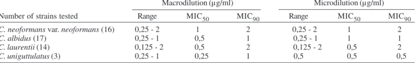
Amphotericin B MIC range values were the smallest among the drugs tested and within the group C. laurentii
was the largest (Table I). For itraconazole the widest MIC range was seen with C. neoformans var. neoformans, and
C. albidus in both methods. The highest MIC50 and MIC90 were shown by C. albidus (Table II). Fluconazole had the highest MIC ranges in susceptibility tests with C. neoformans var. neoformans. The micro and macrodilution methods showed the highest MIC50 values for C. albidus; MIC90 values results were higher than 64 µg/ml for all strains, except C. laurentii, in both methods (Table III). 5-fluorocytosine showed the widest MIC ranges for all strains, the smallest being with C. uniguttulatus (Table IV).
Table V shows agreement percentages (AP) for the results obtained by the micro and macrodilution meth-ods. It can be seen that for C. neoformans var. neoformans
the index was 100% against amphotericin B, fluconazole and 5-fluorocytosine, considering ± 1 dilution, while for itraconazole the AP was 56.3 or 81.3% considering ± 1 or ± 2 dilutions respectively. For the other strains, AP was 100% against amphotericin B; APs of 82.3 and 100% were shown by C. albidus against itraconazole and 71.5 and 100% with C. laurentii considering ± 1 and ± 2 dilutions, respectively. To 5-fluorocytosine the AP was 88.2 and 94.1% for C. albidus, and 85.7 and 100% for C. laurentii,
TABLE I
In vitro susceptibility of 50 environmental Cryptococcus isolates to antifungal amphotericin B Macrodilution (µg/ml) Microdilution (µg/ml)
Number of strains tested Range MIC50 MIC90 Range MIC50 MIC90
C. neoformans var. neoformans (16) 0,25 - 2 1 2 0,25 - 2 1 2
C. albidus (17) 0,25 - 1 0,5 1 0,25 - 1 1 1
C. laurentii (14) 0,125 - 2 0,5 2 0,125 - 2 0,5 2
241 241 241 241 241 Mem Inst Oswaldo Cruz, Rio de Janeiro, Vol. 101(3), M ay 2006
TABLE II
In vitro susceptibility of 50 environmental Cryptococcus isolates to the antifungal itraconazole Macrodilution (µg/ml) Microdilution (µg/ml)
Number of strains tested Range MIC50 MIC90 Range MIC50 MIC90
C. neoformans var. neoformans (16) 0,06 - 4 0,25 1 0,06 - 8 0,5 2
C. albidus (17) 0,125 - 8 2 8 0,06 - 8 4 8
C. laurentii (14) 0,06 - 1 0,25 1 0,06 - 1 0,5 1
C. uniguttulatus (3) 0,25 - 8 0,25 8 0,5 - 4 0,5 4 See Table I footnote
TABLE III
In vitro susceptibility of 50 environmental Cryptococcus isolates to the antifungal fluconazole Macrodilution (µg/ml) Microdilution (µg/ml)
Number of strains tested Range MIC50 MIC90 Range MIC50 MIC90
C. neoformans var. neoformans (16) 0,5 - > 64 4 > 64 0,5 - > 64 4 > 64
C. albidus (17) 4 - > 64 16 > 64 2 - > 64 32 > 64
C. laurentii (14) 0,5 - 32 8 16 1 - 16 4 16
C. uniguttulatus (3) 1 - > 64 8 > 64 2 - > 64 4 > 64 See Table I footnote
TABLE IV
In vitro susceptibility of 50 environmental Cryptococcus isolates to the antifungal 5-fluorocytosine Macrodilution (µg/ml) Microdilution (µg/ml)
Number of strains tested Range MIC50 MIC90 Range MIC50 MIC90
C. neoformans var. neoformans (16) 1 - > 64 > 64 > 64 1 - > 64 > 64 > 64
C. albidus (17) 0,5 - > 64 32 > 64 0,5 - > 64 > 64 > 64
C. laurentii (14) 2 - > 64 32 > 64 1 - > 64 32 > 64
C. uniguttulatus (3) 16 - > 64 > 64 > 64 16 - > 64 32 > 64 See Table I footnote
TABLE V
Agreement percentages between in broth micro and macrodilution methods for Cryptococcus spp. strains isolated from the environment in Ribeirão Preto, SP, Brazil
Species 0 dilution ± 1 dilution ± 2 dilution
(number of strains) Antifungal % % %
C. neoformans var. neoformans Amphotericin B 62.5 100 100
(16) Itraconazole 6.3 56,3 81.3
Fluconazole 43.8 100 100
5-fluorocytosine 93.8 100 100
C. albidus Amphotericin B 35.3 100 100
(17) Itraconazole 41.2 82.3 100
Fluconazole 47.1 100 100
5-fluorocytosine 58.8 88.2 94.1
C. laurentii Amphotericin B 50 100 100
(14) Itraconazole 28.6 71.5 100
Fluconazole 21.4 100 100
5-fluorocytosine 64.3 85.7 100
C. uniguttulatus Amphotericin B 33.3 100 100
(3) Itraconazole 0 100 100
Fluconazole 33.3 100 100
242 242 242 242
242 Invitro susceptibility of Cryptococcus • Reginaldo dos Santos Pedroso et al.
± 1 and ± 2 dilutions respectively. AP was 100% for both dilutions when C. uniguttulatus was tested against the four antifungals studied. The smallest APs were found for itraconazole and the strains C. neoformans var.
neoformans and C. laurentii in dilution ± 1.
DISCUSSION
Antifungal susceptibility data of non C. neoformans
strains are scarce in the literature, even if one considers strains isolated from patients (Kordossis et al. 1998, Bauters et al. 2001, Chen et al. 2001, McCurdy & Morrow 2001, Averbuch et al. 2002, Garcia-Martos et al. 2002, Quindos et al. 2004). The amphotericin B results obtained in both methods tested in this study indicated a similar MIC range. A small number of C. neoformans var.
neoformans strains showed MIC values of 2 µg/ml sug-gesting in vitro resistance to the antifungal. Amphoteri-cin B high MIC values for environmental C. neoformans
var. neoformans have not been reported previously (Franzot & Hamdan 1996, Garcia-Martos et al. 2002, Moraes et al. 2003, Souza et al. 2005). For C. albidus, and C. uniguttulatus the MICs confirm the values of 1 µg/ml reported by Garcia-Martos et al. (2002). However, the same is not true for C. laurentii, which showed MICs of 2 µg/ ml in our tests suggesting again a primary resistance to this antifungal. Based on these results, the testing of greater numbers of clinical and environmental isolates should be considered in order to not only profile the spe-cies behavior against the antifungal, but also to be atten-tive to resistance developments and future research on its mechanisms.
There are no reports of resistance to amphotericin B by C. albidus and C. laurentii even considering the dif-ferent methodologies employed by several authors (Bauters et al. 2001, Chen et al. 2001, Averbuch et al. 2002). All strains tested showed MIC90 values ≥ 1 µg/ml for itraconazole, suggesting primary resistance in environ-mental strains. The values were lower for C. laurentii and high for C. uniguttulatus. The highest MIC50 value was found for C. albidus, indicating that these strains are more resistant to itraconazole when compared to C. neoformans
var. neoformans and C. laurentii. Environmental C. neo-formans var. neoformans resistance to itraconazole is sel-dom reported in the literature (Garcia-Martos et al. 2002). Also, Brazilian studies (Franzot & Hamdan 1996, Alves et al. 2001, Moraes et al. 2003) did not show the presence of environmental C. neoformans strains resistant to this an-tifungal. However, 80% of C. albidus isolates were resis-tant to itraconazole as were C. uniguttulatus strains. Most strains of C. laurentii were sensitive to the drug confirm-ing results by Garcia-Martos et al. (2002). Other reports indicate different resistance levels to itraconazole in clini-cal C. laurentii isolates (Bauters et al. 2001, Chen et al. 2001, Averbuch et al. 2002, Serena et al. 2004).
Fluconazole is one of the main drugs used in the treat-ment of meningeal cryptococcosis and frequently also in maintenance therapy. Most cases of C. neoformans men-ingitis resistant to the drug were reported in AIDS pa-tients after long treatment periods and prophylaxis (Franzot & Hamdan 1996, Alves et al. 2001). MIC ranges for fluconazole were quite wide for all strains, but were smaller
for C. laurentii suggesting that the antifungal is more active in this strain, which confirms previous results re-ported by Garcia-Martos et al. (2002) and Serena et al. (2004).
Most strains were resistant in vitro to 5-fluorocytosine, the drug with the highest MIC50 for all strains tested. Furthermore, clinical use of 5-fluorocytosine in the treat-ment of C. neoformans infections has been frequently associated to in vivo resistance development, despite in vitro variable results from different studies (Franzot & Hamdan 1996, Alexander & Perfect 1997, Alves et al. 2001, Moraes et al. 2003). Also, clinical isolates of C. laurentii
and C. albidus show variable resistance to the antifungal (Kordossis et al. 1998, Bauters et al. 2001, Averbuch et al. 2002). Quindos et al. (2004) in a study in Spain, reported resistance indexes as high as 80%, with MIC values above 128 µg/ml.
Barchiesi et al. (1994) and Espinel-Ingroff et al. (1992) in comparative studies confirmed the correlation of broth macrodilution and microdilution techniques for in vitro antifungal susceptibility testing. The APs for broth macro and microdilution techniques in this report were consid-ered adequate, since all combinations had percentages above 81% for ± 2 dilutions. Aller et al. (2000) consider that APs above 80%, in ± 2 dilutions is indeed an indica-tion of a good correlaindica-tion.
Previous reports by Barchiesi et al. (1994) concerning
C. neoformans and antifungals fluconazole, 5-fluorocytosine and amphotericin B were confirmed in this study, showing APs above 92%, in ± 2 dilutions. The lower APs found for itraconazole suggest the necessity of fur-ther and better evaluation of the applied methods. It is possible that method optimization by a strict control of interfering variables, such as antifungal solubility, inocu-lum concentration and time of incubation might improve these percentages. It is important to emphasize the in vitro and in vivo correlation studies, that allow analysis and understanding of clinical data and to determine MIC ranges for C. neoformans and other emergent clinical strains.
According to the reported results, it is concluded that environmental isolates of C. neoformans var. neoformans, C. albidus, C. laurentii, and C. uniguttulatus may show high MICs for certain drugs, which preliminarily suggest in vitroprimary resistance. Amphotericin B and fluconazole showed the best APs for all strains tested, while for itraconazole and 5-fluorocytosine, APs were dependent on the strain.
REFERENCES
Alexander BD, Perfect JR 1997. Antifungal resistance trends towards the year 2000. Implications for therapy and new approaches. Drugs54: 657-678.
Aller AI, Martin-Mazuelos E, Gutierrez MJ, Bernal S, Chavez M, Recio FJ 2000. Comparison of the Etest and microdilution method for antifungal susceptibility testing of Cryptococcus neoformans to four antifungal agents. J Antimicrob Chemother46: 997-1000.
243 243 243 243 243 Mem Inst Oswaldo Cruz, Rio de Janeiro, Vol. 101(3), M ay 2006
Averbuch D, Boekhout T, Falk R, Engelhard D, Shapiro M, Block C, Polacheck I 2002. Fungemia in a cancer patient caused by fluconazole-resistant Cryptococcus laurentii. Med Mycol40: 479-484.
Barchiesi F, Colombo AL, McGough DA, Rinaldi MG 1994. Comparative study of broth macrodilution and microdilution techniques for in vitro antifungal susceptibility testing for yeasts by using the National Committee for Clinical Labo-ratory Standards proposed standard. J Clin Microbiol32: 2494-2500.
Bauters TGM, Swinne D, Boekhout T, Noens L, Nelis HJ 2001. Repeated isolation of Cryptococcus laurentii from the oropharynx of an immunocompromized patient.
Mycopathologia153: 133-135.
Chen MF, Chiou CC, Liu YC, Wang HZ, Hsieh KS 2001. Cryp-tococcus laurentii fungemia in a premature neonate. J Clin Microbiol39: 1608-1611.
Espinel-Ingroff A, Kish CWJr, Kerkering TM, Fromtling RA, Bartizal K, Galgiani JN, Villareal K, Pfaller MA, Gerarden T, Rinaldi MG 1992. Collaborative comparison of broth macrodilution and microdilution antifungal susceptibility tests. J Clin Microbiol30: 3138-3145.
EUCAST-European Committee on Antimicrobial Susceptibil-ity Testing 2002. Discussion Document E. Dis. 7.1: Method for the determination of minimum inhibitory concentration (MIC) by broth dilution of fermentative yeasts, EUCAST, Muniz, p. 1-21.
Franzot SP, Hamdan JS 1996. In vitro susceptibilities of clini-cal and environmental isolates of Cryptococcus neoformans
to five antifungal drugs. Antimicrob Agents Chemother40: 822-824.
Garcia-Martos P, Noval JF, Garcia-Tapia A, Marin P, Puerto JL, Sepulveda A 2002. Sensibilidad a antifúngicos de especies de Cryptococcus de interés clínico. Med Clin119: 211-213.
Johnson LB, Bradley SF, Kauffman CA 1998. Fungaemia due to C. laurentii and a review of non-neoformans crypto-coccaemia. Mycoses41: 277-280.
Kamalam A, Yesudian P, Thambiah AS 1977. Cutaneous infec-tion by Cryptococcus laurentii. Br J Dermatol97: 221-223. Kordossis T, Avlami A, Velegraki A, Stefanou L, Georgakopoulos G, Papalambrou C, Legakis N 1998. First report of Crypto-coccus laurentii meningitis and a fatal case of Cryptococcus albidus cryptococcaemia in AIDS patients. Med Mycol36: 335-339.
Kunova A, Krcmery V 1999. Fungaemia due to thermophilic cryptococci: 3 cases of Cryptococcus laurentii bloodstream infections in cancer patients receiving antifungal. Scand J Infect Dis31: 328.
Loison J, Bouchara JP, Guiho E, Gentile L, Cimon B, Chennebault JM, Chabasse D 1996. First report of Crypto-coccus albidus septicaemia in an HIV patient. J Infect33: 139-140.
Mccurdy MD, Morrow JD 2001. Ventriculitis due to Crypto-coccus uniguttulatus. South Med J94: 65-66.
Moraes EM, Primola NS, Hamdan JS 2003. Antifungal suscep-tibility of clinical and environmental isolates of Cryptococ-cus neoformans to four antifungal drugs determined by two techniques. Mycoses46: 164-168.
NCCLS-National Commitee For Clinical Laboratory Standards 2002. Publication of M27-A2 2002: Reference Method for Broth Dilution Antifungal Susceptibility Testing of Yeasts:
approved standard, 2nd ed., Villanova, p. 1-30.
Pfaller MA, Barry AL 1994. Evaluation of a novel colorimetric broth microdilution method for antifungal susceptibility testing of yeast isolates. J Clin Microbiol32: 1992-1996. Quindos G, Ruesga MT, Martin-Mazuelos E, Salesa R,
Alonso-Vargas R, Carrillo-Munoz AJ, Brena S, San-Millan R, Ponton J 2004. In vitro activity of 5-fluorocytosine against 1,021 Spanish clinical isolates of Candida and other medically important yeast. Rev Iberoam Micol21: 63-69.
Rodriguez-Tudela JL, Rodero L, Córdoba S 1998. Estado ac-tual de las pruebas de sensibilidad a los antifúngicos: nuevas aproximaciones. I Curso Hispano-Argentino de Micologia Médica, Instituto Nacional de Enfermidades Infecciosas Dr. Carlos G. Malbran, Buenos Aires.
Rodriguez-Tudela JL, Rodero L, Cuenca-Estrela M, Cordoba S 2000. Determinación de la resistencia a los antifúngicos en el laboratório. III Curso Hispano-Argentino de Micologia Médica, Instituto de Salud Carlos III, Espanha.
Serena C, Pastor FJ, Ortoneda M, Capilla J, Nolard N, Guarro J 2004. In vitro antifungal susceptibilities of uncommon basidiomycetous yeasts. Antimicrob Agents Chemother 48: 2724-2726.