Cloning and endogenous expression of a
Eucalyptus grandis
UDP-glucose
dehydrogenase cDNA
Mônica T. Veneziano Labate, Ana L. Ferreira Bertolo, Daniela Defávari do Nascimento, Gunta Gutmanis,
Alexander de Andrade, Maria J. Calderan Rodrigues, Eduardo L.O. Camargo, Luis Felipe Boaretto,
David H. Moon, Juliano Bragatto and Carlos A. Labate
Laboratório Max Feffer de Genética de Plantas, Departamento de Genética,
Escola Superior de Agricultura “Luiz de Queiroz”, Universidade de São Paulo, Piracicaba, SP, Brazil.
Abstract
UDP-glucose dehydrogenase (UGDH) catalyzes the oxidation of UDP-glucose (UDP-Glc) to UDP-glucuronate (UDP-GlcA), a key sugar nucleotide involved in the biosynthesis of plant cell wall polysaccharides. A full-length cDNA fragment coding for UGDH was cloned from the cambial region of 6-month-old E. grandis saplings by RT-PCR. The 1443-bp-ORF encodes a protein of 480 amino acids with a predicted molecular weight of 53 kDa. The recombinant protein expressed inEscherichia coli catalyzed the conversion of UDP-Glc to UDP-GlcA, confirming that the cloned cDNA encodes UGDH. The deduced amino acid sequence of the cDNA showed a high degree of identity with UGDH from several plant species. The Southern blot assay indicated that more than one copy ofUGDH is present inEucalyptus. These results were also confirmed by the proteomic analysis of the cambial region of 3- and 22-year-oldE. grandis trees by 2-DE and LC-MS/MS, showing that at least two isoforms are present. The cloned gene is mainly expressed in roots, stem and bark of 6-month-old saplings, with a lower expression in leaves. High ex-pression levels were also observed in the cambial region of 3- and 22-year-old trees. The results described in this pa-per provide a further view of the hemicellulose biosynthesis during wood formation inE. grandis.
Key words:UDP-glucose, UDP-glucuronate, hemicellulose, pectin, cell wall.
Received: July 7, 2009; Accepted: March 17, 2010.
Introduction
The biosynthesis of hemicelluloses and pectins in higher plants is mainly regulated by the cytosolic enzyme UDP-glucose dehydrogenase (UGDH) (EC 1.1.1.22), that catalyzes the sugar interconversion involving the
four-electron, NAD+-linked, oxidation of UDP-glucose to
UDP-glucuronate (Aminoet al., 1985; Gibeaut, 2000). The reaction is essentially irreversible, resulting in a unidirec-tional flow of UDP-glucuronate (UDP-GlcA) into a pool of sugar nucleotides, predominantly used for the biosynthesis of plant cell wall polysaccharides. UDP-GlcA is the precur-sor of many sugar nucleotides, including UDP-galacturonic acid (UDP-GalA), UDP-xylose (UDP-Xyl), UDP-arabi-nose (UDP-Ara) and UDP-apiose (UDP-Api), which are the substrates for the polymer synthases involved in the for-mation of pectins and hemicelluloses (Gibeaut and Carpita, 1994). UDP-glucuronate is the dominant sugar nucleotide precursor in the biosynthesis of hemicelluloses and pectins, providing half of the biomass of the primary cell wall
(Zablackiset al., 1995; Reiter and Vanzin, 2001; Seifert, 2004).
The presence of isoforms for UGDHs was ignored in previous studies (Tenhaken and Thulke, 1996; Stewart and Copeland, 1998; Seitz et al., 2000). In Arabidopsis, the
UGDHgene family is represented by four highly similar
UGDHisoforms:UGDH1,earlier described by Seitzet al.
(2000), and three others (UGDH2-4), located on distinct chromosomes (Reiter and Vanzin, 2001). More recently, a
fifthUGDHpseudogene (partial sequence) with a weaker
similarity was detected (Klinghammer and Tenhaken, 2007). Moreover, in poplar, at least two isoforms were re-ported (Johanssonet al., 2002). A developmentally
regu-lated and often transient expression pattern for UGDH
transcripts was observed by Seitzet al.(2000), suggesting that the isoforms are expressed in cells only when sugar nu-cleotides derived from the UDP-GlcA are needed for the synthesis of new cell wall polymers. Developmental regu-lation was also suggested by Carvalhoet al.(2008) inE. grandiswood-forming tissue, where one UGDH isoform is preferentially expressed.
The biosynthesis of UDP-GlcA in plants can also oc-cur by the inositol oxygenation pathway. In young
seed-Send correspondence to Carlos A. Labate. Laboratório Max Feffer de Genética de Plantas, Departamento de Genética, Escola Supe-rior de Agricultura “Luiz de Queiroz”, Universidade de São Paulo, 13418-900 Piracicaba, SP, Brazil. E-mail: [email protected].
lings, this alternative pathway operates and oxidizes inositol directly to GlcA, as shown by isotope-labeling ex-periments (Loewuset al., 1973; Roberts and Loewus, 1973; Sasaki and Taylor, 1984). The first irreversible step in the oxidation of myo-inositol is catalyzed by myo-inositol oxy-genase (EC 1.13.99.1), generating D-glucuronate that is phosphorylated by glucuronokinase (EC 2.7.1.43) to
D-glucuronate 1-P (Biswas et al., 1984), and immediately
transformed into UDP-glucuronate by UDP-glucuronate-1-phosphate uridylyltransferase (EC 2.7.7.44). Studies on enzymes participating in the sugar nucleotide intercon-version showed that UGDH is often the least active enzyme of the pathway, and is also present in low amounts, leading to the conclusion that this enzyme might be rate-limiting for the synthesis of cell wall precursors, since there is a de-mand for UDP-glucuronate in all plant tissues and organs at all stages of development (Dalessandro and Northcote, 1977a,b; Robertsonet al., 1995; Gibeaut, 2000).
In wood-forming tissues, the role of UGDH in the regulation of hemicellulose biosynthesis is still poorly un-derstood, particularly in fast growing trees such as Euca-lyptus spp. In the present study, we cloned a
UGDH-encoding cDNA from the cambial zone of 6-month-oldE.
grandis saplings and analyzed the expression pattern of transcripts from different developing tissues and plant ages by semi-quantitative RT-PCR. The cDNA sequences for
UGDH from woody species have previously been
docu-mented for poplar (Johanssonet al., 2002) and cinnamon
(NCBI, AY496079), and recently forEucalyptus gunnii, al-though incomplete (EMBL, CT982226). Therefore, this is
the first complete sequence of aUGDHcDNA reported for
E. grandis. Moreover, this work led to the conclusion, based on Southern blot analysis, that there are at least 2
copies of theUGDHgene inE. grandis. We also isolated
and identified three UGDH protein isoforms from the cambial region of 3- and 22-year-old trees, using 2-DE gels and LC-MS/MS.
Material and Methods
Plant material
Six-month-old saplings from a commercial clone of
E. grandis, kindly provided by Suzano Papel e Celulose, were maintained in growth chambers under controlled con-ditions: 500 mol m-2s-1irradiance, 16/8 h light/dark photo-period, and a 24 °C day/18 °C night temperature regime. The cambial region of 3- and 22-year-old-trees, containing the differentiating xylem and phloem tissues, was sampled by removing the bark and scrapping the inner parts of the stems with a razor blade, and immediately freezing it in liq-uid nitrogen, as described by Celedonet al.(2007).
Design of degenerate primers
The UGDH-encoding cDNA of E. grandis was
cloned, using the RT-PCR technique employing degenerate
primers to highly conserved 5’ and 3’ends (Kunihiroet al.,
2002). To accomplish this, a BLAST search (Altschulet
al., 1977) was performed for plant orthologous sequences encoding UGDH protein. A preliminary analysis of the highly conserved N and C terminal regions among amino acid sequences of UGDH was made by means of the
Clus-talW2 program (Thompson et al., 1994), using soybean
(AAB58398),Arabidopsis(BAB02581), rice (XP468764),
cinnamon (AAR84297), poplar (AAF04455 and
AAR32717), and taro (AA062313). The corresponding
nu-cleotide sequences from soybean (U53418), Arabidopsis
(AP001309), cinnamon (AY496079), poplar (AF0539973 and AY466400), taro (AY222335), rice (AK103919), wheat (BT009444) and maize (AY103689) were used to
construct the degenerate primers flanking theE. grandis
UGDHORF. The forward (5’ATGGTGAAGATHTGYT
GYATY3’) and reverse (5’TTADGCVAYVGCRGGCA
TGTC3’) primers were forced to flank the completeUGDH
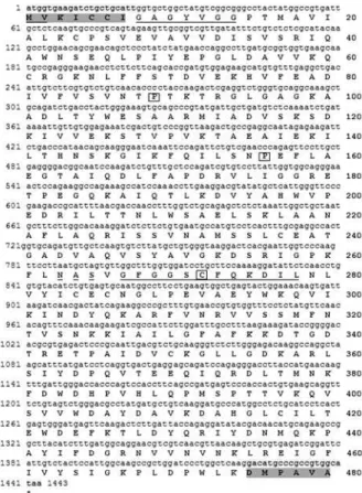
open reading frame. The alignment of the 21 nucleotides from the 5’and 3’ ends of the nine sequences showed that the differences among them were mainly in the third base of the codon, not changing the amino acid, with few excep-tions (Figure 1). These minor differences were overcome by the use of degenerate primers. Primer sequences are rep-resented in standard IUB/IUPAC amino acid and nucleic acid codes.
Cloning and sequence analysis of theUGDHcDNA
Total RNA was isolated from the cambial region of
6-month-old saplings of E. grandisusing the method of
Salzmanet al. (1999), and poly(A) mRNA was purified
from 75mg of total RNA, using the Dynabeads mRNA
Pu-rification kit (Dynal) as specified by the manufacturer, and
eluted in 20mL Tris-HCl 10 mM.
A UGDH encoding cDNA was obtained by RT-PCR, using the SuperScript One-step RT-PCR with Platinum Taq (Invitrogen), and the specific flanking primers
(UGDH_ Forward:_5’CACCATGGTGAAGATHTG
YTGYATY3’;_UGDH_Reverse: 5’TTADGCVAYVGCR
GGCATGTC3’) for the corresponding open reading frame. The directional sequence CACC was added to the sense primer to facilitate the cloning orientation of the cDNA. For
cDNA synthesis, a 50 mL RT-PCR reaction containing
25mL reaction mix (0.4 mM of each dNTP, 2.4 mM of
MgSO4), 100 pg of mRNA, 0.2mM of each primer, and
1mL of RT/PlatinumTaqmix was prepared. The cDNA
was synthesized at 50 °C for 30 min and denatured at 94 °C for 2 min, and amplification was performed using 38 cycles of 15 s at 94 °C, 30 s at 60 °C, 1 min and 30 s at 72 °C, fol-lowed by 7 min at 72 °C. The 1443-bp blunt-end amplifica-tion product was cloned into the pENTR-Direcamplifica-tional-
pENTR-Directional-TOPO®Cloning Vector, for entry into the Gateway System
(Invitrogen), according to the manufacturer’s instructions.
(50mg mL-1), was purified and screened by PCR for the
presence of full-length UGDH cDNA, using degenerate
primers and also by sequencing in both directions with the universal M13 primers and theBigDye Terminator Cycle sequencing kit (Perkin Elmer), to ensure the proper reading frame. The samples were loaded onto an ABI 3100 se-quencer (Perkin Elmer). The complete nt sequence of theE. grandis UGDHwas deposited in the NCBI GenBank under accession nº EF179384.
Expression of UGDH protein inE. coli
The RT-PCR product corresponding to theE. grandis
UGDHcDNA was sub-cloned into the Gateway®pDEST17
vector (Invitrogen) in frame with a N-terminal 6x His tag, for expression of the recombinant protein, using theE. coli
Expression System with Gateway®technology.
For the protein expression, theUGDHrecombinant
pDEST17 vector was introduced into chemically compe-tentE. coliBL21-AI (Invitrogen) by heat shock. A recom-binant clone, previously screened by PCR for the presence
of theUGDHinsert, was grown in LB medium containing
ampicillin (100mg mL-1) at 37 °C and 200 rpm. This starter culture (1 mL, OD600nm= 0.7) was used to inoculate 50 mL
of LB medium supplemented with ampicillin (100mg mL-1) and left to grow at 37 °C (200 rpm) until an OD600nmof 0.4
was reached. After 3 h of induction with 0.2% (w/v) L-ara-binose, the bacterial cells were harvested by centrifugation at 13,000gfor 1 min. The pellet was resuspended in 5 mL
of lysis buffer (100 mM NaH2PO4, pH 8.0; 10 mM Tris,
containing 0.1% (w/v) lysozyme, 1 mM PMSF, 10 mM
ß-mercaptoethanol, 10 mg RNAseA mL-1, 5 mg DNAse
mL-1) and sonicated (three times of 10-second bursts at me-dium intensity). The resulting lysate was separated into sol-uble and insolsol-uble fractions by centrifugation (3,000g) for 15 min.
Purification of the recombinant protein
The histidine fusion protein was purified from the soluble fraction using a purification column prepared with ProBondNickel-Chelating Resin (Invitrogen), according to the manufacturer’s instructions. The purity andMrof the
proteins in the eluted fractions collected were analyzed by 12.5% (w/v) SDS-PAGE, using Coomassie brilliant blue G-250 staining (Candianoet al., 2004).
Protein concentration was determined using the Brad-ford assay (BradBrad-ford, 1976) and bovine serum albumin as a standard with protein dye reagent (Bio-Rad), following the supplier’s instructions.
Enzyme assay
The activity of the purified Poly-His-Tagged euca-lyptus UDP-GDH was measured at 30 °C (Stewart and Copeland, 1999) in a continuous assay, by monitoring, with a Hitachi U3300 spectrophotometer, the increase in absor-bance at 340 nm due to the UDP-Glc-dependent formation
of NADH. Reaction mixtures for the standard assay con-tained, in a final volume of 1 mL, 20 mM Tris-HCL, pH = 8.0, and 2 mM MgCl2. The kinetic data for NAD+or
UDP-Glc as substrates were determined in triplicate assays, keeping one of the substrates at saturating concentrations. The assay was initiated by the addition of UDP-Glc, and ac-tivity was calculated from linear initial reaction rates, based on the assumption that 2 mol of NADH were formed per mole of oxidized UDP-Glc.
Protein extraction for the two-dimensional gel electrophoresis
Total proteins from the cambial region of 3- and 22-year-oldE. grandistrees were extracted as described by Hurkman and Tanaka (1986), with few modifications intro-duced by Celedonet al.(2007).
Two-dimensional gel electrophoresis
Protein samples (containing 2 mg of protein mL-1) were applied onto a 4-7 linear immobilized pH gradient strip (18 cm, GE HealthCare) and focalized using the IPGphor apparatus (GE HealthCare). Strips were rehy-drated for 12 h at 20 °C and 50 V. The proteins were prefocused at 100 V for 1 h and then at 200 V for 1 h, 400 V for 1 h , 700 V for 1 h, 1000 V for 1 h, and finally focused for a total of 70 KVh. After isoeletric focusing (IEF), the strips were kept at -80 °C until needed.
For the second-dimension analysis, the strips were kept at room temperature for 15 min in equilibration buffer [6 M urea, 2% (w/v) SDS, 50 mM Tris-HCl pH 6.8, 30% (v/v) glycerol] containing 1% (w/v) DTT, followed by in-cubation in the same buffer added with 2.5% (w/v) iodo-acetamide and 0.001% bromophenol blue. The second-dimension electrophoresis was performed in 12% (w/v) polyacrylamide gels at 30 mA, until the dye reached the bottom of the gel. Three replicates were performed for each sample. Proteins were detected using Coomassie Brilliant
Blue G-250 (Candianoet al., 2004), with modifications.
Gels were incubated twice for 1 h each in a solution con-taining 3% (v/v) phosphoric acid and 50% (v/v) ethanol, and once for 1 h in 2% (v/v) phosphoric acid. Protein detec-tion was done after leaving the gels overnight in staining solution [17% (v/v) methanol, 15% (w/v) ammonium sul-fate, 2% (v/v) phosphoric acid plus 0.1% (w/v) dye], fol-lowed by 3 washes in water (10 min each). The gels were then stored in 15% (w/v) ammonium sulfate for image anal-ysis and spot selection.
In-gel protein digestion
com-pletely dehydrated with 100% (v/v) ACN for 10 min, rehy-drated with 50 mM ammonium bicarbonate plus 20 mM dithiothreitol (DTT), and maintained for 40 min at 60 °C. This solution was discarded and replaced by 50 mM ammo-nium bicarbonate with 55 mM iodoacetamide, before keep-ing the tubes in darkness for 30 min. Then the gel pieces were dehydrated again with 100% (v/v) ACN and let to air-dry for complete removal of the solvent. Protein diges-tion was carried out with a soludiges-tion of 10 ngmL-1 trypsin (Promega) in 25 mM ammonium bicarbonate, for 12 h at 37 °C. Gel pieces were extracted twice with 50mL of 60% (v/v) ACN containing 1% (v/v) formic acid (FA), and once
with 50mL of 100% ACN. All supernatants were combined
and vacuum-dried. The peptides were then resuspended in
12mL 1% (v/v) FA for MS analysis.
Protein identification by LC-MS/MS
The peptides were separated and identified by on-line chromatography, using a Cap-LC coupled to a Q-TOF Ul-tima API mass spectrometer (Waters, UK). 5 mL of sample were loaded onto a 0.18 mm x 23.5 mm NanoEase Trap-ping Column (Waters, UK) for pre-concentration and desa-lination, followed by peptide separation on a C18, 3.5mm,
75mm x 100 mm NanoEase Symmetry 300 LC column
(Waters, UK). Peptides were eluted in a 60 min linear gra-dient of solvent B [95% (v/v) ACN containing 0.1% (v/v) formic acid in water] and solvent A [5% (v/v) ACN con-taining 0.1% (v/v) formic acid in water], at a flow rate of 250 nL min-1.
MS spectra analysis
All analyses were performed using a positive ion mode at a 3 kV needle voltage. The mass range was set from 300 to 2000 m/z, and the MS/MS spectra acquired for the most intense peaks (³15 counts). Multiple charged pre-cursor ions were selected for fragmentation and peptide se-quencing, using automated data-dependent-acquisition (DDA) MassLynx software (Waters), switching from the MS to the MS/MS mode and then returning to MS. The re-sulting fragmented spectra were processed using the Pro-teinLynx v. 4.0 software (Waters), and the MASCOT MS/MS Ion Search was used to blast the sequences against the SwissProt and NCBI databank. Combined MS-MS/MS searches were conducted with parent ion mass tolerance at 50 ppm, MS/MS mass tolerance of 0.1 Da, carbamidome-thylation of cysteine (fixed modification) and methionine oxidation (variable modification). According to MASCOT probability analysis, only significant hits (p < 0.05) were accepted.
Southern blot analysis
Genomic DNA (15mg) was isolated fromEucalyptus
grandisleaves, as described by Doyle and Doyle (1987), and digested withHindIII,EcoRI, BamHI,EcoRV,NcoI
andSacI restriction enzymes at 37 °C for 16 h. The DNA
fragments were separated on 1% (w/v) agarose gel in 0.5 x TBE buffer (45 mM Tris-borate, 1 mM EDTA, pH 8.0), stained with ethidium bromide, visualized under ultraviolet light, and denaturated before being transferred onto a
Hy-bond N+ nylon membrane (GE HealthCare) by capillary
transfer (Southern, 1975; Sambrook et al., 1989). The
membrane was probed using the AlkPhos Direct Labeling Module (GE HealthCare). The alkaline-phosphatase-labeled-PCR product (698-bp) resulting from the amplifi-cation of a conserved internal region (nt# 188-886, Figure 1) of the eucalyptus cDNA (1443-bp) cloned with the
fol-lowing primers (ugdhDForward: 5’-GAAGAACCTCTT
CTTCAGCA-3’ugdhDReverse:5’-AGTACTCAGCCAC
TTCAGGA-3’) synthesized for the UGDH domain (pfam03721, pfam00984, pfam03720), used as probe, was
detected with the Gene Images CDP-Star Detection Mod-ule (GE HealthCare). Hybridization was carried out at 60 °C for 16 h, followed by stringent washes: twice in fresh primary wash buffer [2 M urea, 50 mM NaH2PO4, pH 7.0,
150 mM NaCl, 1 mM MgCl2containing 0.1% (w/v) SDS
and 0.2% (w/v) blocking reagent] at 55 °C for 10 min, and once in secondary wash buffer [50 mM Tris, 100 mM NaCl
containing 2 mM MgCl2] at room temperature for 5 min.
The hybridization signal was recorded on X-ray film (MXG/PLUS, Kodak) after 1 h exposure between intensi-fying screens (GE HealthCare), at room temperature.
The resulting exposed X-ray film was used as refer-ence for localization of the bands.
Semi-quantitative real-time PCR (RT-PCR)
Tissue samples from 6-month-old saplings and from 3-and 22-year-old plants were used for total RNA isolation, as
described by Salzmanet al.(1999). Poly(A) mRNAs were
purified from 75 mg of total RNA, using the Dynabeads
mRNA Purification kit (Dynal) as specified by the
manufac-turer, and eluted in 20 mL Tris-HCl 10 mM. First-strand
cDNAs were generated using the SuperScript III First-Strand Synthesis SuperMix in a 20mL reaction mixture, con-taining 3/10 of the eluted mRNA, 50 ng random hexamers,
and 1 mL annealing buffer. After 5 min of incubation at
65 °C, 10mL of 2x First-Strand Reaction Mix plus 2mL Su-perScript III/RNase OUT Enzyme Mix, which includes the SuperScript III Reverse Transcriptase, were added, and the final reaction mixture was incubated for 10 min at 25 °C, fol-lowed by 50 min at 50 °C, and 5 min at 85 °C, following the manufacturer’s instructions (Invitrogen). Semi-quantitative RT-PCR assays were performed, using 1/10 of the cDNA preparation per PCR, based on a preliminary semi-quan-titative RT-PCR (data not shown). Primer sequences and
amplicon sizes were the following:UGDH(247-bp; primers
designed specifically for the cloned eucalyptus cDNA for-ward GCCCGTATGATTGCTGATGTC -3’, reverse
5’-TCAAGGTTTGGATGGCTTTC-3’); ubiquitin (223-bp,
primers to theE. grandis constitutive ubiquitin gene, for-ward CGATTGATTCTCAGCAAGC-3’, reverse
5’-GGATGTTGTAGTCAGCCAAGG-3’). Amplification
specificity was checked by melting-curve analysis, and PCR efficiency determined using standard curves for each primer pair constructed with serial dilutions (1:10, 1:100 and 1:1000) of the cDNA preparation.
Results and Discussion
PCR amplification of the full-length cDNA encoding
E. grandisUGDH
In this study, the degenerate primers designed to an-neal highly conserved 5’ and 3’ regions of already known genes, in combination with the RT-PCR technique, proved to be a potential strategy to overcome the difficulties in
cloning a particular novel gene, when cDNA or a genomic library are not available. The RT-PCR reaction produced a 1443-nt-long cDNA sequence, with an open reading frame of 480 amino acids encoding UGDH (Figure 1).
Nucleotide and deduced amino acid sequence analysis
The deduced amino acid sequence for eucalyptus
shows conserved motifs (pfam03721, pfam00984,
pfam03720). Figure 1 (underlined) presents the translation initiation nt motif (aa# 1 and 2), the NAD cofactor binding site (aa# 8-14), the catalytic site (aa# 267-278, with a Cys residue, boxed). A putative glycosylation site (aa# 263-265), also found in poplar (Johanssonet al., 2002), is indicated with a broken line, and Pro residues at aa# 89 and 156 are also boxed, representing the main chain bends in the protein structure (Hempelet al., 1994). The derived amino acid se-quence from the database search BLASTX analysis (Altschulet al., 1997) showed a high degree of identity to the
UGDH of soybean (92%), cinnamon (90%), Arabidopsis
(90%), taro (89%), poplar (89%), and rice (85%), among several plant species, including some putatives UDP-glucose dehydrogenases, such as that of tobacco (91%). The high identity among UGDHs of higher plants suggests strict structural requirements for proper functioning of the protein. It is interesting to observe the high degree of amino acid se-quence identity of the eucalyptus UGDH, even along the N and C terminal regions (Figure 1, highlighted in dark grey), where the differences in the first 25 aa, were less than 4% and 16%, respectively, compared to the leguminosa soybean and gramineae such as rice, maize and, particularly, wheat. The analysis of the phylogenetic tree, generated using the CLC Main Work Bench 5.5 (UPGMA algorithm with Boot-strap analysis, 100 replicates) for the eucalyptus UGDH pro-tein sequence and eight other plant species, shows a high similarity of theE. grandisprotein sequence to various plant sequences, particularly soybean andArabidopsis(Figure S1, supplementary data).
Expression of recombinant eucalyptus UGDH
The recombinant UGDH has been found to be often present in protein inclusion bodies (Tenhaken and Thulke, 1996; Hinterberget al., 2002). It is known that eukaryotic proteins expressed inE. colioften form protein inclusion bodies, due to differences in the protein-folding systems between prokaryotes and eukaryotes (Oka and Jigami, 2006). Therefore, polyhistidine recombinant eucalyptus UGDH protein was produced in BL21-AI cells, and puri-fied under a narrow range of optimized conditions, which included a low centrifugation of the lysate to recover, under native conditions, enough enzymatically active protein in the soluble fraction. The protein was purified at 4 °C, and immediately used for the enzyme assay.
E. coli(Figure 2). The identification of the protein was con-firmed by LC-MS/MS, and the peptides sequenced covered 23.33% of the total protein, with an estimated molecular weight of 52.91 kDa, and a 100% probability that the pep-tides are related to the expected protein (Table S1, supple-mentary data).
The activity of the purified Poly-His-Tagged-UGDH measured at 30 °C (Stewart and Copeland, 1999), in a con-tinuous assay, by spectrophotometrically monitoring the absorbance increase at 340 nm due to the UDP-Glc-dependent formation of NADH, showed that the recombi-nant protein expressed inE. colicatalyzed the conversion of UDP-Glc to UDP-GlcA (Figure 3), thus confirming that the cloned cDNA encodes the UGDH enzyme.
Enzyme kinetics
The recombinant protein expressed inE. coli
cata-lyzed the conversion of UDP-Glc to UDP-GlcA, confirm-ing that the cloned cDNA encodes the UGDH enzyme (Figure 3). The kinetic data were then fitted to the
Michae-lis-Menten equation to obtain the Km and Vmax values
(Segel, 1976). For both substrates, UDP-Glc and NAD+,
hyperbolic saturation curves were observed and theKm
val-ues calculated, using the Origin-software®, 60.7±8.5mM for UDP-Glc (Vmax= 67.9±9.2mmol min-1mg protein-1)
and 67.3±17.9mM for NAD+(Vmax= 171.8±34.8mmol
min-1mg protein-1) (Figures 3A and 3B, respectively). The estimatedKmof the eucalyptus enzyme for NAD+was
simi-lar to the value described for the recombinant protein from
soybean (70± 5 mM), whereas the Kmfor UDP-Glc was
around 3 times higher (22±2mM) (Hinterberget al., 2002). A characterization of three isoforms of UGDH (UGDH2,
UGDH3 and UGDH4) fromArabidopsisevidenced a
simi-lar affinity value for the cofactor NAD+as that observed for
eucalyptus (40-45mM), while major differences were
ob-served in theKmfor UDP-Glc: 123±9mM, 335±16mM
and 171±9 mM, respectively (Klinghammer and
Tenha-ken, 2007). The high affinity of the enzyme for NAD+was also observed in eucalyptus, confirming previous
observa-tions that the UGDHs are not limited by NAD+ levelsin
vivo, while UDP-Glc affinity might be isoform-dependent
(Hinterberg et al., 2002; Klinghammer and Tenhaken,
2007).
Genomic copies ofUGDH
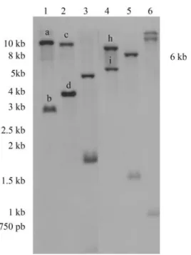
Southern blot analysis indicated the existence of a
least two copies of theUGDHgene within the eucalyptus
genome (Figure 4). The existence of a single-copyUGDH
gene has been described for soybean (Tenhaken and
Thul-ke, 1996) andArabidopsis(Tenhaken and Thulke, 1996;
Seitzet al., 2000). In poplar (Johanssonet al., 2002), two highly homologous genes were found, as also observed in maize (Kärkönenet al.2005). However, a later evaluation
of theArabidopsisand poplar genome projects indicated
multiple copies. The presence of four UGDH isoforms in
Arabidopsis, highly similar to each other, reported by Rei-ter and Vanzin (2001), was confirmed by Klinghammer and
Tenhaken (2007), who found a fifthUGDH pseudogene,
located on chromosome III, with a weaker similarity to the
others. In thePopulus trichocarpa genome (Populus
ge-nome project), fourUGDHgenes were identified, one in
Figure 2- Coomassie-stained SDS-PAGE of recombinant UGDH protein. The eucalyptusUGDHcDNA cloned into a His-tagged pDEST expres-sion vector was used to transform the BL21E. colicells. (1) Extracts from uninduced cells; (2) L-arabinose-induced cells; and (3) the purified re-combinant eucalyptus UGDH from the L-arabinose-inducedE. coli cul-ture. MW: Molecular weight marker (Sigma).
linkage group IV (224080232), one in linkage group VIII (224098951), one in linkage group X (224112137), and one in linkage group XVII (224141486).
We further analyzed the possible number of copies
for UGDH in the Eucalyptus genome by performing a
tblastx analysis of the eucalyptus ESTs sequences available in the GenBank database. The results showed that the
clonedUGDHsequence fromE. grandisis highly similar
(~99%) to the amino acid sequences of seven ESTs pro-duced from the differentiating xylem ofEucalyptus gunnii
(103476385, 103475842, 103479156, 103478292,
103476672, 103476952, 103480206) and fiveEucalyptus
globulusEST sequences, using mRNA isolated from leaf tissue under low temperature conditions (162324959, 162326130, 162329222, 162329166, 162326954). The phylogenetic tree generated using CLC Main Work Bench 5.5 (UPGMA algorithm with Bootstrap analysis, 100 repli-cates) for the complete nucleotide sequence of the clonedE. grandis UGDH mRNA (144926038) with these seven
cambial E. gunnii EST and the five leaf E. globulus
se-quences (Figures 5A and 5B, respectively), suggests that there are at least two groups of nucleotide sequences, most probably indicating two isoforms of this gene in the RNA extracted from the cambial and leaf tissues.
The amplification product obtained from the
eucalyp-tus genomic DNA against the primers flanking theUGDH
open reading frame, corresponds exactly to the same size
(1443-bp) of the eucalyptus UGDH cDNA (Figure 6),
showing the lack of introns along the clonedUGDHgene
sequence. The restriction map of the UGDH ORF
(Figure S2, supplementary data) shows that the restriction enzymesEcoRI,HindIII andEcoRV do not cut the cloned fragment internally. This indicates that the bands observed in the Southern blot assay (Figure 4, lanes 1, 2 and 4), which are all larger than 1.6 kb, most probably contain the
complete clonedUGDHORF.
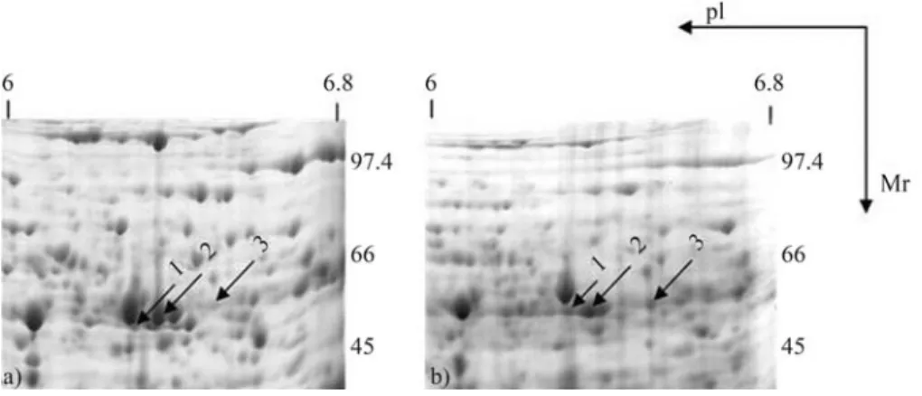
Moreover, in order to investigate the existence of UGDH protein isoforms, we produced 2DE-PAGE gels with proteins isolated from the cambial region of 3- and 22-year-oldE. grandistrees. Figure 7 shows the identifica-tion of three distinct spots (1, 2 and 3) with the peptide data used to identify the proteins shown in Table S2 (supple-mentary data). Spot 3 showed aMrof ~53.0 kDa and a pI of
approximately 6.5, which is in accordance with the
calcu-lated values for the Mr and pI of the cloned sequence
(53.09 kDa and 6.52, respectively). These values are simi-lar to those obtained for soybean (52.94 kDa and 5.81) and poplar (52.99 kDa and 6.20). Thus, spots 1 and 2 represent proteins with different characteristics (lower pI and lower
Mr), indicating the existence of at least one more copy of
UGDHin theE. grandisgenome. The differences between
spots 1 and 2 might be explained by posttranslational modi-fication, such as phosphorylation, although further analysis is needed to confirm this hypothesis.
UGDHexpression in different tissues of 6-month-old saplings and in the cambial region of juvenile and matureE. grandiswood
The expression of UGDH in different tissues of
6-month-old saplings and in the cambial region of 3- and
22-Figure 4- Genomic Southern blot ofE. grandisDNA, restricted with
HindIII (1),EcoRI (2),BamHI (3),EcoRV (4),NcoI (5), andSacI (6). The sizes of the fragments are indicated in kilobase pairs (kb). The alka-line-phosphatase-labeled PCR product (698-bp) resulting from the ampli-fication of a conserved internal region (nt# 188-886, Figure 1) of the clonedE. grandiscDNA (1443-bp) was used as probe.
Figure 5- Phylogenetic tree generated using CLC Main Work Bench 5.5 (UPGMA algorithm with Bootstrap analysis, 100 replicates) for the com-plete nucleotide sequence of the cloned E. grandis UGDH mRNA (144926038): A) using seven E. gunnii EST sequences (103476385, 103475842, 103479156, 103478292, 103476672, 103476952, 103480206) of mRNA isolated from differentiating xylem; B) using five
E. globulus EST sequences (162324959, 162326130, 162329222,
year-old trees was analyzed by semi-quantitative RT-PCR. In the 6-month-old saplings the maximum expression was observed in roots, whereas in stem and bark it was interme-diate, and the leaves displayed the lowest expression (Fig-ure 8). Similar results were observed in tobacco plants by Bindschedleret al.(2005), who compared the expression of
UGDHandNtADH2(the dual specific UGDHsimilar to
ADH), suggesting that the isoforms of UGDH might be
preferentially involved in primary rather than in secondary growth, since they were strongly expressed in roots and in the youngest internodes of the stem, with the lowest level of expression observed in buds and in tobacco leaves, similar to the expression pattern observed by Seitzet al.(2000) in
Arabidopsis.
We then checked the role ofUGDH in the
wood-forming tissues of juvenile and mature trees, by comparing the level of expression relative to the stem of 6-month-old
saplings. Figure 9 shows that the expression ofUGDHin
the cambial region of 3- and 22-year-old trees was signifi-cantly higher than in the stem of 6-month-old saplings, in-dicating that the enzyme may have an important role in controlling hemicellulose biosynthesis during wood forma-tion.
In this study, the strategy of using degenerate primers in combination with the RT-PCR technique, proved to be
efficient to overcome the difficulties in cloning a particular novel gene, when cDNA or a genomic library are not avail-able. We were able to clone and express the recombinant UGDH protein ofE. grandisand to confirm its activity and sequence. The role of UGDH in wood-forming tissues is
still poorly understood, and the isolation of E. grandis
cDNA offers an opportunity to further understand the im-portance of this enzyme in trees.
Figure 6- Amplification product (1443-bp) of the eucalyptus genomic DNA (1), and the corresponding cDNA (2) for eucalyptusUGDH, against the degenerate primers flanking theUGDHopen reading frame.l: 1 kb DNA ladder (Invitrogen), kb: kilobase pair.
Figure 7- 2-DE gels from the cambial region of 3-year-old (A) and 22-year-old (B)E. grandistrees. Arrows indicate the spot numbers of UGDH isoforms. Ip: Isoeletric-point,Mr:Molecular weight in kiloDaltons (kDa).
Figure 8- Semi-quantitative RT-PCR ofUGDHtranscript accumulation in roots, stem, bark and leaves of 6-month-oldE. grandissaplings. Tran-script levels were normalized relative to the ubiquitin expression level as internal standard. Results are expressed as mean of three replicates and standard deviation relative to the leaf expression level, to which the value 1 on the linear scale was assigned.
Acknowledgments
This work was financially supported by the Innova-tion Technology Program funded by FAPESP (01/11080-8) and Suzano Papel e Celulose S.A. Further support was provided by MEC-CAPES with fellowships to Daniela
Defávari do Nascimento and Gunta Gutmanis,CNPq with
fellowship to Alexander de Andrade, and by FAPESP with fellowships to Ana Letícia Ferreira Bertolo, Luis Felipe Boaretto and Eduardo Leal de Oliveira Camargo. We would like to thank Dr. Siu Mui Tsai and Fabiana Can-navan from CENA/ESALQ/USP and Dr Maria Helena de Souza Goldman and Andrea Carla Quiapim from FFCLRP/USP, for their help with DNA sequencing. We would also like to thank Livia M. Franceschini for helping with the configuration of all figures.
References
Altschul SF, Madden TL, Schaffer AA, Zhang J, Zhang Z, Miller WE and Lipman DJ (1997) Gapped BLAST and PSI-BLAST: A new generation of protein database search pro-grams. Nucleic Acids Res 25:3389-3402.
Amino S, Takeuchi Y and Komamine A (1985) Changes in en-zyme activities involved in formation and interconversion of UDP-sugars during the cell cycle in a synchronous culture of Catharanthus roseus. Physiol Plant 64:111-117.
Bindschedler LV, Wheatley E, Gay E, Cole J, Cottage A and Bolwell GP (2005) Characterisation and expression of the pathway from UDP-glucose to UDP-xylose in differentiat-ing tobacco tissue. Plant Mol Biol 57:285-301.
Biswas BB, Ghosh B and Majumder AL (1984) myo-Inositol polyphosphates and their role in cellular metabolism: A pro-posed cycle involving glucose-6-phosphate and myo-ino-sitol phosphates. In: Roodyn DB (ed) Subcellular Biochem-istry. Plenum, London, pp 237-280.
Bradford MM (1976) A rapid and sensitive method for the quan-titation of microgram quantities of protein utilizing the prin-ciple of protein-dye binding. Anal Biochem 72:248-254. Candiano G, Bruschi M, Musante L, Santucci L, Ghiggeri GM,
Carnemolla B, Orecchia P, Zardi L and Righetti PG (2004) Blue silver: A very sensitive colloidal Coomassie G-250 staining for proteome analysis. Electrophoresis 25:1327-1333.
Carvalho MCCG, Caldas DGG, Carneiro RT, Moon DH, Salva-tierra GR, Franeschini LM, Andrade A, Celedon PAF, Oda S and Labate CA (2008) SAGE transcript profiling of the ju-venile cambial region ofEucalyptus grandis. Tree Physiol 28:905-919.
Celedon PAF, Andrade A, Meireles KGX, Carvalho MCCG, Caldas DGG, Moon DH, Carneiro RT, Franceschini LM, Oda S and Labate CA (2007) Proteomic analysis of the cambial region in juvenileEucalyptus grandisat three ages. Proteomics 7:2258-2274.
Dalessandro G and Northcote DH (1977a) Possible control sites of polysaccharide synthesis during cell growth and wall ex-pansion of pea seedlings (Pisum sativum L.). Planta 134:39-44.
Dalessandro G and Northcote DH (1977b) Changes in enzymatic activities of nucleoside diphosphate sugar interconversions
during differentiation of cambium to xylem in sycamore and poplar. Biochem J 162:267-279.
Doyle JJT and Doyle JL (1987) Isolation of plant DNA from fresh tissue. Focus 12:13-15.
Gibeaut DM and Carpita NC (1994) Biosynthesis of plant cell wall polysacharides. FASEB J 8:904-915.
Gibeaut DM (2000) Nucleotide sugars and glycosyltransferases for synthesis of cell wall matrix polysaccharides. Plant Phy-siol Biochem 38:69-80.
Hempel J, Perozich J, Romovacek H, Hinich A, Kuo I and Fein-gold DS (1994) UDP-glucose dehydrogenase from bovine liver primary structure and relationship to other dehydro-genases. Protein Sci 3:1074-1080.
Hinterberg B, Klos C and Tenhaken R (2002) Recombinant UDP-glucose dehydrogenase from soybean. Plant Physiol Biochem 40:1011-1017.
Hurkman WJ and Tanaka CK (1986) Solubilization of plant mem-brane proteins for analysis by two-dimensional gel electro-phoresis. Plant Physiol 81:802-806.
Johansson H, Sterky F, Amini B, Lundeberg J and Kleczkowski LA (2002) Molecular cloning and characterization of a cDNA encoding poplar UDP-glucose dehydrogenase, a key gene of hemicellulose/pectin formation. Biochem Biophys Acta 1576:53-58.
Kärkönen A, Murigneux A, Martinant JP, Pepey E, Tatout C, Dudley BJ and Fry SC (2005) UDP-glucose dehydrogenases of maize: A role in cell wall pentose biosynthesis. Biochem J 391:409-415.
Klinghammer M and Tenhaken R (2007) Genome-wide analysis of the UDP-glucose dehydrogenase gene family in Arabi-dopsis, a key enzyme for matrix polysaccharides in cell walls. J Exp Bot 58:3609-3621.
Kunihiro S, Kawanishi Y, Sano M, Naito K, Matsuura Y, Tateno Y, Gojobori T, Yamagata Y, Abe K and Machida M (2002) A polymerase chain reaction-based method for cloning novel members of a gene family using a combination of de-generate and inhibitory primers. Gene 289:177-184. Loewus F, Chen MS and Loewus MW (1973) Themyo-inositol
oxidation pathway to cell wall polysaccharides. In: Lowes F (ed) Biogenesis of Plant Cell Wall Polysaccharides. Aca-demic Press, New York, pp 1-27.
Oka T and Jigami Y (2006) Reconstruction ofde novopathway for synthesis of UDP-glucuronic acid and UDP-xylose from intrinsic UDP-glucose inSaccharomyces cerevisiae. FEBS J 273:2645-2657.
Reiter WD and Vanzin GF (2001) Molecular genetics of nucleo-tide sugar interconversion pathways in plants. Plant Mol Biol 47:95-113.
Roberts RM and Loewus F (1973) The conversion of D-glucose-6-14C to cell wall polysaccharide material inZea maysin presence of high endogenous levels of myoinositol. Plant Physiol 52:646-650.
Robertson D, Beech I and Bolwell GP (1995) Regulation of the enzymes of UDP-sugar metabolism during differentiation of French bean. Phytochemistry 39:21-28.
Sasaki K and Taylor IEP (1984) Specific labeling of cell wall polysaccharides with myo-[2-3H] inositol during
germina-tion and growth ofPhaseolus vulgarisL. Plant Cell Physiol 25:989-997.
tissues containing high levels of phenolic compounds or car-bohydrates. Plant Mol Biol Rep 17:11-17.
Sambrook J, Fritsch EF and Maniatis T (1989) Molecular Clon-ing: A Laboratory Manual. 2nd edition. Cold Spring Harbor Laboratory Press, Cold Spring Harbor.
Segel IH (1976) Biochemical Calculations. 2nd edition. John Wiley & Sons, New York, 441 pp.
Seifert GJ (2004) Nucleotide sugar interconvertions and cell wall biosynthesis: How to bring the inside to the outside. Curr Opin Plant Biol 7:277-284.
Seitz B, Klos C, Wurm M and Tenhaken R (2000) Matrix poly-saccharide precursors inArabidopsiscell walls are synthe-sized by alternative pathways with organ-specific expres-sion patterns. Plant J 21:537-546.
Stewart DC and Copeland L (1998) Uridine-5’-diphosphate dehy-drogenase from soybean nodules. Plant Physiol 116:349-355.
Stewart DC and Copeland L (1999) Kinetic properties of UDP-glucose dehydrogenase from soybean nodules. Plant Sci 147:119-125.
Southern EM (1975) Detection of specific sequences among DNA fragments separated by gel eletrophoresis. J Mol Biol 98:503-517.
Tenhaken R and Thulke O (1996) Cloning of an enzyme that syn-thesizes a key nucleotide-sugar precursor of hemicellulose biosynthesis from soybean: UDP-glucose dehydrogenase. Plant Physiol 112:1127-1134.
Thompson JD, Higgins DG and Gibson TJ (1994) Clustal W: Im-proving the sensitivity of progressive multiple sequence alignment through sequence weighting, position-specific gap penalities and weigh matrix choice. Nucleic Acids Res 22:4673-4680.
Zablackis E, Huang J, Muller B, Darvill AG and Albersheim P (1995) Characterization of the cell-wall polysaccharides of Arabidopsis thalianaleaves. Plant Physiol 107:1129-1138.
Internet Resources
Mascot search engine for rapid protein identification using mass spectrometry data. MASCOT MS/MS Ion Search, http://www.matrixscience.com/ (November 11, 2009). Multiple sequence alignment program ClustalW2,
http://www.ebi.ac.uk/Tools/clustalw2/index.html (Novem-ber 11, 2009).
National Center for Biotechnology Information,
http://www.ncbi.nlm.nih.gov/ (November 11, 2009). Populus genome Project,
http://ge-nome.jgi-psf.org/Poptr1_1/Poptr1_1.home.html/ (Novem-ber 11, 2009).
Supplementary Material
The following online material is available for this ar-ticle:
Table S1 - Peptide sequencing data of the recombi-nant UGDH protein shown in Figure 2, obtained by LC-MS/MS.
Table S2 - Peptide sequencing data of UGDH pro-teins shown in Figure 7, identified by LC-MS/MS.
Figure S1 - Phylogenetic tree generated for the euca-lyptus UGDH protein sequence and eight other plant spe-cies.
Figure S2 - Restriction map of the clonedE. grandis
UGDHcDNA.
This material is available as part of the online article from http://www.scielo.br/gmb.
Associate Editor: Carlos F.M. Menck
and gi numbers are: Eucalyptus grandis, 144926039; Cinnamomum
osmophloeum, 40317278;Populus tomentosa, 39939262;Glycine Max,
1518540;Arabidopsis thaliana, 11994517;Sorghum bicolor, 242047160;
Nicotiana tabacum, 48093457; Colocasia esculenta, 29028306;Oryza
sativaJaponica, Group 215737390.
Name EC 1.1.1.22
% Probability Peptide matches Coverage (%) Mr1) pI2)
AAB58398 100 12 23.33 52.90816 5.93
Submitted mass Experimental mass Peptide sequences
600.304 1198.592 (K)RAFFSTDVEK(H)
951.99 1901.964 (K)HVFEADIVFVSVSQPTK(T)
677.302 1352.588 (K)AADLTYWESAAR(M)
693.825 1385.634 (K)DVYAHWVPEDR(I)
738.401 1474.786 (R)ILTTNLWSAELSK(L)
752.393 1502.77 (R)ILTTNLWSAELSR(L)
537.79 1073.564 (K)LAANAFLAQR(I)
809.881 1617.746 (K)FLNASVGFGGSCFQK(D)
816.898 1631.78 (K)FLNASVGFGGSCFQK(D)
554.344 1106.672 (K)KIAILGFAFK(K)
731.863 1461.71 (K)KVSVVWDAFEATK(D)
740.368 1478.72 (K)KVSVVWDAYDAVK(D)
1)
Molecular weight in kDa.
Name EC 1.1.1.22
Probability (%) Peptide matches Coverage (%) Theoretical1)Mr/pI2) Experimental1)Mr/pI2)
AAR32717 100 4 9 52.963/5.92 57.907/6.39
Submitted mass Charge Experimental mass Peptide sequences
490.3026 2 490.3026 (K)IAILGFAFK(K)
537.7942 2 1073.5738 (K)LAANAFLAQR(I)
677.3171 2 1352.6196 (K)AADLTYWESAAR(T)
738.3889 2 1474.7632 (R)ILTTNLWSAELSK(L)
Spot 2
Name EC 1.1.1.22
Probability (%) Peptide matches Coverage (%) Theoretical1)Mr/pI2) Experimental1)Mr/pI2)
AAO62313 99.99 6 12 52.947/6.06 57.625/6.44
Submitted mass Charge Experimental mass Peptide sequences
382.2243 2 762.4340 (R)MIADVSK(S)
398.2232 2 794.4318 (K)TLDYQR(I)
537.8185 2 1073.6224 (K)LAANAFLAQR(I)
677.3301 2 1352.6456 (K)AADLTYWESAAR(M)
462.9116 3 1385.7130 (K)DVYAHWVPEDR(I)
738.4020 2 1474.7894 (R)IITTNLWSAELSK(L)
Spot 3
Name EC 1.1.1.22
Probability (%) Peptide matches Coverage (%) Theoretical1)Mr/pI2 Experimental1)Mr/pI2)
AAR32717 99.99 3 7 53.543/5.92 59.101/6.61
Submitted mass Charge Experimental mass Peptide sequences
490.3130 2 978.6114 (K)IAILGFAFK(K)
677.3294 2 1352.6442 (K)AADLTYWESAAR(T)
738.4080 2 1474.8014 (R)ILTTNLWSAELSK(L)
1)
Molecular weight in kDa.
2)