387 387387 387387 Mem Inst Oswaldo Cruz, Rio de Janeiro, Vol. 94(3): 387-394, May/Jun. 1999
Increased Pro-inflammatory Cytokines (TNF-
a
and IL-6) and
Anti-inflammatory Compounds (sTNFRp55 and sTNFRp75)
in Brazilian Patients during Exanthematic Dengue Fever
Luzia MO Pinto, Solange A Oliveira*, Elzinandes LA Braga, Rita MR Nogueira,
Claire F Kubelka/
+Departamento de Virologia, Instituto Oswaldo Cruz, Av. Brasil 4365, 21045-900 Rio de Janeiro, RJ, Brasil *Disciplina de Doenças Infecto-Parasitárias, HUAP, UFF, Niterói, RJ, Brasil
Pro-inflammatory cytokines, tumor necrosis factor (TNF-a), interleukin-6 (IL-6) and interleukin-1b
(IL-1b) as well as anti-inflammatory compounds, soluble TNF-Receptor p55 (sTNFRp55), sTNFRp75
and IL-1 receptor antagonist (sIL-1Ra), were investigated in 34 Brazilian cases of dengue fever (DF) originated from a study of exanthematic virosis. The presence of pro-inflammatory cytokines was de-tected in sera from these patients by ELISA. TNF-a and IL-6 levels were significantly higher than
con-trol subjects in 32% and 52% patients, respectively. To our knowledge this was the first time a receptor antagonist and soluble receptors for cytokines were detected in sera obtained during exanthematic DF without hemorrhagic manifestations. Both sTNFRp55 and sTNFRp75 were consistently elevated in 42% and 84% patients, respectively. Most patients had IL-1b levels not different from those of normal
sub-jects, except for one case. Only 16% patients had altered levels of IL-1Ra. Previous studies in dengue hemorrhagic fever patients demonstrated production of these soluble factors; here we observed that they are found in absence of hemorrhagic manifestations. The possible role of these anti-inflammatory compounds in immune cell activation and in regulating cytokine-mediated pathogenesis during dengue infection is discussed.
Key words: dengue - tumor necrotis factor a - interleukin 6 - soluble tumor necrosis - factor receptor
Dengue disease is caused by single-stranded positive sense RNA arboviruses, belonging to the
Flaviviridae family and classified as four antigeni-cally distinct dengue virus serotypes (Sabin & Schlesinger 1945, Hamon et al. 1960, Brown 1986). Infection can be asymptomatic or lead to different forms of disease. Dengue fever (DF) is characterized by mild systemic manifestations such as fever, retro-orbital headache, severe myalgias, and rash. Some patients develop a more severe and life-threatening syndrome termed dengue hemor-rhagic fever (DHF) where plasma leakage takes place into interstitial spaces, resulting in hypovolaemia, thrombocytopenia and hemorrhage; circulatory collapse leading to shock may occur and is then referred as dengue shock syndrome (DSS) (Halstead et al. 1988, Halstead 1990, PAHO 1994). The worldwide number of annual DF cases is estimated to be more than 100 million, with
Financial support: Fiocruz, CNPq and Colab, Brazil. +Corresponding author. Fax: +55-21-564 7638. E-mail: claire@gene.dbbm.fiocruz.br
Received 1 September 1998 Accepted 15 January 1999
250,000 reported cases of DHF (Monath 1994). In Brazil, about 400,000 cases of dengue were noti-fied this year during the first six months (Brazilian National Health Foundation, Heath Ministry 1998). The pathogenesis of dengue disease is not fully understood and is considered an immunopathologic process associated with prior immune sensitization by a heterotypic virus (Halstead 1980, Monath 1986, Kurane et al. 1991). Dengue infection pro-vides lifelong homotypic immunity, but only tran-sient cross-protection against other serotypes is achieved, making sequential infection possible (Pang 1987, Kliks et al. 1989). The relative risk of experiencing most severe forms of disease has been considered to be several fold higher after second-ary infection (Monath 1986).
Mononuclear phagocytes appear to be princi-pal target cells for dengue virus replication (Halstead 1980, Anderson et al. 1997). The under-lying mechanism is believed to involve activation of virus infected macrophages and producion of cytokines such as tumor necrosis factor-a (TNF-a), interleukin-6 (IL-6) and interleukin-1b (IL-1b)
388 388 388 388
388 Cytokines and Soluble Receptors in Dengue Luzia MO Pinto et al.
of endothelial cells leading to vascular permeabil-ity which may be the phenomenon involved in the pathogenesis of dengue infection (Anderson et al. 1997). The production of pro-inflammatory cytokines (TNF-a, IL-6 and IL-1b) and the extent
of the inflammatory response are partially modu-lated by anti-inflammatory compounds. Soluble extracellular domains of 55-kDa and 75-kDa, sTNFRp55 and sTNFRp75, are liberated extracel-lularly from membrane-bound TNF receptors (Kornelisse et al. 1996). IL-1 receptor antagonist (IL-1Ra) may function as an inhibitor by antago-nizing IL-1 binding to IL-1 cell surface receptor in a competitive interaction (Pereda et al. 1995).
Several cytokines were detected in sera from patients undergoing viral infections. Their exacer-bated induction is associated with pathological changes such as hepatic lesions in acute (Torre et al. 1994) and chronic (Yoshoka et al. 1989) viral hepatitis or HIV-1 replication in AIDS (Poli & Fauci 1993). They play also a role in septic shock which has some clinical features similar to DSS (Dinarello 1996a).
Pro-inflammatory cytokines were found to be increased in Asiatic and American patients with DHF/DSS (Hober et al. 1993, 1998, Kuno & Bailey 1994, Iyngkaran et al. 1995, Kubelka et al. 1995) and recently soluble TNF receptor was detected in DHF/DSS patients (Hober et al. 1996b, Bethell et al. 1998). Nevertheless, these investigations are far from elucidating the complex mechanisms of im-munopathology during dengue disease; more cases in different countries and several immunological parameters deserve to be studied. Moreover, none of these works discuss the cytokine profile for DF. During the present work we observed the pres-ence of pro-inflammatory cytokines (TNF-a, IL-6
and IL-1b) and anti-inflammatory compounds
(sTNFRp55, sTNFRp75 and sIL-1Ra) in sera of Brazilian patients with exanthematic DF without hemorrhagic manifestations and infected with Den-gue-1 or -2 viruses.
MATERIALS AND METHODS
Patients and laboratory diagnostics - Thirty four cases of DF included in this work originated from a study on exanthematic virosis with patients attended at the Antônio Pedro University Hospi-tal, Niterói in 1995-1996 (22 women, 12 men; 1-58 years old, average 29 ± 14.9). All patients were diagnosed based on clinical manifestations of DF and confirmed serologically by the presence of IgM in MAC-ELISA test (Nogueira et al. 1992). Also, all patients were seronegative for measles and rubeolla specific IgMs. Hemagglutination inhibi-tion test (HI) (Clarke & Casals 1958) was carried out and titers equal or greater than 1/160 were
considered as secondary infection for a post-epi-demic period. Nine healthy individuals (3 women, 6 men; 22-45 years old, average 32.4 ± 7.9) were used as negative controls.
Cytokine and receptor cytokine assays - Serum samples were obtained during appointment and were stored in aliquots at -20°C until use for different assays. Serum levels of TNF-a, IL-6 and IL-1b were
assayed in High Sensitivity ELISA kits (QuantikineTM HS, R&D Systems) and sTNFRp55, sTNFRp75 and IL-1Ra in ELISA kits (QuantikineTM, R&D Systems) according to the manufacturer’s in-structions. They presented the following limits of sensitivity achieved in standard curves: TNF-a, 0.5
pg/ml; IL-6, 0.156 pg/ml; IL-1b, 0.125 pg/ml;
sTNFRp55, 7.8 pg/ml; sTNFRp75, 7.8 pg/ml and IL-1Ra, 46.9 pg/ml.
Statistical analysis - Statistical analysis was performed by calculating a t=2.306value, found
in the table of percentage points of thet
Distribu-tion: nnnnn=8 is the number of control samples (9)
mi-nus 1 and aaaaa=0.025 is the degree of significance
used for the test. A referential limit value for posi-tivity was calculated according to the following formula:
Average of values from control samples + [Standard Deviationof values from control samples X t
(n=8;a=0.025)].
Determinations above referential limit values were considered positive.
The correlation coefficient (r)was calculated
between levels of different factors.
The Fisher exact test was applied to determine if the frequency of positive patients for circulating soluble factors was significative and to associate arthropathy or the type of infection (primary/sec-ondary) with the production of soluble factors. A
p value of £ 0.05 was required for differences to
be considered significant.
RESULTS
389 389389 389389 Mem Inst Oswaldo Cruz, Rio de Janeiro, Vol. 94(3), May/Jun. 1999
Pro-inflammatory cytokines levels - The
TNF-a concentration in plasma was increased in one
third (10 out of 31) of tested patients with DF (Table I), in comparison with healthy individuals. The maximal value achieved was 17.5 pg/ml, in an adult patient on day 8 of disease. Approximately half the patients (16 out of 31) showed a rise in levels of IL-6 (Table I). The maximal value was 102 pg/ml in the same patient whose value for TNF-a was the highest.
Only one adult patient out of 31 was positive for IL-1b and exhibited a high level of the
cytokine, 80 pg/ml (Table II). Again this was the same patient who had high levels of TNF-a and
IL-6.
Soluble receptor and receptor antagonist levels - Five patients out of 31 (16%) showed increased levels of sIL-1Ra (Table II). The maximal value detected was approximately 5 ng/ml, in a patient during the first day of disease.
Among all factors sTNFRp75 was found at high-est frequency, 84% (26 of 31), in the sera of dengue patients. The concentration of sTNFRp55 in plasma was elevated in 42 % (13 of 31) DF (Table III).
Age-dependent incidence of soluble factors -Pro-inflammatory cytokines and anti-inflammatory compounds were present in children as well as in adults. Due to the low number of child patients (seven), though, no statistics was performed among age groups.
TABLE I
Determination of TNF-a and IL-6 in sera from patients with dengue fever
TNF-a (pg/ml)a IL-6 (pg/ml)
Days of Controls=2.69±0.62 Controls=0.66±0.22
disease Referential limit of positivity=4.16 Referential limit of positivity=1.79
1 3.63 1.62
2 2.92 3.43 1.98b
0.71
3 2.15 2.1 13.94 0.96 2.96 10.88
4 2.28 4 7.52 4.07 1.19 1.71 1.88 3.25
5 3.96 3.35 3.14 5.39 15.26 4.57 10.36 3.88 3.77 0.63 1.37 1.1 0.66 1.52 1.1 1.59
6 4.38 3.03 2.94 1.22
7 3.35 3.35 10.8 4.74 3.43 0.57 0.53 0.46
8 3.0817.48 2.8 2.61 3.25 0.6102.21 0.62 0.68 0.59
12 3.14 0.85
14 2.9 0.75
a: patients with dengue had a higher frequency of positive TNF-a levels (P=0.0004) and IL-6 (P=0.001) when compared with controls in Fisher exact test (one-sided; a=0.05); b: bold numbers represent values above referential limit of positivity.
TABLE II
Determination of IL-1b and IL-1Ra in sera from patients with dengue fever
IL-1ba (pg/ml) IL-1Ra (pg/ml)
Days of Controls= 0.33±0.10 Controls= 439±226
disease Referential limit of positivity=0.56 Referential limit of positivity= 962
1 0.55 4839b
2 0.18 0.51 322 350
3 0.32 0.23 0.4 647 1392
4 0.4 0.45 0.25 0.19 753 584 427 367 220
5 0.28 0.26 0.27 0.23 0.43 0.36 0.32 0.3 350 394 483 1934 833 443 164
6 0.15 0.41 460 2373 831
7 0.25 0.41 0.28 0.22 209 168 276 300 268
8 0.39 79.95 0.37 0.33 0.27 448 190 295 600 969
12 0.2 286
14 0.22
390 390 390 390
390 Cytokines and Soluble Receptors in Dengue Luzia MO Pinto et al.
Association and correlation between soluble factors present in serum from dengue patients
-Soluble receptors for TNF-a (sTNFRp55 and
sTNFRp75) appeared simultaneously in 12 out of 30 patients studied. The association of TNF-a with
sTNFRp75 wase more frequent than TNF-a and
sTNFRp55. Concomitant TNF-a and IL-6 was
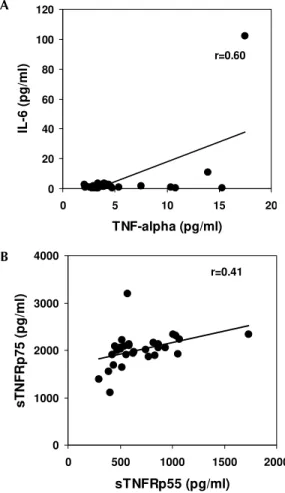
observed in only six patients (Fig. 1).
If the correlation coefficient is calculated,
TNF-a and IL-6 show a significant correlation; a weaker
correlation was also found for sTNFRp55 and sTNFRp75 (Fig. 2). Other factors could not be as-sociated.
In the only exception for a positive IL-1b, the
patient had levels 255-fold higher than the aver-age of the rest of the patients and no sIL-1Ra was produced in this serum. As mentioned before this patient had the highest values for the three cytokines, but clinical manifestations remained
TABLE III
Determination of sTNF-Rp55and sTNF-Rp75in sera from patients with dengue fever
sTNF-Rp55a(pg/ml) sTNF-Rp75 (pg/ml)
Days of Controls= 4512±96 Controls= 1416± 176
disease Referential limit of positivity=674 Referential limit of positivity=1822
1 1065b 2233
2 434 425 1688 1910
3 829 573 1005 1903 3196 2344
4 634 1731 625 535 1975 2335 1940 2090
5 784 451 747 470 866 1032 930 298 1387 2092 2021 1995 2057 2307 2064
6 559 1056 1907 1897 1919
7 583 864 771 520 2215 1874 2130 2100
8 498 505 392 583 816 2058 2170 2131 1554 2023
12 515 1638
14 404 1106
a: patients with dengue had a higher frequency of positive sTNF-Rp55 (P<0.0001) and sTNF-Rp75 (P<0.0001) when compared with controls in Fisher exact test (one-sided; a=0.05); b: bold numbers represent values above referential limit of positivity.
Fig. 1: association ratio between different soluble factors present in patient sera during dengue fever.
0 20 40 60 80 100 120
0 5 10 15 20
TNF-alpha (pg/ml)
IL-6 (pg/ml)
r=0.60
0 1000 2000 3000 4000
0 500 1000 1500 2000
sTNFRp55 (pg/ml)
sTNFRp75 (pg/ml)
r=0.41
A
B
391 391391 391391 Mem Inst Oswaldo Cruz, Rio de Janeiro, Vol. 94(3), May/Jun. 1999
without outstanding features. All five patients posi-tive for sIL-1Ra produced no IL-1b; thus, no
asso-ciation between these two factors was shown.
Association of soluble factor production with arthropathy or to the type of infection (primary or secondary) during dengue fever - Attempts to as-sociate arthritis and/or arthralgia with pro-inflam-matory cytokines or with anti-inflampro-inflam-matory com-pound production were not successful (Table IV). No significance was found using Fisher exact test in data from all factors measured. Furthermore, no statistical difference was observed between groups of primary and secondary infection in the produc-tion of these soluble factors (Table V).
DISCUSSION
Dysregulated expression of inflammatory cytokines TNF-a, IL-6 and IL-1b is known to be
implicated in immunopathologic mechanisms such as inflammation, necroinflammatory injuries and endotoxic shock (Dinarello 1996a). Viral genic products originating from HTLV-I, Hepatitis B Virus are able to activate promoters from transcrip-tion factors such as NFkB and NF-IL-6 (Kishimoto
et al. 1994) known to induce pro-inflammatory cytokines which are frequently detected circulat-ing durcirculat-ing viral infections (Hober et al. 1989).
During this work, TNF-a, IL-6, sTNFRp55 and
sTNFRp75 were detected in the sera of Brazilian patients with DF, devoid of hemorrhagic manifes-tations and infected with Dengue-1 or -2. In gen-eral, the levels of cytokines in most of our DF pa-tients were lower than those described in literature for DHF/DSS (Hober et al. 1993, 1996a, Kuno & Bailey 1994, Iyngkaran et al. 1995, Kubelka et al. 1995, Bethell et al 1998). This may be explained either by relative disease mildness, virus load, by differences in viral serotype, host genetic factors that may influence in the immunological responses or in test sensitivity.
Our results reveal that cytokines, TNF-a and
IL-6 and soluble TNF receptors were present in significantly increased levels until the eighth day of DF, in several but not in all patients. Factor pro-duction at later stages of disease remains to be de-termined. The levels of TNF-a observed suggest a
production from the third day of disease onwards. IL-6 was already present at significant levels at patients at the first two days and sTNFRp75 ap-peared in the first day and could be detected in most patients (four out of five) at the eighth day. Nevertheless, higher number of patients should be tested to confirm these results.
In contrast, IL-1b seems not to be altered as
DF progresses. This is in agreement with earlier studies on DHF/DSS (Hober et al. 1993). Circu-lating IL-1b levels detected usually in pathologic
conditions are relatively low compared with lev-els of IL-6 and TNF-a. Unlike TNF-a, 6 or
IL-1Ra, a significant amount of proIL-1b remains
in-side the cell. IL-1b also binds to large proteins such
as a2-macroglobulin, complement, and the
sIL-1RII, which in turn binds preferentially to IL-1b
when compared to IL-1a or IL-1Ra (Dinarello
1996a), being not easily reactive in regular ELISA assays.
The biological properties of TNF-a share
re-markable similarities to those of IL-6 and IL-1b:
they are endogenous pyrogens and inducers of acute-phase responses. IL-1-b and TNF-a induce
IL-6 production. It has been claimed that levels of
TABLE IV
Association of soluble factor presence in sera with arthropathy during dengue fever. Frequency of
patients with significative production
Soluble Arthritis
factor Positive Negative Pa
TNF-a 6/15b 4/15 0.3499
IL-6 6/15 3/15 0.2135
IL-1 0/15 1/15 0.5000
TNF-Rp55 6/15 7/15 0.5000
TNF-Rp75 13/15 10/14 0.2908
IL-1Ra 4/13 1/14 0.1862
a: no difference in frequencies was found. P value was calculated in Fisher exact test (one-sided; a=0.05); b: number of patients with positivity for the soluble factor/total number of patients.
TABLE V
Association of soluble factor presence in sera during dengue fever with the type of infection (primary or secondary). Frequency of patients with significative
production
Soluble Type of Infection
factor Primary Secondary Pa
TNF-a 4/14b 4/11 0.5042
IL-6 8/14 6/11 0.6075
IL-1 0/14 1/11 0.4400
TNF-Rp55 6/14 6/11 0.4296
TNF-Rp75 11/13 11/12 0.5313
IL-1Ra 3/15 2/13 0.6038
392 392 392 392
392 Cytokines and Soluble Receptors in Dengue Luzia MO Pinto et al.
IL-6 often may better correlate with severity of an infectious disease (Dinarello 1992, 1996b). In our work IL-6 was the most frequently detected cytokine. Moreover, one patient with all three cytokines elevated had levels of IL-6 six-fold higher than TNF-a and 1,5-fold higher than
IL-1b. Previous work studying DHF patients described
contradictory data relating IL-6 levels and sever-ity of disease (Hober et al. 1993, Kuno & Bailey 1994, Bethell et al. 1998).
Exanthema is not exclusively dependent on the elevation of circulating factors, since all patients developed rash and some were negative for their production; on the other hand, hemorrhagic mani-festations cannot be directly associated with pro-duction of any of the circulating factors. Moreover, the presence of cytokines in circulation seems not to be related to arthropathy. During malignant as-cites (Van Zee et al. 1992) the concentration of sTNFR in plasma was lower than in the synovial fluid where inflammation actually occurred; more-over, differences in factor localization were corre-lated with disease severity. If cytokines and/or soluble receptors play some role in joint manifes-tations during dengue disease, local production should be investigated.
An association found between two soluble TNF receptors (sTNFRp55 and sTNFRp75) could indi-cate that their induction mechanisms might be re-lated. Circulating sTNFRs may provide different regulatory pathways for modulatingTNF-a effects.
sTNFRs can compete for TNF-a with cell surface
receptors and thus reduce the activity of the cytokine; on the other hand they may also enhance TNF-a function regulating its bio-availability, most
likely by stabilizing the active TNF-a oligomer
(Leuwenberg et al. 1994). According to our data TNF-a seems to have a better association with
sTNFRp75 than p55. If this is confirmed, it may be postulated that sTNFRp75 might modulate
TNF-a activity in vivo during DF. Furthermore, we
ob-served in both normal and DF sera that sTNFRp75 was more abundant than sTNFRp55; this is in ac-cord with usual descriptions from in vivo studies during inflammatory processes or in normal indi-viduals (Hart et al. 1996).
IL-1Ra is stimulated under conditions where pro-inflammatory cytokines would be inhibited. IL-10 and IL-4 suppress macrophage release of pro-inflammatory cytokines (TNF-a, IL-6 or IL-1b and
IL-8) and stimulate the secretion of IL-1Ra and nonsignaling type II IL-1 receptor (Tilg et al. 1997). Also, IL-4 inhibits the release of sTNFRs from monocytes. Therefore, besides soluble receptors, other cytokines can antagonize the biological ac-tivity of pro-inflammatory cytokines and could
eventually be acting during dengue infection. This could explain IL-1Ra has increased expression in five patients but attempts to inversely correlate with pro-inflammatory cytokines have failed until now. Moreover, in an earlier work, Hober et al. (1996a) described that in Dengue-3-induced DHF/DSS no apparent correlation between TNF-a and
sTNFRp75 could be made.
Taken together, clinical observations and se-rum titration described here show that pro-inflam-matory cytokines (such as TNF-a and IL-6) are
also produced during mild non hemorrhagic mani-festations of exanthematic DF and it is likely that they play a role in this pathology, as described ear-lier for DHF/DSS. Severity may be related to the amount of circulating cytokines. Soluble receptors, mainly sTNFRp75, are also increased and, con-sidering its association with TNF-a, may be used
as a marker of immunological activation, since it is more stable than pro-inflammatory cytokines. From our findings the question still remains: could sTNFp75 act as modulator of an excessive biologi-cal activity of TNF-a (Tilg et al. 1997) preventing
severe disease during DF? Further studies deserve to be performed to broaden our understanding about the balance among different circulating im-munological factors and their effects in develop-ment of disease.
ACKNOWLEDGMENTS
To Drs Takumi Iguchi and José A Losana for statis-tical advice, Drs Hermann G Schatzmayr and Marilda M Siqueira for continuous encouragement, Dr Marize P Miagostovich and Ms Eliane Saraivo for performing laboratory diagnostics.
REFERENCES
Anderson R, Wang S, Osiowy C, Issekutz AC 1997. Activation of endothelial cells via antibody-enhanced dengue virus infection of peripheral blood mono-cytes. J Virol 71: 4226-4232.
Andus T, Gross V, Holstege A, Ott M, Weber M, David M, Gallati H, Gerok W, Schölmerich J 1992. High concentrations of soluble Tumor Necrosis Factor receptors in ascites. Hepatology 16: 749-55. Bethell DB, Flobe K, Cao XT, Day NP, Pham TP,
Buurman WA, Cardosa MJ, White NJ, Kwiatkowski D 1998. Pathophysiologic and prognostic role of cytokines in dengue hemorrhagic fever. J Infect Dis
177: 778-782.
Brown F 1986. The classification and nomenclature of viruses: summary of Meetings of the International Committee on Taxonomy of Viruses in Sendai, Sep-tember 1984. Intervirol 25: 141-143.
Clarke DH, Casals J 1958. Techniques for hemaggluti-nation and hemagglutihemaggluti-nation inhibition with arthro-pod-borne viruses. Am J Trop Med Hygie 7: 561-573.
393 393393 393393 Mem Inst Oswaldo Cruz, Rio de Janeiro, Vol. 94(3), May/Jun. 1999
diseases. Immunol Rev 127: 119-146.
Dinarello CA 1996a. Cytokines as mediators in the patho-genesis of septic shock, p. 134-165. InET Rietschel & H Wagner (eds), Pathology of the Septic Shock, Springer-Verlag, Heidelberg.
Dinarello CA 1996b. Biologic basis for interleukin-1 in disease. Blood 87: 2095-2147.
Halstead SB 1980. Immunological parameters of Togaviruses disease syndromes, p. 107-174. In RW Schlesinger, The Togaviruses. Biology, Structure,
Replication, Academic Press, New York.
Halstead SB 1988. Pathogenesis of dengue : challenges to molecular biology. Science 239: 476-481. Halstead SB 1990. Dengue and dengue hemorrhagic
fe-ver. Curr Scie 3: 434-438.
Halstead SB, O´Rourke EJ, Allison AC 1977. Dengue viruses and mononuclear phagocytes. J Exp Med 146: 218-229.
Hamon WMcD, Rudnick A, Sather GE 1960. Virus as-sociated with epidemic hemorrhagic fevers of Phil-ippines and Thailand. Science131: 1102-1103. Hart PH, Hunt EK, Bonder CS, Watson CJ, Finlay-Jones
JJ 1996. Regulation of surface and soluble TNF re-ceptor expression on human monocytes and synovial fluid macrophages by IL-4 and IL-10. J Immunol 157: 3672-3680.
Hober D, Delannoy AS, Benyoucef S, Groote DD, Wattré P 1996a. High levels of sTNFRp75 and TNFa in Dengue-infected patients. Microbiol Immunol 40: 569-573.
Hober D, Haque A, Wattré, Beaucaire G, Mouton Y, Capron A 1989. Production of tumor necrosis fac-tor-alpha (TNF-a) and interleukin-1 (IL-1) in pa-tients with AIDS. Enhanced level of TNF is related to higher cytotoxic activity. Clin Exp Immunol 78: 329-333.
Hober D, Poli L, Roblin B, Gestas P, Chungue E, Granic G, Imbert P, Pecarere JL, Vergez-Pascal R, Wattre P, Maniez-Montreuil M 1993. Serum levels of tumor necrosis factor-a (TNF-a), interleukin-6 (IL-6) and interleukin-1b (IL-1b) in Dengue-infected patients.
Am J Med Hyg 48: 324-331.
Hober D, Shen L, Benyoucef S, De Groote D, Deubel V, Wattré P 1996b. TNFa production by monocytic-like cells exposed to dengue virus antigens. Immunol
Lett 53: 115-120.
Hober D, Nguyen TL, Shen L, Ha DQ, Huong VTQ, Benyucef S, Nguyen TL, Bui TMP, Loan HK, Le BL, Bouzidi A, Groote DD, Drouet MT, Deubel V, Wattré P 1998. Tumor necrosis factor alpha levels in plasma and whole-blood culture in dengue-in-fected patients: relationship between virus detection and pre-existing specific antibodies. J Med Virol 54: 210-218.
Iyngkaran N, Yadav, Sinniah M 1995. Augmented in-flammatory cytokines in primary dengue infection progressing to shock. Singapore Med J 36: 218-221. Kishimoto T, Taga T, Akira S 1994. Cytokine signal
transduction. Cell 6: 253-262.
Kliks SC, Nisalak A, Brant WE, Wahl L, Burke DS 1989. Antibody-dependent enhancement of Dengue virus growth in human monocytes as a risk factor for
Den-gue hemorrhagic fever. Am J Trop Hyg 40: 444-451. Kornelisse RF, Savelkoul HFJ, Mulder PHG, Suur MH, van der Straaten PJC, van der Heijden AJ, Sukhai RN, Hählen K, Neijens HJ, de Groot R 1996. Interleukin-10 and soluble tumor necrosis factor in cerebrospinal fluid of children with bacterial men-ingitis. J Infect Dis 173: 1498-502.
Kubelka CF, Borges PA, vonSydow FOF, Lampe E 1995. Analysis of tumor necrosis factor-a serum level in Brazilian patients with Dengue-2. Mem Inst Oswaldo
Cruz 90: 741-42.
Kuno G, Bailey RE 1994. Cytokine responses to Den-gue infection among Puerto Rican patients. Mem Inst
Oswaldo Cruz 89: 179-182.
Kurane I, Mady, BJ, Ennis FA 1991. Antibody-depen-dent enhancement of Dengue virus infection. Med
Virol 1: 211-221.
Leuwenberg JFM, Dentener MA, Buurman WA 1994 Lipopolysaccharide-mediated soluble TNF receptor release and TNF receptor expression by monocytes.
J Immunol 152: 5070-5076.
Monath TP 1986. Pathology of the Flaviviruses, p. 375-440. InM Schlesinger & S Schlesinger (eds), The
Togaviridae and Flaviviridae, Plenum Press, New
York & London.
Monath TP 1994. Dengue : the risk to developed and developing countries. Proc Natl Acad Sci 91: 2395-2400.
Nogueira RMR, Miagostovich MP, Cavalcanti SMB, Marzochi KBF, Schatzmayr HG 1992. Levels of IgM antibodies against Dengue virus in Rio de Janeiro, Brazil. Res Virol 143: 423-27.
PAHO-Pan American Health Organization 1994. Den-gue and denDen-gue hemorrhagic fever in Americas: guidelines for prevention and control. PAHO/WHO (Scientific Publication N#548), Washington, D.C. Pang T 1987. Dengue haemorrhagic fever : virus or
re-sponse? Bio Essays 6: 141.
Pereda MP, Sauer J, Castro CP, Finkielman S, Stalla GK, Holsboer F, Artz E 1995. Corticotropin-releasing hormone differentially modulates the interleukin-1 system according to the level of monocyte activa-tion by endotoxin. Endocrinology 136: 5504-5510. Poli G, Fauci AS 1993. Cytokine modulation of HIV
expression. Sem Immunol 5: 165-173.
Sabin AB, Schlesinger RW 1945. Production of immu-nity to Dengue virus modified by propagation in mice. Science 101: 640-642.
Tilg H, Dinarello CA, Mier JW 1997. IL-6 and APPs anti-inflammatory and immunosuppressive media-tors. Immunol Today 18: 428-432.
Torre D, Zeroli C, Giola M, Ferrario G, Fiori GP, Bonetta G, Tambini R 1994 Serum levels of Interleukin-1a, Interleukin-1b, Interleukin-6, and Tumor Necrosis Factor in patients with Acute Viral Hepatitis. Clin
Infect Dis 18: 194-198.
Van Zee KJ, Kohno T, Fischer E, Rock CS, Moldawer LL, Lowry SF 1992. Tumor necrosis factor soluble receptors circulate during experimental and clinical inflammation and can protect against excessive tu-mor necrosis factor-ain vitro and in vivo. Proc Natl
394 394 394 394
394 Cytokines and Soluble Receptors in Dengue Luzia MO Pinto et al.
Yang KD, Lee CS, Hwang KP, Chu ML, Shaio MF 1995. A model to study cytokine profiles in primary and heterologously secondary dengue-2 virus infections.
Acta Virol 39: 19-21.