205 205205 205205 Mem Inst Oswaldo Cruz, Rio de Janeiro, Vol. 94, Suppl. I: 205-210, 1999
Morphobiological Characterization of
Trypanosoma cruzi
Chagas, 1909 and its Distinction from other Trypanosomes
Maria Auxiliadora de Sousa
Coleção de Tripanosomatídeos, Laboratório de Transmissores de Hematozoários, Departamento de Entomologia, Instituto Oswaldo Cruz, Av. Brasil 4365, 21045-900 Rio de Janeiro, RJ, Brasil
Key words: Trypanosoma cruzi Trypanosoma rangeli Trypanosoma conorhini Trypanosoma lewisi -Trypanosoma minasense - -Trypanosoma freitasi - Chagas disease - morphobiology - characterization
The etiologic agent of the human American trypanosomiasis was discovered by Chagas (1909a) initially in triatomine bugs, Panstrongylus megistus, collected in Lassance, State of Minas Gerais, Brazil. Its infectivity to some laboratory animals was soon confirmed, a peculiar small try-panosome appearing in the blood of these animals, which Chagas (1909a) named Trypanosoma cruzi
in honor of Oswaldo Cruz. The first natural infec-tion of a mammal with T. cruzi was recorded in a cat also by Chagas (1909b), who subsequently found the parasite in the blood of a child, and as-sociated to it the illness found in that child, as well as in other persons living in houses infested with triatomine bugs in Lassance. At that time, Chagas (1909b) also carried out studies on this parasite in human beings, experimental mammal hosts, triatomines and axenic cultures. Believing that T. cruzi presented schizogonic stages in the lungs of vertebrate hosts, Chagas (1909b) temporarily placed it in a new genus, Schizotrypanum, but sub-sequently corrected such mistake (Chagas 1913). Chagas (1912, 1924) also found T. cruzi in wild animals, armadillos (incriminating P. geniculatus
as its vector) and squirrel monkeys. The evolution of T. cruzi in Triatoma infestans and the possibil-ity of this species and T. sordida actuating as vec-tors were also reported by Chagas (1912).
Hartmann (1910) and Vianna (1911) discov-ered the intracellular multiplication of T. cruzi, but the latter correctly interpreted it as occurring by binary fission. Vianna (1911) carried out an exten-sive histopathological study in post-mortem pa-tients and experimental animals, evidencing the parasite multiplication in several tissues, mainly myocardium and skeletal muscles. Brumpt (1912) studied the development of T. cruzi in P. megistus,
Fax: +55-21-290.9339 or 598.4320. E-mail: [email protected] Received 9 June 1999
Accepted 9 August 1999
cimicids and ticks, discovering the infectivity to mammals of the trypomastigotes found in the pos-terior intestine and in the feces (metacyclics). An important revision of T. cruzi biology was under-taken by Dias (1934), being noteworthy his de-tailed study on the parasite development in triatomines. The name Schizotrypanum as subge-nus, T. (S.) cruzi being the type species, became largely accepted after Hoare’s reviews on mam-mal trypanosomes (1964, 1972). More recently, an interesting aspect of T. cruzi biology was described by Deane et al. (1984), who discovered stages typi-cally found in triatomines also in the lumen of anal glands of the opossum Didelphis marsupialis.
subter-206 206 206 206
206 T. cruzi Morphobiological Characterization Maria Auxiliadora de Sousa
minal kinetoplast (Fig. 2: H6, 7); (4) easy growth at about 27oC in several conventional culture me-dia (NNN, LIT, Warren, and so on), presenting stages similar to those found in triatomines (Fig. 2: H); (5) capacity for intracellular multiplication as amastigotes, either in mammal host tissues or cellular cultures, subsequently originating trypomastigotes similar to the blood forms.
Aiming to confirm whether new isolates are or not T. cruzi, several authors have also used or sug-gested the use of “protection assays” (Dias 1935, Barreto & Ribeiro 1979, Marinkelle 1982, Steindel et al. 1998). Otherwise, as the epimastigotes of T. cruzi, either from insect gut or axenic cultures, are susceptible to complement lysis (Muniz & Borriello 1945), this test can be additionally used for its char-acterization. Isolates (from mammals or triatomi-nes) which have features of the subgenus
Schizotrypanum, while partially characterized, have been usually identified as T. cruzi-like organ-isms. The validity of those species proposed to some T. cruzi-like trypanosomes found in non-hu-man primates (T. prowazeki, T. lesourdi, T. sanmartini) is matter of discussion, they not being largely accepted (Deane 1964, Hoare 1972, Marinkelle 1976b).
The occurrence of T. cruzi-like organisms in American bats is a problem to be considered in epidemiological studies on the agent of Chagas disease. Whereas being largely accepted that T. cruzi occurs in bats as well (Clark & Dunn 1932, Deane 1961, 1964, Hoare 1972, Marinkelle 1976a, 1982, Barreto & Ribeiro 1979), new T. ( Schizotry-panum) species/subspecies were proposed to sev-eral isolates from chiropterans mainly based on their apparent lack of infectivity to common labo-ratory animals and/or triatomines, as well as on morphological peculiarities (reviewed by Dias 1935, Hoare 1972, Marinkelle 1976a, Molyneux 1991). However, the state of our knowledge about these organisms is yet unsatisfactory and compara-tive studies using well-characterized T. cruzi strains are necessary to better establish their distinction. Otherwise, the actual host-restriction of some
Schizotrypanum species from bats should be con-firmed, at least by examining for periods longer than 60 days those hemocultures from experimen-tally inoculated laboratory animals.
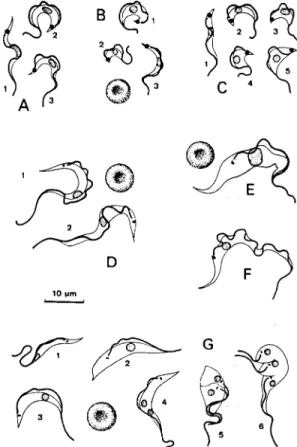
According to Hoare (1972), the mean total length of the bloodstream trypomastigotes in sev-eral T. cruzi isolates, either from natural or experi-mental hosts, ranged from 16.3 to 21.8 µm. The morphological variability of these trypomastigotes was early noted by Chagas (1909b), who described the slender (Fig. 1: A1, B3, C1) and broad (Fig. 1: A2, B1, C3) forms. However, other types, as short (Fig. 1: B2), very broad (Fig.1: C4, 5) and
inter-mediate forms (Fig. 1: A3, C2) can also be found (Silva 1959, Brener & Chiari 1963). The rate of these forms varies according to the parasite strain, the phase of infection, as well as the host species (Silva 1959, Brener 1965, Andrade 1974, Sogayar 1978, Urdaneta-Morales 1983). Brener (1965) and Andrade (1974) grouped some T. cruzi strains ac-cording to the predominance of each form and their behavior in experimentally infected mice, mainly considering the pattern of parasitemia, predomi-nant lesions or tissue parasitism, virulence and le-thality. However, those groupings can not be cor-related to each other at all; for example, those strains predominantly presenting stout forms, pro-ducing increasing parasitemia and high mortality in mice (Brener 1965, Carneiro et al. 1991) do not fit in with the strain types (biodemes) proposed by
Fig. 1 - A, B, and C: Trypanosoma cruzi, Y, FL and CL strains, respectively, forms from the blood of experimentally infected mice. D: T. rangeli; 1: bloodstream trypomastigotes from natu-rally infected Saimiri ustus; 2: blood forms from experimen-tally infected Callithrix jacchus. E: T. minasense, bloodstream stage from naturally infected C. penicillata. F: T. conorhini, bloodstream type trypomastigote from the supernatant of para-site co-culture with L929 cell line at 37oC. G: T. lewisi, stages
207 207207 207207 Mem Inst Oswaldo Cruz, Rio de Janeiro, Vol. 94, Suppl. I, 1999
Andrade (1974). Otherwise, T. cruzi strains deter-mining only subpatent parasitemia in mice can not be considered according to those parameters.
Some studies have suggested that the different
T. cruzi blood forms may have also different bio-logical roles and that, at least partially, the features of a strain may depend on the predominance of one of them (Brener 1965, 1969). Several authors provided evidences that the slender forms would be more fitted for cellular invasion, whereas the broad ones would more promptly develop in triatomines or axenic cultures (Silva 1959, Brener 1969, Howells & Chiari 1975, Deane 1979, Sousa et al. 1982, Schmatz et al. 1983). However, Deane (1979) emphasized the possible occurrence of un-differentiated or ambivalent bloodstream trypomastigotes, perhaps represented by the so-called intermediate forms, which would develop either inside cells or in axenic cultures. Brener and Chiari (1965) also reported that the pattern of early growth in culture (or citrated blood) at 28oC of different T. cruzi strains varied according to their predominant blood form.
Since T. cruzi has a notable range of hosts, it is not infrequent that these can be parasitized by other trypanosomes, and that mixed infections can oc-cur when their geographic distribution overlaps. It is frequently possible to distinguish these trypano-somes from T. cruzi by classical morphobiological approaches, and herein some of them are reported with emphasis on useful features to their differen-tial diagnosis. Aiming at a more complete charac-terization of these trypanosomes or knowledge of other species some important reviews (Hoare 1972, D’Alessandro 1976, Marinkelle 1976a, b, Molyneux 1976, 1991, Wells 1976, D’Alessandro-Bacigalupo & Saravia 1992) and original papers should be consulted. It is worthy mentioning that those morphobiological approaches are not in op-position to the use of biochemical and molecular techniques to characterize and identify these try-panosomes, but rather they greatly contribute for a more wide and comprehensive view of their evo-lution and taxonomy.
T. rangeli Tejera, 1920 is a non-pathogenic try-panosome of human beings and several mammals transmitted through inoculative route by some triatomine species (mainly genus Rhodnius), and frequently shares hosts with T. cruzi (Hoare 1972, D’Alessandro 1976, D’Alessandro-Bacigalupo & Saravia 1992). Although the natural or experimen-tal mammal hosts of T. rangeli (or T. rangeli-like parasites) usually display low or subpatent para-sitemia, its bloodstream trypomastigotes can be distinguished from those of T. cruzi mainly by their larger size and smaller kinetoplast (Fig. 1: D1, 2). According to D’Alessandro (1976) the mean total
length of T. rangeli blood forms from various re-ports ranged from 26.4 to 33.8 µm. No multiplica-tion site of this parasite in mammals has been con-vincingly demonstrated (D’Alessandro-Bacigalupo & Saravia 1992). In triatomine bugs, T. rangeli
develops in the intestine, but unlike T. cruzi, can invade the hemocele, as well as the salivary glands, where large number of metacyclics are formed. These are small and broad trypomastigotes, 8-13 µm in length (D’Alessandro 1976), with an incon-spicuous undulating membrane, a subterminal and a relatively large kinetoplast (Fig. 2: J11). T. rangeli
can also be cultivated in conventional liquid me-dia usually overlaying blood-agar slants, but it is more exigent than T. cruzi to be maintained by se-rial transfers. Either in triatomines or in recently isolated cultures (at about 27oC), T. rangeli can be very polymorphic, presenting small epimastigotes or aflagellate forms (Fig. 2: J1, 2), medium-sized epi- or trypomastigotes (Fig. 2: J5-7), peculiar long epi- and trypomastigotes (which may reach 100 µm or more, according to D’Alessandro, 1976) (Fig. 2: J3, 4, 8), besides metacyclic trypomasti-gotes (Fig. 2: J9-11). Several T. rangeli-like trypa-nosomes were described as separate species (T. diasi, T. mycetae, T. myrmecophagae, T. saimirii, and so on), but their taxonomic status has been dis-cussed by several authors (Deane & Damasceno 1961, Hoare 1972, D’Alessandro 1976, D’Alessandro-Bacigalupo & Saravia 1992, Ziccardi & Lourenço-de-Oliveira 1998).
T. (Herpetosoma) lewisi (Kent 1880) is a com-mon parasite of rats in various parts of the world (Rattus rattus, R. norvegicus and others), being transmitted by fleas; it is normally host-restricted, confined to rats, and non-pathogenic (Hoare 1972, Molyneux 1976). Its bloodstream trypomastigotes are very distinct from those of T. cruzi, usually having the nucleus anteriorly placed, the small rod-like kinetoplast and the poorly-developed mem-brane undulating (Fig. 1: G1-3). During its repro-ductive phase, T. lewisi multiplies in the blood (as epimastigotes) mainly by successive unequal di-visions (Fig. 1: G5, 6). Following this phase only monomorphic trypomastigotes (“adults”) remain in the blood; their mean total length is 30.6 µm (Hoare 1972). T. lewisi does not develop in triatomines, but can growth in axenic cultures at about 27oC (Hoare 1972, Molyneux 1976). There are several T. lewisi-like trypanosomes described under other specific names (T. forattinii, T. coutinhoi, and so on) and which were only par-tially studied (Deane 1961, Hoare 1972).
culti-208 208 208 208
208 T. cruzi Morphobiological Characterization Maria Auxiliadora de Sousa
minal kinetoplast (Fig. 2: I6); they seem to arise from spheromastigotes by an unrolling process (Fig. 2: I5).
Other species belonging to the Megatrypanum
subgenus, as T. minasense (Fig. 1: E) from non-human primates, T. freitasi from opossums, T. leonidasdeanei and T. pessoai from bats, are also easily distinguished from T. cruzi by their large bloodstream forms, non-infectivity for mice, hard or no growth in triatomines and in conventional culture media (Deane & Damasceno 1961, Hoare 1972, Marinkelle 1976a, b, Molyneux 1991, Ziccardi et al. 1996). However, T. freitasi can also be found in the lumen of anal glands of opossums (Deane & Jansen 1990).
ACKNOWLEDGMENTS
To Profs Dayse CB Branco and Glória MC Gonçalves for helping in the illustrations. To Drs Mario Steindel, Mariangela Ziccardi, Neide Thomaz for pro-viding some materials used in the illustrations. To Dr Wladimir Lobato Paraense for reviewing the manuscript.
REFERENCES
Andrade SG 1974. Caracterização de cepas do Trypa-nosoma cruzi isoladas no Recôncavo Baiano. Rev Pat Trop 3: 65-121.
Barreto MP, Ribeiro RD 1979. Reservatórios silvestres do Trypanosoma (Schizotrypanum) cruzi Chagas 1909. Rev Inst Adolfo Lutz 39: 25-36.
Bogliolo AR, Lauria-Pires L, Gibson WC 1996. Poly-morphisms in Trypanosoma cruzi: evidence of ge-netic recombination. Acta Trop 61: 31-40. Brener Z 1965. Comparative studies of different strains
of Trypanosoma cruzi. Ann Trop Med Parasitol 59: 19-26.
Brener Z 1969. The behaviour of slender and stout forms of Trypanosoma cruzi in the blood-stream of nor-mal and immune mice. Ann Trop Med Parasitol 63: 215-220.
Brener Z, Chiari E 1963. Variações morfológicas observadas em diferentes amostras de Trypanosoma cruzi. Rev Inst Med Trop São Paulo 5: 220-224. Brener Z, Chiari E 1965. Aspects of early growth of
different Trypanosoma cruzi strains in culture me-dium. J Parasitol 51: 922-926.
Brumpt E 1912. Le Trypanosoma Cruzi évolue chez Conorhinus megistus, Cimex lectularius, Cimex Boueti et Onithodorus moubata. Cycle évolutif de ce parasite. Bull Soc Pathol Exot 5: 360-367. Carneiro M, Romanha AJ, Chiari E 1991. Biological
characterization of Trypanosoma cruzi strains from different zymodemes and schizodemes. Mem Inst Oswaldo Cruz 86: 387-393.
Carrasco HJ, Frame IA, Valente SA, Miles MA 1996. Genetic exchange as a possible source of genomic diversity in sylvatic populations of Trypanosoma cruzi. Amer J Trop Med Hyg 54: 418-424. Chagas C 1909a. Neue Trypanosomen. Arch Schiffs
Tropenhyg 13: 120-122.
Fig. 2- H: Trypanosoma cruzi, CL strain in axenic culture; 1-3: epimastigotes; 4-5: transitional trypomastigotes; 6-7: metacyclics. I: T. conorhini, Lourenço-de-Oliveira/Ziccardi strain in axenic culture; 1-4: epimastigotes; 5: transitional stage to metacyclic; 6: metacyclic. J: T. rangeli, SC 58 strain; 1-10 stages from a recently isolated culture; 1: small epimastigotes; 2: small aflagellate form; 3-6: epimastigotes; 7-8: trypomastigotes; 9-10: metacyclic-like trypomastigotes; 11: metacyclic from salivary glands of experimentally infected
Rhodnius prolixus. Camera lucida drawings. All cultures were maintained at about 27oC. Giemsa-stained smears after HCl digestion.
ter-209 209209 209209 Mem Inst Oswaldo Cruz, Rio de Janeiro, Vol. 94, Suppl. I, 1999
Chagas C 1909b. Nova tripanozomiaze humana. Estudos sobre a morfolojia e o ciclo evolutivo do Schizotry-panum cruzi n. gen, n. sp., ajente etiolojico de nova entidade morbida do homem. Mem Inst Oswaldo Cruz 1: 11-80.
Chagas C 1912. Sobre um trypanosomo do tatú, Tatusia novemcincta, transmittido pela Triatoma geniculata Latr. (1811). Possibilidade de ser o tatú um depositario do Trypanosoma Cruzi no mundo exte-rior. Brazil-Med I: 305-306.
Chagas C 1913. Revisão do cyclo evolutivo do “Trypa-nosoma Cruzi”. Brazil-Med 27: 225.
Chagas C 1924. Sobre a verificação do “Trypanosoma cruzi” em macacos do Pará (Chrysothrix sciureus). Scien Med 2: 75-76.
Clark HC, Dunn LH 1932. Experimental studies on Chagas disease in Panama. Am J Trop Med 12: 49-77.
D’Alessandro A 1976. Biology of Trypanosoma (Herpetosoma) rangeli Tejera, 1920, p. 327-403. In WHR Lumsden & DA Evans (eds), Biology of the Kinetoplastida, vol 1, Academic Press, London. D’Alessandro-Bacigalupo A, Saravia NG 1992.
Trypa-nosoma rangeli, p. 1-54. In JP Kreier, JR Baker (eds), Parasitic Protozoa, 2nd ed, vol 2, Academic Press, San Diego.
Deane LM 1961. Tripanosomídeos de mamíferos da região Amazônica. I. Alguns flagelados encontrados no sangue de mamíferos silvestres do Estado do Pará. Rev Inst Med Trop São Paulo 3: 15-28.
Deane LM 1964. Animal reservoirs of Trypanosoma cruzi in Brazil. Rev Bras Malariol D Trop 16: 27-48.
Deane LM, Damasceno RG 1961. Tripanosomídeos de mamíferos da Região Amazônica. II. Tripanosomas de macacos da Zona do Salgado, Estado do Pará. Rev Inst Med Trop São Paulo 3: 61-70.
Deane LM, Deane MP, Lourenço-de-Oliveira R 1986. Are asian monkeys the original hosts of Trypano-soma conorhini? Mem Inst Oswaldo Cruz 81: 127-129.
Deane MP 1947. Ocorrência do Trypanosoma conorrhini em “barbeiros” e em rato na cidade de Belém, Pará, e seu cultivo em meio NNN. Rev Ser Esp Saúde Públ 1: 433-442.
Deane MP 1979. Significance of polymorphism in Try-panosoma cruzi, p. A6-7. Anais do Congresso Internacional sobre Doença de Chagas, Rio de Janeiro.
Deane MP, Deane LM 1961. Studies on the life cycle of Trypanosoma conorrhini. “In vitro” development and multiplication of the bloodstream trypanosomes. Rev Inst Med Trop São Paulo 3: 149-160.
Deane MP, Jansen AM 1990. Developmental stages of Trypanosoma (Megatrypanum) freitasi Rego, Magalhães & Siqueira, 1957 in the opossum Didel-phis marsupialis (Marsupialia, Didelphidae). J Protozool 37: 44-47.
Deane MP, Lenzi HL, Jansen A 1984. Trypanosoma cruzi: vertebrate and invertebrate cycles in the same mammal host, the opossum Didelphis marsupialis. Mem Inst Oswaldo Cruz 79: 513-515.
Dias E 1934. Estudos sobre o Schizotrypanum cruzi. Mem Inst Oswaldo Cruz 28: 1-110.
Dias E. 1935. Revisão geral dos hemoflagellados de Chirópteros, p. 10-88. Novena Reunión de la Sociedad Argentina de Patologia Regional, Mendoza.
Dias E, Seabra CAC 1943. Sôbre o Trypanosoma conorrhini, hemoparasito do rato transmitido pelo Triatoma rubrofasciata. Presença do vector infectado na cidade do Rio de Janeiro. Mem Inst Oswaldo Cruz 39: 301-330.
Hartmann M 1910. Notiz über eine weitere art der schizogonie bei Schizotrypanum cruzi (Chagas). Arch Protistenk 20: 361-363.
Hoare CA 1964. Morphological and taxonomic studies on mammalian trypanosomes. X. Revision of the systematics. J Protozool 11: 200-207.
Hoare CA 1972. The Trypanosomes of Mammals. A Zoological Monograph. Blackwell Scientific Publi-cation, Oxford.
Howells RE, Chiari CA 1975. Observations on two strains of Trypanosoma cruzi in laboratory mice. Ann Trop Med Parasitol 69: 435-448.
Marinkelle CJ 1976a. Biology of the trypanosomes of bats, p. 175-216. In WHR Lumsden, DA Evans (eds), Biology of the Kinetoplastida, vol 1, Academic Press, London.
Marinkelle CJ 1976b. The biology of the trypanosomes of non-human primates, p. 217-256. In WHR Lumsden, DA Evans (eds), Biology of the Kinetoplastida, vol 1, Academic Press, London. Marinkelle CJ 1982. Prevalence of Trypanosoma
cruzi-like infection of Colombian bats. Ann Trop Med Parasitol 76: 125-134.
Molyneux DH 1976. Biology of trypanosomes of the subgenus Herpetosoma, p. 285-325. In WHR Lumsden, DA Evans (eds), Biology of the Kinetoplastida, vol 1, Academic Press, London. Molyneux DH 1991. Trypanosomes of bats, p. 195-223.
In JP Kreier, JR Baker (eds), Parasitic Protozoa, 2nd ed, vol 1, Academic Press, San Diego. Muniz J, Borriello A 1945. Estudo sôbre a ação lítica de
diferentes sôros sôbre as formas de cultura e sanguícolas do “Schizotrypanum cruzi”. Rev Brasil Biol 5: 563-576.
Schmatz DM, Boltz RC, Murray PK 1983. Trypanosoma cruzi: separation of broad and slender trypomastigotes using a continuous hypaque gradi-ent. Parasitology 87: 219-227.
Sherlock IA, Carcavallo RU, Girón IG 1997. List of natural and experimental flagellate infections in sev-eral triatominae species, p. 289-298. In RU Carcavallo, IG Girón, J Jurberg, H Lent (eds), Atlas dos Vetores da Doença de Chagas nas Américas, vol I, Fiocruz, Rio de Janeiro.
Silva LHP 1959. Observações sôbre o ciclo evolutivo do Trypanosoma cruzi. Rev Inst Med Trop São Paulo 1: 99-118.
210 210 210 210
210 T. cruzi Morphobiological Characterization Maria Auxiliadora de Sousa
Sousa MA, Brito CMM, Alencar AA 1982. Aspectos do comportamento biológico de tripomastigotas sangüíneos finos e largos de uma mesma cepa de Trypanosoma cruzi, p. 138. Anais da IX Reunião Anual sobre Pesquisa Básica em Doença de Chagas, Caxambu, MG, Brazil.
Steindel M, Grisard EC, Pinto CJC, Cordeiro FD, Ribeiro-Rodrigues R, Romanha AJ 1998. Characterization of trypanosomes from the subgenus Schizotrypanum iso-lated from bats, Eptesicus sp. (Chiroptera: Vespertilionidae), captured in Florianópolis, Santa Catarina State, Brazil. J Parasitol 84: 601-607. Urdaneta-Morales S 1983. Pleomorphism in
trypomastigotes of Trypanosoma cruzi from blood
and cell culture. Tropenmed Parasit 34: 225-228. Vianna G 1911. Contribuição para o estudo da anatomia
patolojica da “Molestia de Carlos Chagas”. Mem Inst Oswaldo Cruz 3: 276-294.
Wells EA 1976. Subgenus Megatrypanum, p. 257-284. In WHR Lumsden, DA Evans (eds), Biology of the Kinetoplastida, vol 1, Academic Press, London. Ziccardi M, Lourenço-de-Oliveira R 1998.
Morphologi-cal features of trypanosomes from squirrel monkeys from the Brazilian Amazon. Mem Inst Oswaldo Cruz 93: 45-55.