CLINICAL SCIENCE
I Faculdade de Ciências Médicas de Minas Gerais - Belo Horizonte/ MG,
Brazil.
III Sports Medicine Group of the Orthopedics Institute, Hospital das
Clíni-cas da Faculdade de Medicina da Universidade de São Paulo - São Paulo/ SP, Brazil.
III Orthopedics Institute, Hospital das Clínicas da Faculdade de Medicina da
Universidade de São Paulo - São Paulo/SP, Brazil.
IV Instituto de Ortopedia e Traumatologia de Roortopedia Porto Velho - Porto
Velho//RO, Brazil.
Email: [email protected] Tel: 55 11 3266.5559
Received for publication on December 29, 2009 First review completed on January 15, 2009 Accepted for publication on January 15, 2010
IMPORTANCE OF THE DIFFERENT POSTEROLATERAL KNEE STATIC STABILIZERS:
BIOMECHANICAL STUDY
Rodrigo Campos Pace Lasmar,I Adriano Marques de Almeida,II José Wilson Serbino Jr.,IV Roberto Freire da Mota
Albuquerque,III Arnaldo José HernandezII
doi: 10.1590/S1807-59322010000400013
Lasmar RCP, Marques de Almeida A,Serbino Jr. JW,Mota Albuquerque RF,Hernandez AJ. Importance of the different posterolateral knee static stabilizers: a biomechanical study. Clinics. 2010;65(4):433-40.
PURPOSE: The purpose of this study was to evaluate the relative importance of the different static stabilizers of the posterolateral corner of the knee in cadavers.
METHODS: Tests were performed with the application of a varus and external rotation force to the knee in extension at 30 and 60 degrees of lexion using 10 cadaver knees. The forces were applied initially to an intact knee and then repeated after a selective sectioning of the ligaments into the following: section of the lateral collateral ligament; section of the lateral collateral ligament and the popliteoibular complex; and section of the lateral collateral ligament, the popliteoibular complex and the posterolateral capsule. The parameters studied were the angular deformity and stiffness when the knees were submitted to a 15 Newton-meter varus torque and a 6 Newton-meter external tibial torque. Statistical analysis was performed using the ANOVA (Analysis of Vari-ance) and Tukey’s tests.
RESULTS AND CONCLUSION: Our indings showed that the lateral collateral ligament was important in varus stability at 0, 30 and 60 degrees. The popliteoibular complex was the most important structure for external rotation stability at all angles of lexion and was also important for varus stability at 30 and 60 degrees. The posterolateral capsule was important for varus stability at 0 and 30 degrees and for external rotation stability in extension. Level of evidence: Level IV (cadaver study).
KEYWORDS: Knee; Ligaments; Joint instability; Biomechanics; Cadaver.
INTRODUCTION
The posterolateral corner of the knee presents complex and controversial anatomy and biomechanics. Its main components are the lateral collateral ligament (LCL), the popliteus tendon, the popliteofibular ligament and the
articular capsule with its reinforcements. These structures function jointly as posterolateral stabilizers, particularly for varus stress and for external rotation. However, there is disagreement about the isolated role that each structure performs.
Several biomechanical studies have been proposed in an attempt to elucidate this information, using the selective section of ligaments and observing the behavior of these knees after each section1-6 or assessing the tension of these ligaments
during the application of deforming forces on the knees.7,8
Posterolateral instability was described by Hughston et al.9 as a posterior rotational subluxation of the lateral tibial
The goal of determining the speciic function of each structure is to provide objective data that should enable surgeons to better understand the articular biomechanics and to apply this knowledge in surgical procedures. New surgical procedures can be developed based on this knowledge, making the treatments of these lesions more effective and reestablishing articular stability.10-11
The aim of this study was to evaluate, in an anatomical specimen, the stabilization function of the different structures of the posterolateral corner of the knee through selective section of the ligaments of this region.
METHODS
This study was conducted after institutional approval of our research protocol. For this survey, we used the knees of cadavers whose lower limbs did not present any sign of osteoarticular disease or sequelae of traumatic lesions in the lower limb during the physical examination and upon joint inspection. Knees of cadavers with metabolic diseases, such as diabetes, or with infectious diseases were excluded. Medial parapatellar arthrotomy was undertaken for the exclusion of any intra-articular pathology, such as lesions on the cruciate ligaments, meniscuses or advanced articular degeneration.
The study inspected 10 knees from 10 cadavers. According to data obtained from the death certiicates, the average age at death was 54 years, ranging from 42 to 65 years. Six cadavers were male and four female, whereas ive cadavers were one from African descent and ive Caucasian descent. Six left and four right knees were used.
Removal and preparation of the anatomical pieces
The femur was osteotomized 20 cm above the articular interline, and the tibia and the ibula were osteotomized 20 cm below the same interline at the same level where the soft parts were sectioned.
The knees were kept at negative 15°C. On the eve of the tests, the pieces were thawed for 12 hours until they were at room temperature prior to the performance of the procedure. The maximum time between freezing of the piece and its thawing was six weeks.
Before the performance of the tests, the soft parts around the knee were removed, preserving all the capsulo-ligamentary structures, the popliteus muscle with its tendon and the peripatellar portion of the quadriceps muscle with its tendon.10-14 The ibula was fastened to the tibia at a distance
of 4 cm distal to the proximal extremity of the ibula, using a 4.5 mm diameter cortical screw13-15, and afterwards the ibula
was sectioned 2 cm distal to the screw.
After preparation of the knee, the LCL was carefully
identified and isolated with white surgical threads, and the popliteous tendon was identiied above the origin of the popliteofibular ligament (hence, we will call it the popliteoibular complex; PFC) and identiied with green surgical threads (Figure 1).
Mechanical trials
The mechanical trials were carried out on a KRATOS® 5002 mechanical testing machine with a load cell of 100 kgf adjusted to the scale of 50 kgf (Figure 2 and 3). The precision for force reading was 0.49 N and 0.01 mm for linear displacement of the mobile crossbeam, and the linear displacement speed was 20 mm/min. The parameters of force and displacement were recorded by the testing equipment and transmitted to the computer via a LYNX® ADS 2000 data acquisition system.
Each knee was evaluated in terms of its angular deformity
Figure 1 - Photograph of the lateral aspect of the left knee. A-
Popliteo-ibular complex (popliteus tendon + popliteoPopliteo-ibular ligament isolated by green surgical threads). B- Collateral lateral ligament (isolated by white surgical threads).
capacity when submitted to a given moment. The moments applied were deined for this study as lexion for the varus movement and torsion for the external rotation movement.
Two repetitions were performed for each type of trial, whereas only the last repetition was registered by the computer. The sequence and type of trial are described below: 1) varus trial with the knee in extension, 30º of lexion and 60º of lexion; and 2) external rotation trial with the knee at 60º of lexion, 30º of lexion and in extension. Each knee was analyzed biomechanically through the trial sequences described above under four different structural conditions. The resulting groups were as followed: intact joint (INT); lateral collateral ligament sectioned (group A); lateral collateral ligament and popliteoibular complex sectioned (group B); and lateral collateral ligament, popliteoibular complex and posterolateral capsule sectioned (group C).
The entire trial was performed with the ligaments intact. Upon completion of the intact knee trial, the lateral collateral ligament was sectioned. This knee then became part of group A, and the whole sequence of tests as already described was executed once again. This methodology was repeated when the knee entered group B and then group C.
Parameters analyzed
The parameters studied were angular deformity and stiffness when the knee was submitted to the lexion moment (varus) of 15 N.m and to the external rotation moment of 6 N.m. Stiffness was measured with a basis on each graph of the varus and external rotation trials and was deined as the ratio of variation of moment (N.m) and the variation of angular deformity (degree) between two points in the elastic region.
Statistical analysis
Each parameter was compared among groups using the repeated measures analysis of variance (ANOVA) test and the Tukey’s test for multiple comparisons. The level of signiicance adopted was 5% (p = 0.05).
RESULTS
The data obtained with the application of force in varus or in external rotation on the knees was captured by the computer and stored up to the second decimal place in Newtons and degrees. During the biomechanical trials, it was not necessary to exclude any knee or to repeat any biomechanical test. The results are presented as angular deformation and stiffness in varus and external rotation in extension and under 30° and 60° of lexion. The statistical analysis compared the four groups with each other in all the positions analyzed.
Angular deformity
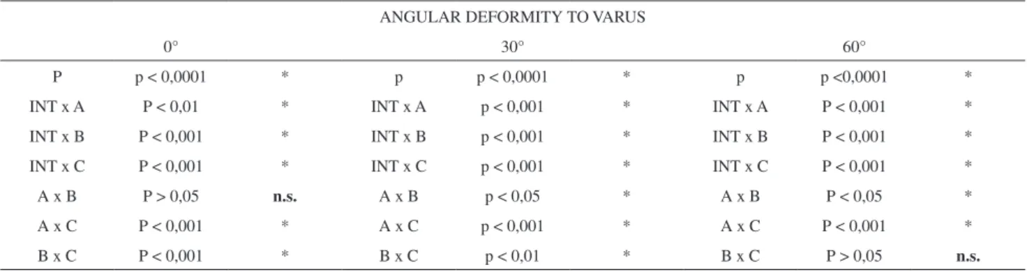
With regard to angular deformity in varus (Tables 1 and 3), we observed that in comparison A (LCL section) x B (LCL and PFC section), there was no signiicant difference between the two situations when the knee was tested in extension. In other words, the PFC section did not increase angular deformity in varus with the knee in extension after the previous section of LCL. At 30 and 60 degrees of lexion, the differences were signiicant.
Figure 3 - KRATOS® testing machine used for the biomechanical study. A.
Mechanical testing device. B. Data acquisition system. C. Testing machine pivo (arrow indicates direction of movement). D. Load cell (100 kgf). E. Knee ixed to the device. F. Base image of the knee ixed to the device.
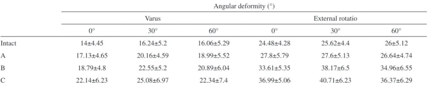
Table 1 - Angular deformity of the knee during the application of force in varus and in external rotation (mean ± standard deviation).
Angular deformity (°)
Varus External rotatio
0° 30° 60° 0° 30° 60°
Intact 14±4.45 16.24±5.2 16.06±5.29 24.48±4.28 25.62±4.4 26±5.12
A 17.13±4.65 20.16±4.59 18.99±5.52 27.8±5.79 27.6±5.13 26.64±4.74
B 18.79±4.8 22.55±5.2 20.89±6.04 33.61±5.35 38.17±6.5 34.96±6.55
We also observed that, in comparison to B x C, there was no signiicant difference among the values obtained with the knee at 60° of lexion. In other words, the PLC section did not increase angular deformity in varus with the knee at 60° of lexion after the previous section of the LCL and of the PFC. In extension and at 30° of lexion, the differences were signiicant (Figure 4).
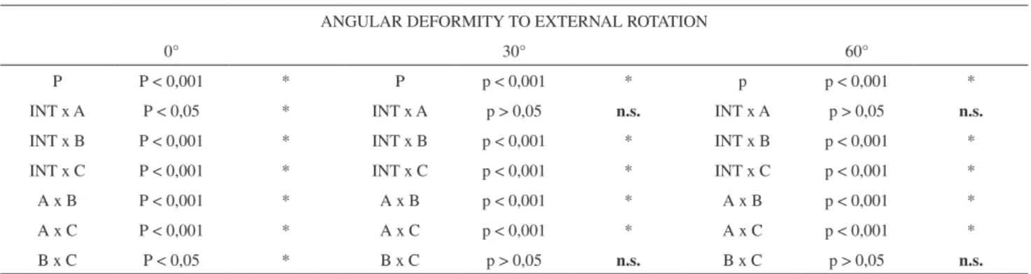
With regard to angular deformity in external rotation (Tables 1 and 5), we observed that in the INT x A comparison, no significant differences occurred when the knees were tested at 30° and at 60° of lexion. Hence, the LCL section did not increase angular deformity for external rotation in the abovementioned positions, despite having increased with the knee in extension. The B x C comparison did not present signiicant differences with the
knee at 30° and at 60° of lexion either. In other words, the PLC section did not increase angular deformity in external rotation with the knee at 30° and at 60° of lexion after the previous section of the LCL and of the PFC. In extension, the differences were signiicant (Figure 5).
Stiffness
With regard to stiffness for varus (Tables 2 and 4), there was no signiicant difference in the stiffness found comparing the INT x A situation when the knee was in extension and at 30° of lexion. At 60° of lexion, the difference was already signiicant, and the LCL section altered stiffness in varus. The B x C comparison did not present signiicant differences at any angle of lexion tested. The PLC section did not exhibit
Table 2 - Stiffness during the application of a deforming force in varus and external rotation (mean ± standard deviation).
Stiffness (N.nm/°)
Varus External rotatio
0° 30° 60° 0° 30° 60°
Intact 1414.63±293.93 1431.32±309.1 1535.69±313.02 522.65±48.48 544.97±62.32 562.44±94.21
A 1356.04±232.76 1381.52±268.92 1340.33±271.03 505.63±46.89 518.83±44.22 521.68±62.11
B 1276.52±193.03 1267.38±190.63 1179.53±206.25 446.33±31.75 421.14±43.74 342.71±62.52
C 1208.52±209.45 1186.19±200.02 1078.4±237.92 412.92±34.03 380.15±48.64 318.5±61.77
Table 3 - Comparative table and p values for the situations in varus testing to angular deformity.
ANGULAR DEFORMITY TO VARUS
0° 30° 60°
P p < 0,0001 * p p < 0,0001 * p p <0,0001 *
INT x A P < 0,01 * INT x A p < 0,001 * INT x A P < 0,001 *
INT x B P < 0,001 * INT x B p < 0,001 * INT x B P < 0,001 *
INT x C P < 0,001 * INT x C p < 0,001 * INT x C P < 0,001 *
A x B P > 0,05 n.s. A x B p < 0,05 * A x B P < 0,05 *
A x C P < 0,001 * A x C p < 0,001 * A x C P < 0,001 *
B x C P < 0,001 * B x C p < 0,01 * B x C P > 0,05 n.s.
Table 4 - Comparative table and p values for the situations in varus testing to stiffness.
STIFFNESS TO VARUS
0° 30° 60°
p p < 0,0001 * P p < 0,0001 * p P <0,0001 *
INT x A P > 0,05 n.s. INT x A p > 0,05 n.s. INT x A P < 0,001 *
INT x B p < 0,001 * INT x B p < 0,001 * INT x B P < 0,001 *
INT x C p < 0,001 * INT x C p < 0,001 * INT x C P < 0,001 *
A x B P < 0,05 * A x B p < 0,01 * A x B P < 0,01 *
A x C p < 0,001 * A x C p < 0,001 * A x C P < 0,001 *
altered stiffness in varus after the previous section of the LCL and of the PFC (Figure 6).
With regard to the stiffness for external rotation (Table 2 and 6), there was no signiicant difference in the INT x A comparison at any angle of lexion tested. The LCL section did not alter stiffness in external rotation in extension, at 30°, or at 60° of knee lexion. We failed to identify any signiicant difference in the B x C comparison at any angle
of lexion tested. The PLC section did not alter stiffness in external rotation after the previous section of the LCL and of the PFC (Figure 7).
DISCUSSION
Ligament lesions of the posteolateral corner of the knee continue to represent a challenge to orthopedic surgeons. In spite of all the papers that have already been published, there is still a great deal of uncertainty concerning the function
Figure 4 - Comparison of angular deformity to varus among groups with
0, 30 and 60 degrees of lexion. All values were signiicant, except where notated N.S. (not signiicant).
Figure 5 - Comparison of angular deformity to external rotation among
groups with 0, 30 and 60 degrees of lexion. All values were signiicant, except where notated N.S. (not signiicant).
Table 5 - Comparative table and p values for the situations in external rotation testing to angular deformity.
ANGULAR DEFORMITY TO EXTERNAL ROTATION
0° 30° 60°
P P < 0,001 * P p < 0,001 * p p < 0,001 *
INT x A P < 0,05 * INT x A p > 0,05 n.s. INT x A p > 0,05 n.s.
INT x B P < 0,001 * INT x B p < 0,001 * INT x B p < 0,001 *
INT x C P < 0,001 * INT x C p < 0,001 * INT x C p < 0,001 *
A x B P < 0,001 * A x B p < 0,001 * A x B p < 0,001 *
A x C P < 0,001 * A x C p < 0,001 * A x C p < 0,001 *
B x C P < 0,05 * B x C p > 0,05 n.s. B x C p > 0,05 n.s.
Figure 6 - Comparison of stiffness to varus among groups with 0, 30 and
60 degrees of lexion. All values were signiicant, except where notated N.S. (not signiicant).
Figure 7 - Comparison of stiffness to external rotation among groups with
and biomechanics of structures of the posterolateral corner of the knee.
The loads used, namely 15 N.m for varus and 6 N.m for external rotation, did not induce any lesion on the part during the tests.
Results for angular deformity in varus
After the tests conducted in varus to measure angular deformation, we veriied that the lateral collateral ligament was important in restricting this movement at all the lexion angles, particularly when the knee was at 30 and at 60 degrees of lexion. Several papers1,2,5,8 have reported similar
results, concluding that the LCL was important in the stabilization in varus at all degrees of lexion.
We also observed that the popliteoibular complex was not important for restriction in varus with the knee in extension. However, the popliteofibular complex section showed signiicant alteration in stabilization in varus with the knee at 30 and 60 degrees of lexion. Gollehon et al.1 observed that
the popliteus tendon section associated with that of the arcuate ligament generated varus increases at 90 degrees of lexion. Shahane et al.5 tested the “popliteus complex” in varus,
dividing it into two components: the popliteoibular ligament and the popliteus tendon. They concluded that the popliteus tendon section was not important in restriction for varus, but the popliteoibular ligament section was important at 60 and 90 degrees of lexion.
The two components were not isolated separately in our study, and we performed the unique section of what we call the popliteoibular complex. This complex involves two structures, and, when it is sectioned, both the popliteus tendon and the popliteofibular ligament are considered sectioned. Accordingly, our results should be compared with the section of the two associated structures (popliteus tendon and popliteoibular ligament).
Gollehon et al.1 and Shahane et al.5 argue that the
popliteus tendon presents a static and dynamic stabilization
function, and Shahane et al.5 declare that static stabilization
is produced by the popliteofibular ligament. When we section the popliteoibular complex, we are evaluating in a more objective manner the popliteoibular ligament, which loses its function in this situation. The study by Shahane et al.5 shows data that contributes toward this reasoning. In his
study, the popliteus tendon section did not alter stabilization in varus, whereas the popliteofibular ligament section remained important. In our study, because we sectioned these two components (popliteoibular complex) together, a signiicant difference occurred. We noted that this difference was to the detriment of the popliteoibular ligament section.
Pasque et al.6 and LaPrade et al.8 in turn reported that
the popliteus tendon and the popliteoibular ligament were not important in the stabilization of varus at any degree of lexion, contradicting our results and those obtained by Gollehon et al.1 and Shahane et al.5 We note that in the study
of Pasque et al.6 these structures were evaluated in knees that
had the LCL intact, whereas in our study, the LCL had been previously sectioned. Knowing that the LCL is the main knee stabilizer for varus1,2,5,8 and with it intact, the isolated
section of the popliteus tendon (in its joint portion with the popliteoibular ligament) did not lead to any increase in angular deformity. In the work of LaPrade et al.8 the
measurement of force on the popliteus tendon and on the popliteoibular ligament with varus application was also performed with the LCL intact, which limited deformity in varus and prevented these structures from being exposed to a greater deforming load.
From the data above, we conclude that, during the application of a deforming force in varus, the LCL would exhibit a lesion before the popliteoibular complex. A rupture of the PFC would only occur after the rupture of the LCL.
The results also show that the PLC was important as a restrictor for varus, particularly with the knee in extension; however, this importance was lost as the knee was lexed. In extension and at 30 degrees, there were significant alterations in the angular deformity, but the differences
Table 6 - Comparative table and p values for the situations in external rotation testing to stiffness.
STIFFNESS TO EXTERNAL ROTATION
0° 30° 60°
p P < 0,001 * p p < 0,001 * p p < 0,001 *
INT x A P > 0,05 n.s. INT x A p > 0,05 n.s. INT x A p > 0,05 n.s.
INT x B P < 0,001 * INT x B p < 0,001 * INT x B p < 0,001 *
INT x C P < 0,001 * INT x C p < 0,001 * INT x C p < 0,001 *
A x B P < 0,01 * A x B p < 0,001 * A x B p < 0,001 *
A x C P < 0,001 * A x C p < 0,001 * A x C p < 0,001 *
among the mean values were greater with the extended knee than at 30 degrees. With the knee lexed at 60 degrees, the alterations with the PLC section were not signiicant. This discovery can be explained by the fact that the PLC relaxes as the knee is lexed.
Results obtained for stiffness in varus
The results show that the LCL has no inluence on the stiffness of the posterolateral complex at 0 and 30 degrees of lexion. At 60 degrees of lexion, the stiffness of the LCL becomes significant. The PLC and the popliteus tendon relax, which impacts the decrease in stiffness of the posterolateral complex, thus making the LCL important for this property.
The PLC alone did not prove important for stiffness in the varus application tests at any degree of lexion, although it did interfere with the angular deformity for varus at 0 and 30 degrees of knee lexion.
Results obtained for angular deformity in external rotation
We observed that the lateral collateral ligament was important for posterolateral stabilization of the knee under external rotation when the latter was in extension. As the knee was lexed to 30 and 60 degrees, the lateral collateral ligament was not important for this function. Our results resemble those published by LaPrade et al.8
with respect to the function of the LCL as stabilizer for external rotation in extension, although they observed that the LCL also acts in the limitation of external rotation at 30 degrees of lexion. Wrobe et al.2 also observed that there
was an increase of external rotation with the LCL section, particularly when the knee was close to extension, in knees that previously had undergone sectioning of the ACL. The popliteofibular complex was important for stabilization under external rotation at all degrees of lexion when the LCL was previously ruptured. In extension, even after the LCL section (group A) was signiicant when compared with the intact knee in external rotation, the posterior section of the popliteoibular complex was signiicant once again. We conclude that the popliteoibular complex was important for external rotation in extension.
In the comparison of group A (LCL section) with the intact knee, there was no signiicant increase at 30 and 60 degrees of lexion, and in comparing groups A and B (LCL section + popliteus tendon), we observed a significant increase. We conclude that this increase occurred to the detriment of the popliteoibular complex section, which is also important in stabilization for external rotation at 30
and 60 degrees. These discoveries corroborate the study of Shahane et al.,5 who, after the popliteus tendon section,
observed an increase in external rotation at 60 and 90 degrees and, after the popliteofibular ligament section, observed an increase in external rotation at all the degrees of lexion tested. In our study, we evaluated whether the popliteoibular complex section involves the joint section of the popliteus tendon and of the popliteoibular ligament. For this reason, our results should be compared with groups where the two structures were sectioned, such as in the study by Shahane et al.,5 who obtained results similar to
ours. LaPrade et al.8 discussed the interaction between the
function of the LCL and that of the popliteus tendon and of the popliteoibular ligament, where the LCL acts mainly close to extension, and the latter two acquire importance as the knee is lexed. This synchronism was also observed in our experiment.
The PLC proved important in the restriction of external rotation only with the knee in extension. We can justify this fact through the analysis of articular biomechanics; as the knee is lexed, relaxation occurs. In this manner, the PLC would cease to play an important role already at 30 degrees of lexion, and continuing similarly at 60 degrees of lexion.
Results obtained for stiffness in external rotation
The results show that after the popliteoibular complex was already sectioned, the associated section of PLC did not show any difference at any degree of lexion. We assume that the popliteoibular complex is the most important structure in external rotational stabilization of the knee. Jointly observing the properties of angular deformity and stiffness, we identified that the only situations where significant differences occurred were those in which we were testing an intact popliteoibular complex with another in which this complex had been sectioned.
CONCLUSIONS
The results of this biomechanical study to evaluate the relative importance of the different static stabilizers of the posterolateral corner of the knee allow us to conclude that the following:
1. The lateral collateral ligament was important in knee stabilization for varus at all the lexion angles tested (0°, 30° and 60°);
3. The posterolateral capsule during extension was impor-tant for stabilizing the varus and for external rotation.
However, at 30 degrees of lexion, it was only important for varus.
REFERENCES
1. Gollehon DL, Torzilli PA, Warren RF. The role of the posterolateral and cruciate ligaments in the stability of the human knee: A biomechanical study. J Bone Joint Surg Am. 1987;69:233-42.
2. Wroble RR, Grood ES, Cummings JS, Henderson JM, Noyes FR. The role of the lateral extraarticular restraints in the anterior cruciate ligament-deicient knee. Am J Sports Med 1993;21:257-63.
3. Veltri DM, Deng XH, Torzilli PA, Maynard MJ, Warren RF. The role of the popliteoibular ligament in stability of the human knee: a biomechanical study. Am J Sports Med. 1996;24:19-27.
4. Kaneda Y, Moriya H, Takahashi K, Shimada Y, Tamaki T. Experimental study on external tibial rotation of the knee. AmJ Sports Med 1997;25:796-800.
5. Shahane SA, Ibbotson C, Strachan R, Bickerstaff DR. The popliteoibular ligament: An anatomical study of the posterolateral corner of the knee. J Bone Joint Surg (Br) 1999;81:636-42.
6. Pasque C, Noyes FR, Gibbons M, Levy M, Grood E. The role of the politeoibular ligament and the tendon of popliteus in providing stability in the human knee. J Bone Joint Surg Br. 2003;85:292-8.
7. Sugita T, Amis AA. Anatomic and biomechanical study of the lateral collateral and popliteoibular ligaments. Am J Sports Med. 2001;2:466-72.
8. LaPrade RF, Tso A, Wentorf FA. Force measurements on the ibular collateral ligament, popliteoibular ligament, and popliteustendon to applied loads. Am J Sports Med. 2004;32:1695-701.
9. Hughston JC, Andrews JR, Cross MJ, Moschi A. Classiication of knee ligament instabilities. Part II: The lateral compartment. J Bone Joint Surg Am. 1976;58:173-9.
10. Yanasse RH, Cavallari CE, Chaud FL, Hernandez AJ, Mizobuchi RR, Laraya MH. Measurement of tibial slope angle after medial opening wedge high tibial osteotomy: case series. Sao Paulo Med J. 2009;127:34-9.
11. Favano A, Santos-Silva PR, Nakano EY, Pedrinelli A, Hernandez AJ, Greve JM. Peptide glutamine supplementation for tolerance of intermittent exercise in soccer players. Clinics. 2008;63:27-32
12. Hernandez, AJ. Correlação das propriedades biomecânicas dos ligamentos do joelho com seus parâmetros antropométricos [tese]. São Paulo: Faculdade de Medicina, Universidade de São Paulo;1994.
13. Kokron AEV. Avaliação biomecânica da estabilidade da reconstrução na lesão isolada do ligamento cruzado posterior com um e dois feixes de enxerto. Estudo experimental com tendão do músculo quadríceps da coxa e com tendões dos músculos semitendíneo e grácil [tese]. São Paulo: Faculdade de Medicina, Universidade de São Paulo; 2000.
14. Pereira JARM. Estudo biomecânico da inluência da espessura do enxerto e da técnica de dois feixes na reconstrução do ligamento cruzado posterior [dissertação]. São Paulo: Faculdade de Medicina, Universidade de São Paulo;2004.