112
Revista da Sociedade Brasileira de Medicina Tropical 49(1):112-114, Jan-Feb, 2016 http://dx.doi.org/10.1590/0037-8682-0125-2015
Short Communication
Corresponding author: Dr. Pedro Eduardo Almeida da Silva.
e-mail: [email protected]
Received 13 April 2015
Accepted 1 June 2015
The BACTEC MGIT™ 320 system as a laboratory tool
to diagnose tuberculosis in a Brazilian hospital
with a high prevalence of HIV infection
Clarice Brinck Brum
[1], Daniela Fernandes Ramos
[1], Fernanda de Souza Abilleira
[1],
Ana Bárbara Scholante Silva
[1], Andrea von Groll
[1]and Pedro Eduardo Almeida da Silva
[1][1]. Faculdade de Medicina, Núcleo de Pesquisa em Microbiologia Médica, Universidade Federal do Rio Grande, Rio Grande, Rio Grande do Sul, Brasil.
ABSTRACT
Introduction: The World Health Organization endorses the BACTEC Mycobacterial Growth Indicator Tube (MGIT)™ system as a rapid, sensitive, and specifi c method to diagnostic of tuberculosis.Here, we compared the performance of this system against Ogawa-Kudoh cultures and microscopy. Methods: A total of927 samples were obtained between December 2011 and December 2013 from 652 cases of suspected tuberculosis at the School Hospital of the Federal University of Rio Grande in Brazil. Results: The MGIT system confi rmed tuberculosis in more cases in less time. Conclusions: The MGIT system is an effective tool for early diagnosis of tuberculosis, especially in patients with HIV/AIDS.
Keywords: Diagnosis. HIV coinfection. Tuberculosis.
A major factor driving the spread of tuberculosis is the considerable delay in diagnosis and treatment(1). Thus, early detection is essential(2). Tuberculosis can be diagnosed by sputum smear microscopy for acid-fast bacilli. However, this method has poor sensitivity, especially in samples with low bacterial load(3). On the other hand, bacilli cultures are more sensitive than microscopy, are highly specifi c, and are thus the gold standard for detecting Mycobacterium tuberculosis. Moreover, isolation of the microorganism allows identifi cation to species, determination of susceptibility to antimicrobials, and molecular studies. However, culture on solid media requires 20-40 days. In contrast, systems such as the Mycobacterial Growth Indicator Tube (MGIT), which are commercially available and are based on liquid media, require only 10-15 days(2).
Despite its advantages as high accuracy and fastness to obtain the results, the BACTEC MGITTM 960 system is typically only used in reference laboratories with the required infrastructure. BACTEC MGITTM 320, a more compact version more suitable for satellite laboratories, has also been released. However, any new method or approach, regardless of sophistication, commercial availability, or in-house testing and development, should be evaluated through well-designed and well-controlled clinical trials in endemic, low-resource settings, where such methods are most needed(4) (5). One such setting is the coastal lagoon micro-region in the State of Rio Grande do Sul in Brazil, a high-priority city for tuberculosis
control, with estimated population over 240,000. In 2013, the incidence of tuberculosis was 57.4/100,000 in the city(6) and 43.2/100,000 in the state, whereas the national incidence was 35.4/100,000(7). Additionally, the incidence of patients living with HIV/AIDS was 9.1/100,000 in the state, the highest in the country(8), with 19.3% of HIV patients co-infected with
M. tuberculosis in 2013(7).
In this study, we evaluated BACTEC MGITTM 320 as a routine diagnostic tool for tuberculosis at the Dr. Miguel Riet Corrêa Jr. Hospital of the Federal University of Rio Grande/ Rio Grande do Sul in Brazil, a 204-bed reference hospital for the care of patients living with HIV/AIDS. Between December 2011 and December 2013, we obtained 447 pulmonary and 480 extrapulmonary samples from 652 patients with suspected tuberculosis, of whom 291 were HIV-positive. The patient population consisted of 253 females and 399 males 1-96 years. This study was grounded in ethical principles, and was approved by the Research Ethics Committee in the Area of Health of the Federal University of Rio Grande (Protocol 76/2012).
113
Brum CB et al. - Diagnosing tuberculosis in low-resource settings by BACTEC MGITTM 320
The authors declare that there is no confl ict of interest. CONFLICT OF INTEREST
FINANCIAL SUPPORT
REFERENCES
TABLE 1 - Ogawa-Kudoh and Mycobacterial Growth Indicator
Tube 320 cultures of 119 tuberculosis cases stratifi ed by microscopy
results.
MGIT OK
n % n %
Positive microscopy
positive culture 44 91.7 42 87.5
negative culture 4 8.3 6 12.5
total 48 100.0 48 100.0
Negative microscopy
positive culture 71 100.0 50 70.4
negative culture 0 0.0 21 29.6
total 71 100.0 71 100.0
MGIT: Mycobacterial Growth Indicator Tube. OK: Ogawa-Kudoh.
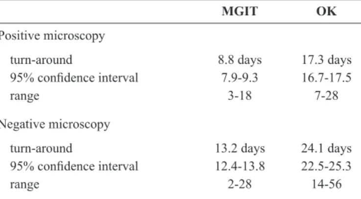
TABLE 2 - Days required to confi rm microscopy diagnosis by
Ogawa-Kudoh and Mycobacterial Growth Indicator Tube cultures.
MGIT OK
Positive microscopy
turn-around 8.8 days 17.3 days
95% confi dence interval 7.9-9.3 16.7-17.5
range 3-18 7-28
Negative microscopy
turn-around 13.2 days 24.1 days
95% confi dence interval 12.4-13.8 22.5-25.3
range 2-28 14-56
MGIT: Mycobacterial Growth Indicator Tube. OK: Ogawa-Kudoh.
growth once per week up to eight weeks, while MGIT tubes were monitored hourly up to 42 days in the automated BACTEC MGITTM 320 system. Growing isolates were confi rmed to be
M. tuberculosis by PCR of the IS6110 insertion region using
the primers INS-1 (5'-CGTGAGGGCATCGAGGTGGC) and INS-2 (5'-GCGTAGGCGTCGGTGACAAA)(11). Culture methods were compared in BioEstat version 5.0 using kappa index and turn-around time calculated at 95% confi dence interval.
Of 927 samples, 8.8% of Ogawa-Kudoh cultures were contaminated, as were 7.7% of MGIT cultures. Of 652 patients, 119 were confi rmed to have tuberculosis, of whom 60 were co-infected with HIV. Microscopy was positive to presence of acid-fast bacilli in 48 (40%) confi rmed cases. On the other hand, 92 (77.3%) were positive on Ogawa-Kudoh cultures, and two (1.7%) samples were contaminated. Finally, 115 (96.7%) of confi rmed cases were also positive on MGIT cultures. Of the 48 samples that were positive by microscopy, 42 and 44 were confi rmed by Ogawa-Kudoh and MGIT cultures, respectively. Notably, all 71 patients who were negative by microscopy were positive by MGIT culture, and 50 were also positive by Ogawa-Kudoh culture (Table 1). Among patients co-infected with
M. tuberculosis and HIV, MGIT was more sensitive (58/60)
than Ogawa-Kudoh culture (45/60) and microscopy (22/60). In general, cultures are used to increase diagnostic sensitivity or to test the sensitivity of other diagnostic techniques. Thus, cultures are expected to contribute to enhance the diagnosis of tuberculosis. Accordingly, we found that Ogawa-Kudoh and MGIT cultures accounted for 51% and 60% of positive diagnoses. respectively, as assessed by published methods(12) (13) (14). Notably, the kappa index was 0.9 between Ogawa-Kudoh and MGIT cultures, indicating excellent agreement (p < 0.0001).
Nevertheless, the results indicate that MGIT cultures were more sensitive than Ogawa-Kudoh cultures, and detected tuberculosis in 23 more patients. This result is consistent with previous studies of MGIT 960 in Brazil and other countries(12) (13). MGIT 320 and MGIT 960 require the same sample processing, as well as the same additional tests to confi rm infection with M. tuberculosis. However, the MGIT 320 compact system does not require stable electricity to maintain constant temperature during incubation, and is also more suitable for laboratories with lower caseload.
An important aspect of culture methods is turn-around, which has been reported to be shorter for MGIT (10-15 days) than for solid media (20-40 days)(2). In line with these observations, the gap between a positive test on microscopy and a confi rmed diagnosis was 8.8 days for MGIT and 17.3 days for Ogawa-Kudoh cultures (Table 2). Similarly, a negative result on microscopy was typically confi rmed in 13.2 days by MGIT culture, and 24.1 days by Ogawa-Kudoh. These data show that the time to diagnosis by MGIT is shorter than Ogawa-Kudoh culture by an average of 10 days.
In summary, our data demonstrate that use of mycobacterial cultures as a routine diagnostic tool may signifi cantly enhance and accelerate detection of M. tuberculosis, especially among patients living with HIV/AIDS. However, cost-effectiveness will have to be analyzed to fully assess the suitability of BACTEC MGITTM systems in satellite laboratories.
The study was supported by FAPERGS/MS/CNPq/SESRS process number: 1193-2551/13-6 and MCTI/CNPq/MS-SCTIE - Decit process number: 40/2012.
1. Dye C, Williams BG, Espinal MA, Raviglione MC. Erasing the world's slow stain: strategies to beat multidrug-resistant tuberculosis. Science 2002; 295:2042-2046.
2. Schito M, Peter TF, Cavanaugh S, Piatek AS, Young GJ,
Alexander H, et al. Opportunities and challenges for cost-effi cient
114
Rev Soc Bras Med Trop 49(1):112-114, Jan-Feb, 2016
3. Valença MS, Rocha JR, Ramis IB, Carrion LL, Madruga C, Macedo MB, et al. Improving tuberculosis control through the partnership between university and the health system. Rev Soc Bras Med Trop 2012; 45:491-495.
4. Palomino JC. Nonconventional and new methods in the diagnosis
of tuberculosis: feasibility and applicability in the fi eld. Eur Respir
J 2005; 26:339-350.
5. Huf G, Kritski A. Evaluation of the clinical utility of new diagnostic tests for tuberculosis: the role of pragmatic clinical trials. J Bras Pneumol 2012; 38:237-245.
6. Secretaria de Estado da Saúde do Rio Grande do Sul. Secretaria da Saúde - SES/RS. Plano Estadual de Saúde: 2012/2015 - 1ª edição - 2013. p 68.
7. Brasil. Ministério da Saúde. Secretaria da vigilância em Saúde -. Boletim Epidemiológico Nº 2, vol 44. 2014.
8. Ministério da Saúde. Boletim Epidemiológico - Aids e DST. Ano II - nº 1. 2013. p17.
9. Ministério da Saúde (MS). Brasil. Secretaria de Vigilância em Saúde. Departamento de Vigilância Epidemiológica. Manual de recomendações para o controle da tuberculose no Brasil / Ministério
da Saúde, Secretaria de Vigilância em Saúde, Departamento de Vigilância Epidemiológica. Brasília: MS; 2011.
10. Ministério da Saúde (MS). Manual Nacional de Vigilância Laboratorial da Tuberculose e outras Micobactérias. Brasília: MS; 2008.
11. Hermans PWM, Soolingen D van, Dale JW, Schuitema ARJ, Mcadam RA, Catty D, et al. Insertion Element IS986 from Mycobacterium tuberculosis: a Useful Tool for Diagnosis and Epidemiology of Tuberculosis. J Clin Microbiol 1990; 28:2051-2058.
12. Moreira ASR, Huf G, Vieira MA, Fonseca L, Ricks M, Kritski AL. Performance comparison between the mycobacteria growth indicator tube system and Löwenstein-Jensen medium in the routine detection of Mycobacterium tuberculosis at public health care facilities in Rio de Janeiro, Brazil: preliminary results of a pragmatic clinical trial. J Bras Pneumol 2013; 39:365-367.
13. Chihota VN, Grant AD, Fielding K, Ndibongo B, Zyl A van, Muirhead D, et al. Liquid vs. solid culture for tuberculosis: performance and cost in a resource-constrained setting. Int J Tuberc Lung Dis 2010; 14:1024-1031.