Prototype of a new tip developed to be coupled to dental light-curing
units for optimizing bonding of orthodontic brackets and accessories
Sergio Luiz Mota Júnior1, Márcio José da Silva Campos2, Marco Abdo Gravina3, Marcelo Reis Fraga4,
Robert Willer Farinazzo Vitral5
How to cite this article: Mota Júnior SL, Campos MJS, Gravina MA, Fraga MR, Vitral RWF. Prototype of a new tip developed to be coupled to dental light-curing units for optimizing bonding of orthodontic brackets and accessories. Den-tal Press J Orthod. 2013 Nov-Dec;18(6):112-6.
Submitted: September 19, 2011 - Revised and accepted: October 03, 2011
» The author reports no commercial, proprietary or financial interest in the prod-ucts or companies described in this article.
Contact address: Sergio Luiz Mota Júnior
Rua Humaitá, 10 – Apto 303 – Juiz de Fora /MG – CEP: 36.016-150 – Brazil E-mail: [email protected]
1 PhD Student in Health, Federal University of Juiz de Fora (UFJF). 2 Post-Doctorate Student in Health,UFJF. Professor of the graduate program in
Orthodontics, UFJF.
3 MSc in PhD in Orthodontics, State University of Rio de Janeiro (UERJ).
Associate professor of Orthodontics, School of Dentistry, and professor of the graduate program in Orthodontics, UFJF.
4 MSc in Orthodontics, Pontifical Catholic University of Minas Gerais
(PUC-MG). Doctorate student in Health, UFJF. Professor of the graduate program in Orthodontics, UFJF.
5 Associate professor and program director of the graduate program in
Orthodontics, UFJF.
Objective:development of a new device to be coupled to light-curing units for bonding orthodontic brackets and accessories, and test its efficacy in an in vitro mechanical trial. The inner surface of the device is mirrored and is based on physical concepts of light refraction and reflection. The main advantage of such device is the reduced clinical time needed for bonding and the low possibility of contamination during the process. Methods: One hun-dred and twenty specimens were used for testing the shear bond strength of brackets bonded with the device. The Adhesive Remnant Index (ARI) was also determined. The sample was divided into 2 groups. In group 1 a halogen light-curing unit was used while in group 2 a led light-curing unit was used. Each group was then subdivided. In subgroups H1 and L1, a conventional light guide rod was used while in subgroups H2 and L2 bonding was per-formed with the mirrored device coupled to the tip of the guide light rod. Results: The values obtained for the shear bond strength and the ARI in the subgroups were compared. Results showed that there was no statistically significant difference for the shear strength (p > 0.05) and the ARI (p > 0.05) between the subgroups. Conclusion: The tests of mechanical trials and the ARI analysis showed that the new device fulfilled the requirements for bonding orth-odontic accessories, and that the time for bonding was reduced to half, being necessary only one light exposure.
Keywords:Light-curing of dental adhesives. Orthodontic brackets. Patents. Shear strength.
Objetivo:desenvolver uma nova ponteira para ser acoplada aos aparelhos fotopolimerizadores utilizados para cola-gem de braquetes e acessórios ortodônticos, e testar sua da efetividade em ensaio mecânico in vitro. A ponteira é es-pelhada na superfície interna e baseia-se em conceitos físicos de refração e reflexão de luz. Apresenta como principal vantagem o menor tempo clínico durante o procedimento de colagem, reduzindo a possibilidade de contaminação durante o processo. Métodos: por meio de ensaio de resistência ao cisalhamento e determinação do índice rema-nescente de adesivo (IRA), testou-se a ponteira desenvolvida em 120 corpos de prova. A amostra foi dividia em dois grupos. No grupo 1, foi utilizado aparelho fotopolimerizador de fonte de luz halógena e, no grupo 2, fonte de LED. Cada grupo foi subdividido. Nos subgrupos H1 e L1, utilizou-se a ponteira convencional. Nos subgrupos H2 e L2 a colagem foi feita utilizando a ponteira desenvolvida para a polimerização do material de colagem. Resultados: os va-lores dos testes de cisalhamento e IRA para os subgrupos foram comparados entre si. Os resultados mostraram que não houve diferença estatisticamente significativa para o ensaio de resistência ao cisalhamento (p > 0,05) nem para o IRA (p > 0,05) entre os subgrupos. Conclusão: os testes de ensaio mecânico, assim como a análise do IRA, mostraram que a nova ponteira desenvolvida cumpriu os requisitos necessários à colagem dos acessórios ortodônticos, e que o tempo de colagem foi reduzido pela metade, sendo necessária uma só incidência.
INTRODUCTION
The light-emitting sources for light curing of composite resins that are available in the market are halogen lamps, blue light-emitting diodes (LEDs),
argon laser and xenon plasma arc.3,20 Although the
latter reduce curing time to one quarter23 and one
third,24 respectively, in comparison to that afforded
by conventional curing units, the machines are costly,
which hampers their use in private dental offices.23,24
LED is the acronym for Light-Emitting Diode, a semiconductor diode which, when energized, emits visible light through luminescence and not through a heated filament, consuming less power and
produc-ing less heat.18,19,20 Halogen light curing units have
been replaced by LED units2,18 due to its small
num-ber of components and lower power consumption.6,18
In Orthodontics, bonding brackets and accessories
is time-consuming3 and humidity control is difficult,
which may lead to contamination of the enamel
sur-face and consequent bonding failure.21
Based on the present conditions for bonding brackets and orthodontic accessories, combined with
concepts of Optical Physics, this study aimed at de-veloping and testing a new model of curing unit-ad-justed tip, with the purpose of reducing time, achiev-ing better quality and reducachiev-ing power consumption during the bonding procedure in Orthodontics.
MATERIAL AND METHODS
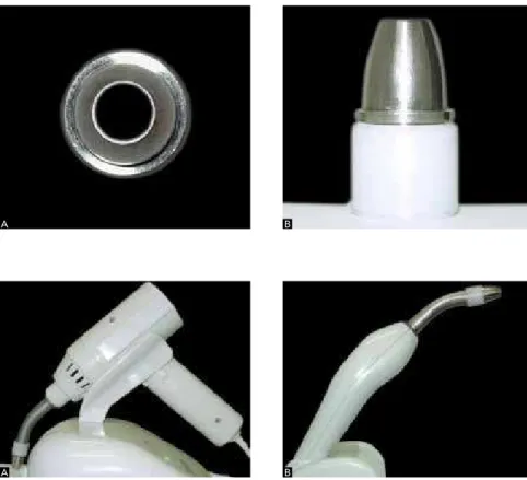
Based on the existing models of curing units, of which tip contains an optical fiber filament, we de-veloped a mirrored concave tip, based on the fact that spherical concave mirrors are polished caps which are reflective in their inner surfaces. The tip we devel-oped (Fig 1A – upper view, 1B – lateral view) is ad-justed to the tips of conventional curing units (Fig 2). The filing of the patent application was registered with the National Institute of Industrial Property (Instituto Nacional da Propriedade Industrial – INPI) un-der the number 020110074054.
The Committee of Ethics in Animal Experi-mentation of the Federal University of Juiz de Fora (UFJF) approved this research project (protocol nº 025/2010).
Figure 1 - A) Upper view, B) Lateral view of the new developed tip.
Figure 2 - A) Halogen light curing unit with
coupled tip; B) LED curing unit with coupled tip.
A
A
B
In order to assess the performance of the tip in orthodontic bonding, we carried out the mechanical trial of shear bond strength and the analysis of the adhesive remaining index (ARI) due to their mor-phological and histological similarity to human teeth, 120 bovine mandibular incisors were used in this in
vitro study.11,17,22 The teeth were selected with a
ste-reomicroscope (Stemi 2000 – C, Zeiss), at the Mod-ern Physics Department of the Sciences Institute of the Federal University of Juiz de Fora (UFJF).
Two groups of 60 teeth each were formed. Group 1 comprised teeth of which bracket bonding was per-formed with a halogen-light curing unit (Dabi Atlan-te Ultralux), while for group 2, a LED-source curing unit (Dabi Atlante Ultraled) was used. Each group was subdivided in 2 subgroups, with 30 teeth each. In subgroups H1 and L1, the conventional tip was used, while in subgroups H2 and L2 the experimental tip we developed was used.
Bracket bonding was performed according to the instructions of the manufacturer of the composite
resin (3M Transbond XT®). 120 brackets were used
(Dental Morelli, reference 10.30.201).
In subgroup H1, the mesial and the distal surfaces were exposed to light for 10 seconds each. In sub-group L1, the mesial and distal surfaces were exposed to light for 5 seconds each. Light exposure was 10 sec-onds in subgroup H2 and 5 secsec-onds in subgroup L2. Bonding in group 1 happened with the light held 5 mm away from the bracket-tooth interface, with an incidence parallel to the bonding surface. In group 2, the tip was positioned so as to embrace the bracket in a single incidence.
The bodies of proof for testing shear resistance
were assembled15 in a Universal Testing Machine,
EMIC – DL 10000, equipped with a 50Kgf Trd 21 load cell, software Tesc version 3.04 of the Military Institute of Engineering– Rio de Janeiro – LEM DE/4. After the mechanical trial was performed, the ARI for each body of proof was determined with the Stemi 2000 – C stereomicroscope (Zeiss).
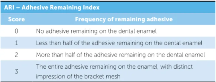
The ARI indices were classified into 4 scores,1 as
shown in Table 1.
RESULTS
Table 2 shows mean values and standard deviation of each subgroup (H1, H2, L1 and L2).
Student’s t test was used for comparison between subgroups. The p value for comparison between H1-H2 and L1-L2 was 0.365 and 0.176, respectively, thus, showing no statistically significant difference between the subgroups.
Figure 3 shows the distribution of values regard-ing the shear resistance test between groups.
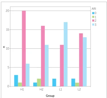
The ARI distribution for each group is shown in Figure 4. The Kruskal-Wallis test was used for ARI statistical analysis. With p > 0.05, no statistically sig-nificant difference between groups was found.
ARI – Adhesive Remaining Index
Score Frequency of remaining adhesive
0 No adhesive remaining on the dental enamel
1 Less than half of the adhesive remaining on the dental enamel
2 More than half of the adhesive remaining on the dental enamel
3 The entire adhesive remaining on the enamel, with distinct
impression of the bracket mesh
Table 1 - Criteria for using ARI, according. (Source: Årtun et al,1 1984).
Table 2 - values of shear resistance tests in MPa.
Subgroup n Mean ± SD
H1 30 9.89 ± 5.36
H2 30 9.01 ± 5.61
L1 30 7.01 ± 4.26
L2 30 7.63 ± 5.48
Total 120
Figure 3 - Box plot diagram of shear resistance in MPa for each subgroup.
T
ension
Group
0.00 5,00 10.00 15,00 20.00 25,00
H1 H2 L1 L2
98 107 111 64
during the procedure.5,7,10,12,13,14,16 With the new tip, bonding time was reduced, consequently reducing errors caused by saliva contamination.
For subgroups H1 and L1, variation of light expo-sure time was in accordance with the recommenda-tions given by the manufacturer of the 3M Transbond
XT® composite resin, while for subgroups H2 and L2
it was half the time proposed by the manufacturer. The recommended times are: 20 seconds for an halo-gen source (10 seconds for the mesial surface and 10 seconds for the distal surface) and 10 seconds for a LED source (5 seconds for the mesial surface and 5 seconds for the distal surface).
Bond resistance corresponds to bond strength divided by the bonding interface area (mm²). The bracket/adhesive system must resist to a minimum force of 6 MPa (megapascal) to be considered
clini-cally successful.4,8,9,14 All groups in this study had a
mean value greater than what is required for orth-odontic success.
As for the ARI assessment, the Kruskal-Wallis test showed no statistically significant difference between groups (p > 0.05). The majority of bodies of proof from each subgroup scored 2 or 3, with most of the adhesive remaining on the enamel surface, thus indi-cating that rupture occurred in the adhesive/bracket interface and not in the enamel/adhesive interface, as expected after adequate light curing.
Therefore, the shear test results and the ARI re-sults proved both bonding techniques, either carried out with the conventional tip or with the tip devel-oped in this study to be efficient.
CONCLUSIONS
1 – Both the mechanical trials and the ARI analy-sis showed that the new tip met the necessary require-ments for bonding orthodontic accessories;
2 – Bonding time was reduced by half, and only a single incidence was necessary.
DISCUSSION
Aiming at reducing orthodontic bonding time, we developed a tip that can be coupled to curing units. This device is based on the concepts of curing unit light refraction and relection. When visible light reaches the end of the optical iber of the curing unit, it changes from one medium (optical iber) to another (air), and the light ray is refracted into a medium with a lower refractive index. This refracted ray is no longer dissipated across the medium, but relected by the in-ner mirrored wall of the new device. Because the new tip converges towards the bracket-tooth end, the light rays reach the composite resin in the mesial, distal, in-cisal/occlusal and gingival directions simultaneously, which reduces curing time up to ¼ of the time recom-mended by the manufacturer of traditional tips.
One of the disadvantages of using light-curing composite resins is the high number of steps that are necessary during bonding, which increases working time and the likelihood of errors and contamination
Figure 4 - Bar chart for each group, with regard to ARI incidence.
Group
n
H1 H2 L1 L2
ARI 0 1 2 3 20
15
10
5
1. Årtun J, Bergland S. Clinical trials with crystal growth conditioning as an alternative to acid-etch enamel pretreatment. Am J Orthod. 1984;85(4):333-40.
2. Barghi N, Fischer DE, Pham T. Revisiting the intensity output of curing lights in private dental oices. Compend Contin Educ Dent. 2007;28(7):380-4.
3. Bishara SE, Gordan VV, VonWald L, Olson ME. Efect of an acidic primer on shear bond strength of orthodontic brackets. Am J Orthod Dentofacial Orthop. 1998;114(3):243-7.
4. Bishara SE, VonWald L, Lafoon JF, Jakobsen JR. Efective of altering the type of enamel conditioner on the shear bond strength of a resin-reinforced glass ionomer adhesive. Am J Orthod Dentofacial Orthop. 2000;118(3):288-94.
5. Bishara SE, Lafoon JF, Vonwald L, Warren JJ. The efect of repeated bonding on the shear bond strength of diferent orthodontic adhesives. Am J Orthod Dentofacial Orthod. 2002;121(5):521-5.
6. Bullough JD. LED lighting systems. Lighting Answers. NLPIP. 2003 [Acesso 15 set 2011];7(3):1-23. Disponível em: http://www.lrc.rpi.edu/ programs/nlpip/lightingAnswers/led/abstract.asp.
7. Cacciafesta V, Sfondrini MF, Baluga L, Scribante A, Klersy C. Use of a self-etching primer in combination with a resin-modiied glass ionomer: efect of water and saliva contamination on shear bond strength. Am J Orthod Dentofacial Orthop. 2003;124(4):420-6.
8. Cacciafesta V, Sfondrini MF, Barina E, Scribante A, Garino F, Klersy C. Efect of diferent light sources and guide on shear bond strength of brackets bonded with 2 adhesives systems. Am J Orthod Dentofacial Orthop. 2005;128(1):99-102.
9. Cacciafesta V, Sfondrini MF, Calvi D, Scribante A. Efect of luoride application on shear bond strength of brackets bonded with a resin-modiied glass-ionomer. Am J Orthod Dentofacial Orthop. 2005;127(5):580-3.
10. Cacciafesta V, Sfondrini MF, Scribante A, De Angelis M, Klersy C. Efect of blood contamination on shear bond strength of brackets bonded with a self-etching primer combined with a resin-modiied glass ionomer. Am J Orthod Dentofacial Orthop. 2004;126(6):703-8.
11. Campos MIC, Campos CN, Vitral RWF. O uso de dentes bovinos como substitutos em dentes humanos em pesquisas odontológicas: uma revisão de literatura. Pesq Brasil Odontop Clin Odontol. 2008;8(1):127-32.
12. Correr Sobrinho L, Correr GM, Consani S, Sinhoreti MAC, Consani RLX. Inluência do tempo pós-ixação na resistência ao cisalhamento de bráquetes colados com diferentes materiais. Pesqui Odontol Bras. 2002;16(1):43-9.
REFERENCES
13. Dominguez GC, Tortamato A, Carvalho PAL, Bomfim RA, Horliana RF, Vigorito JW. Self-etching primer: resistência confiável na colagem de acessórios ortodônticos? Estudo clínico. Ortodontia SPO. 2005;38(1):10-5.
14. Dorminey JC, Dunn WJ, Taloumis LJ. Shear bond strength of orthodontic brackets bonded with a modiied 1-step etchand-and-primer technique. Am J Orthod Dentofacial Orthop. 2003;124(4):410-3.
15. Dutra GAA, Rocha JM, Fraga MR, Vitral RWF. Avaliação comparativa in vitro da resistência à força de cisalhamento apresentada pelo bráquete ceramico InVu. Pesq Bras Odontoped Clin Integr. 2009;9(2):173-9. 16. Eliades T, Eliades G, Brantley WA, Johnston WM. Polymerization
eiciency of chemically cured and visible light-cured orthodontic adhesives: degree of cure. Am J Orthod Dentofacial Orthop. 1995;108(3):294-301.
17. Fonseca RB, Haiter-Neto F, Fernandes-Neto AJ, Barbosa GA, Soares CJ. Radiosensity of enamel and dentin of human, bovine and swine teeth. Arch Oral Biol. 2004;19(11):912-22.
18. Onofre NML, Retamoso LB, Marchioro EM, Berthold TB. Atuação da luz halógena e do LED (light emitting diode) na resistência de união de brackets colados no esmalte dentário humano. Rev Odonto Ciênc. 2007;22(57):238-42.
19. Owens BM, Rodriguez KH. Radiometric and spectrophotometric analysis of third generation light-emitting diode (LED) light-curing units. J Contemp Dent Pract. 2007;8(2):43-51.
20. Rueggeberg FA. State-of-the-art: Dental photocuring – A review. Dent Mater. 2011;27(1):39-52.
21. Sayinsu K, Isik F, Sezen S, Aydemir B. Efect of blood and saliva contamination on bond strength of brackets bonded with a protective liquid polish and a light-cured adhesive. Am J Orthod Dentofacial Orthop. 2007;131(3):391-4.
22. Saleh F, Taymour N. Validity of using bovine teeth as a substitute for human counterparts in adhesive tests. East Mediterr Health J. 2003; 9(1-2):201-7.
23. Talbot TQ, Blankenau RJ, Zobitz ME, Weaver AL, Lohse CM, Rebellato J. Efect of argon laser irradiation on shear bond strength of orthodontic brackets: an in vitro study. Am J Orthod Dentofacial Orthop. 2000;118(3):274-9.