Interhemispheric Functional and Structural
Disconnection in Alzheimer
’
s Disease: A
Combined Resting-State fMRI and DTI Study
Zhiqun Wang1☯, Jianli Wang2☯, Han Zhang3, Robert Mchugh2, Xiaoyu Sun2, Kuncheng Li1,4,5*, Qing X. Yang2,6*
1Department of Radiology, Xuanwu Hospital of Capital Medical University, Beijing, China,2Center for NMR Research, Department of Radiology, Pennsylvania State University College of Medicine, Hershey, Pennsylvania, United States of America,3Center for Cognition and Brain Disorders and the Affiliated Hospital, Hangzhou Normal University, Hangzhou, Zhejiang, China,4Key Laboratory for Neurodegenerative Diseases (Capital Medical University), Ministry of Education, Beijing, China,5Beijing Key Laboratory of Magnetic Resonance Imaging and Brain Informatics, Beijing, China,6Department of Neurosurgery (George M. Leader Foundation Alzheimer’s Laboratory), The Pennsylvania State University College of Medicine, Hershey, Pennsylvania, United States of America
☯These authors contributed equally to this work.
*[email protected](KL);[email protected](QXY)
Abstract
Neuroimaging studies have demonstrated that patients with Alzheimer’s disease presented disconnection syndrome. However, little is known about the alterations of interhemispheric functional interactions and underlying structural connectivity in the AD patients. In this study, we combined resting-state functional MRI and diffusion tensor imaging (DTI) to inves-tigate interhemispheric functional and structural connectivity in 16 AD, 16 mild cognitive im-pairment (MCI), as well as 16 cognitive normal healthy subjects (CN). The pattern of the resting state interhemispheric functional connectivity was measured with a voxel-mirrored homotopic connectivity (VMHC) method. Decreased VMHC was observed in AD and MCI subjects in anterior brain regions including the prefrontal cortices and subcortical regions with a pattern of AD<MCI<CN. Increased VMHC was observed in MCI subjects in posterior
brain regions with patterns of AD/CN<MCI (sensorimotor cortex) and AD<CN/MCI
(occipi-tal gyrus). DTI analysis showed the most significant difference among the three cohorts was the fractional anisotropy in the genu of corpus callosum, which was positively associated with the VMHC of prefrontal and subcortical regions. Across all the three cohorts, the diffu-sion parameters in the genu of corpus callosum and VMHC in the above brain regions had significant correlation with the cognitive performance. These results demonstrate that there are specific patterns of interhemispheric functional connectivity changes in the AD and MCI, which can be significantly correlated with the integrity changes in the midline white matter structures. These results suggest that VMHC can be used as a biomarker for the degenera-tion of the interhemispheric connectivity in AD.
OPEN ACCESS
Citation:Wang Z, Wang J, Zhang H, Mchugh R, Sun
X, Li K, et al. (2015) Interhemispheric Functional and Structural Disconnection in Alzheimer’s Disease: A Combined Resting-State fMRI and DTI Study. PLoS ONE 10(5): e0126310. doi:10.1371/journal. pone.0126310
Academic Editor:Yong Liu, & National Laboratory of
Pattern Recognition, CHINA
Received:August 19, 2014
Accepted:March 31, 2015
Published:May 4, 2015
Copyright:© 2015 Wang et al. This is an open
access article distributed under the terms of the Creative Commons Attribution License, which permits unrestricted use, distribution, and reproduction in any medium, provided the original author and source are credited.
Data Availability Statement:The data are available
upon request because of constraints imposed by the local ethical committee. Readers may contact the corresponding author to request the data.
Funding:This work was supported by the NSF of
Introduction
Alzheimer's disease (AD), the most common form of dementia, presents with memory and cognitive decline. Mild cognitive impairment (MCI) is considered a precursor of AD; people with MCI are diagnosed with AD at a rate of 10–15% per year [1] compared to 2–3% per year for the general population of the same age range. It is unclear, however, how the AD pathologi-cal lesions initiated in the medial temporal lobes lead to prominent functional deficits in memory and ultimately to dementia. Deciphering such structural-functional relationships could shed light into the trajectory of the functional pathogenesis of AD and MCI from normal aging. It has been shown that AD patients can perform normally for some tasks that relied on intrahemispheric processing but perform poorly for the tasks that required interhemispheric communication [2], which suggests a plausible hypothesis that AD patients may present a deficit in the interhemispheric integration of information. This hypothesis is supported by morphologic MRI findings that the corpus callosum, the most important fiber tract for inter-hemispheric connectivity, has consistently exhibited marked atrophy in AD and MCI [3,4]. Diffusion tensor imaging (DTI) studies of the corpus callosum have demonstrated significant changes in fractional anisotropy (FA), mean diffusivity (MD), radial diffusivity (λ┴) as well as axial diffusivity (λk) in early AD and MCI [5–7]. Yet, the relationship between these structural changes and the functional deficits remains to be determined [8].
To begin to address this issue, one must have a quantitative measure for inter-hemispheric functional connectivity. Recently, resting-state functional connectivity (RSFC) has been ap-plied to investigate AD and MCI [9–15]. Using regions-of-interest-based functional connectiv-ity approach, reduced functional connectivconnectiv-ity related to the hippocampus [9,10] and posterior cingulate cortex (PCC) [11–13] has been observed in AD and MCI patients. By employing the independent component analysis (ICA) method, researchers have reported widespread disrup-tions of selective brain networks such as the default mode network (DMN) and attention net-work in AD and MCI [14,15]. Furthermore, functional connectivity is consistently found across homotopic sites in the two hemispheres [16,17]. Strong interhemispheric RSFC is a common characteristic of the brain’s intrinsic functional networks such as the DMN, memory and sensorimotor networks [9,18,19]. Thus, the interhemispheric RSFC can be potentially used for assessing the integration of information from the two hemispheres. At present, interhemi-spheric functional connectivity in AD and MCI remains barely explored.
Recently, a novel voxel-wise image analysis method called voxel-mirrored homotopic con-nectivity (VMHC) was proposed to assess RSFC between the two hemispheres. VMHC quanti-fies the RSFC between each voxel in one hemisphere and its mirrored counterpart in the other. Using this method, interhemispheric RSFC was found to increase in the sensorimotor regions and decrease in higher-order cognitive regions during normal aging [20]. Clinically, character-istic patterns of significant VMHC disruptions have been found in autism [21], cocaine addic-tion [22], schizophrenia [23], multiple sclerosis [24], and depression [25]. These findings suggested that specific patterns of interhemispheric disconnection could reflect the functional consequences of pathologic damages in the associated diseases. Thus, a specific pattern of inter-hemispheric connectivity change is anticipated in MCI and AD when cognitive impairments are significantly progressed. We hypothesize that there are measureable differences in inter-hemispheric connectivity in AD and MCI patients compared to the age-matched cognitive nor-mal (CN) healthy subjects. Furthermore, since there should be strong links between structural and functional connectivity in the human brain, we speculate that interhemispheric structural connectivity is disrupted by AD pathology, which will result in the loss of functional connectiv-ity. Recent studies indicate myelin can be directly damaged by amyloid-βplaques that are an important early event in the pathogenesis of AD [26]. To test this hypothesis, we examined the
Competing Interests:The authors have declared
interhemispheric connectivity in AD and MCI functionally using VMHC and structurally using DTI. We aimed to find a progressive VMHC disruption in the specific brain areas that cognitive and memory functions are depended on.
Materials and Methods
Subjects
Forty-eight right-handed subjects (16 AD, 16 MCI, and 16 age/gender-matched CN) were re-cruited and participated in the study at the memory clinic of a neurological institute. The study was approved by local Medical Research Ethics Committee. All subjects provided written in-formed consent prior to participation, consistent with the Declaration of Helsinki. All the AD and MCI patients underwent a complete physical and neurological examination standard labo-ratory tests, and an extensive battery of neuropsychological assessments.
The AD patients were clinically diagnosed by the AD specialists in the memory clinic and met the criteria for dementia from the Diagnostic and Statistical Manual of Mental Disorders 4th Edition [27], as well the criteria for possible or probable AD from the National Institute of Neurological and Communicative Disorders and Stroke/Alzheimer Disease and Related Disor-ders Association (NINCDS-ADRDA) [28].
Participants with MCI had memory impairment but did not meet the criteria for dementia. The criteria for the identification and classification of subjects with MCI were as follows: (a) impaired memory performance on a normalized objective verbal memory delayed-recall test; (b) recent history of symptomatic worsening in memory; (c) normal or near-normal perfor-mance on cognitive tests, including a Mini-Mental State Examination (MMSE) score>24, as well as normal activities described in a daily living scale; and (d) global rating of0.5 on the Clinical Dementia Rating (CDR) score [29], with a score of at least 0.5 on the memory domain [30,31]. The diagnosis of MCI fulfilled the new criteria of the MCI due to AD recommended by the National Institute on Aging-Alzheimer‘s Association workgroups [32].
The criteria for CN were as follows: (a) no neurological or psychiatric disorders; (b) no neu-rological deficiencies; (c) no significant abnormal findings in conventional brain MRI; (d) no cognitive complaints; (e) MMSE score of 28 or higher; and (f) CDR score of 0.
Data acquisition
Resting state fMRI data processing and statistical analysis
The rs-fMRI data were processed using statistical parametric mapping (SPM8) and Data Pro-cessing Assistant for Resting-State fMRI (DPARSF) toolkits [33]. The first four images of each rs-fMRI data set were discarded to remove the initial transient signal fluctuations. Subsequent images were corrected for slice-timing and re-aligned within the session to remove any minor head movements. The T1-weighted image was co-registered to the mean rs-fMRI image using rigid-body transformation, and then spatially normalized to the Montreal Neurological Insti-tute (MNI) space using nonlinear transformation and the SPM8 T1 template. The rs-fMRI images were spatially normalized in a spatial resolution of 3 × 3 × 3 mm using the same nor-malization parameters as the T1 image, then smoothed with an 8 × 8 × 8 mm full width at half maximum (FWHM) Gaussian kernel. Linear detrending and temporal bandpass filtering (0.01–0.08 Hz) were applied to reduce the effect of low-frequency drifts and high-frequency physiological noise. Finally, the effects from several nuisance variables including six head mo-tion parameters, global mean signal, cerebrospinal fluid signal and white matter signal were re-moved by multiple linear regression analysis.
For VMHC analysis, a left-right hemisphere symmetric brain template was generated from the 48 subjects to remove the geometric differences between the two hemispheres. First, a mean T1 image was generated by averaging the 48 spatially normalized T1 images. Next the symmetric brain template was obtained by flipping the left and right hemispheres along the midline of the x-axis and averaged with the original image to create the final template. Then the T1 image from each individual subject that had been normalized to the MNI space was co-registered nonlinearly to this group-specific symmetric brain template. The same transforma-tion was then applied to the rs-fMRI images. Homotopic RSFC between each pair of symmetric voxels in left-right hemispheres was calculated with a Pearson’s correlation and then Fisher-Z transformed for further statistical analysis. The connectivity values in each pair of voxels were the same. For group analysis, the voxels within a range of x = ± 4.5 mm from the midline were excluded from the VMHC maps to reduce the blurring artifacts near the midline. The average VMHC map from each cohort was evaluated with a voxel-based one-sample t-test [Family-wise error (FWE) corrected,p<0.001, extent threshold = 10]. Group comparisons of VMHC were conducted using a one-way analysis of variance (ANOVA) with age and total grey matter volume as the nuisance covariates (p<0.01, corrected using the AFNI AlphaSim program (http://afni.nimh.nih.gov/pub/dist/doc/manual/AlphaSim.pdf) with a cluster size>97 voxels, FWHM = 8 mm, and cluster radius connection = 5). The total grey matter volume that used in the data analysis was the ratio between the brain grey matter volume and the intracranial vol-ume from each subject. The brain and intracranial volvol-umes were measured from the segmented T1 images. The intracranial volume equals the total volume of grey matter, white matter, and cerebrospinal fluid.
DTI data processing and statistical analysis
subject and then used in further post-hoc analysis with IBM SPSS Statistics for Windows Ver-sion 21. Correlation analyses were conducted among regional diffuVer-sion parameters, regional VMHCs and MMSE scores for all the subjects and for each subject group. Since age is both a significant risk factor for AD and correlates with diffusion parameters in normal aging [34], age was used as a confounding factor in the correlation analysis.
Results
Clinical and neuropsychological examination
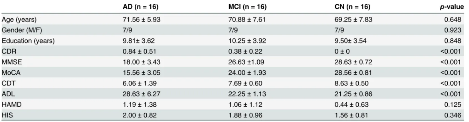
Clinical and demographic data of the three cohorts were shown inTable 1. The MMSE, CDR, Montreal Cognitive Assessment (MoCA), Clock Drawing Task (CDT) and Activity of Daily Living scale (ADL) scores were significantly different among the three groups. No significant differences were found in gender, age, education, Hamilton Depression Scale (HAMD), or Hachinski Ischemic Score (HIS) among cohorts.
Interhemispheric functional connectivity in the CN subjects
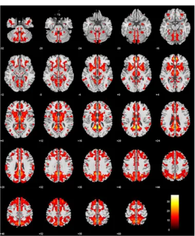
Fig 1shows the VMHC map of the CN group representing the interhemispheric connectivity in the brain. Each pair of voxels symmetrically located on each hemisphere in the VMHC map represents the correlation coefficient between the time series of the rs-fMRI data from the two voxels. The VMHC map from CN revealed strong interhemispheric connectivities in the brain regions known to be important for cognition, including the anterior and posterior cingulate cortex (ACC and PCC), dorsolateral prefrontal cortex (DLPFC), orbitofrontal cortex (OFC), sensorimotor cortex (SMC), parietal and occipital cortices, hippocampus and various temporal cortices, insula, basal ganglia, and thalamus. This VMHC map from the CN cohort is consis-tent with those obtained by a previous study [25].
Differences in interhemispheric functional connectivity among AD, MCI
and CN
Fig 2andTable 2show the differences among the three cohorts by voxel-based comparisons of the VMHC maps. Compared to the CN subjects, the AD patients had significantly weaker VMHC in the OFC, ACC, nucleus accumbens (NAcc), primary olfactory cortex (POC),
Table 1. Demographic information and neuropsychological test scores.
AD (n = 16) MCI (n = 16) CN (n = 16) p-value
Age (years) 71.56±5.93 70.88±7.61 69.25±7.83 0.648
Gender (M/F) 7/9 7/9 7/9 0.923
Education (years) 9.81±3.62 10.25±3.92 9.50±3.54 0.848
CDR 0.84±0.51 0.38±0.22 0±0 <0.001
MMSE 18.00±3.43 26.63±1.09 28.63±0.72 <0.001
MoCA 15.56±3.05 24.00±1.93 28.56±0.81 <0.001
CDT 6.06±1.39 7.69±0.60 8.63±0.50 <0.001
ADL 28.63±6.27 22.25±1.13 21.25±0.86 <0.001
HAMD 1.19±1.38 1.06±1.12 0.44±0.63 0.125
HIS 2.00±0.82 1.88±0.96 1.56±0.81 0.346
Data are presented as mean±std. p-value indicates the significance of the difference among the three study cohorts (one-way ANOVA). Abbreviations: CDR, Clinical Dementia Rating; MMSE, Mini-Mental State Examination; MoCA, Montreal Cognitive Assessment; CDT, Clock Drawing Task; ADL, Activity of Daily Living Scale; HAMD, Hamilton Depression Scale; HIS, Hachinski Ischemic Score.
putamen, caudate, and insula, suggesting a reduction of interhemispheric connectivity in these structures in AD (Fig 2A). The AD patients also had significantly weaker VMHC than the MCI subjects in the OFC, putamen, caudate, insula, sensorimotor cortex (SMC), and occipital gyrus (OcG) (Fig 2B). In contrast, the MCI subjects had significantly stronger VMHC in the SMC than the CN subjects (Fig 2C).
To further demonstrate the above variations in AD and MCI, we extracted the VMHC from those regions that showed significant differences among the three cohorts (Fig 2D–2H). The OFC, ACC and NAcc showed a trend of gradual decreasing VMHC from CN to MCI and then to AD. However, in the regions of SMC and OcG, VMHC in MCIs appeared significantly stronger than those in both ADs and CNs.
DTI and volume changes among AD, MCI and CN
Voxel-based analysis detected significant difference in FA between CNs and ADs in the genu of the corpus callosum (Fig 3A). The reduction of FA in the genu of the corpus callosum pre-sented a similar trend as seen in VMHC, i.e. AD<MCI<CN (Fig 3B). This trend of degener-ation of the corpus callosum genu was supported by the significantly increased local MD and λ┴(Fig 3C and 3D). There was no significant difference inλkamong any of the three groups (Fig 3E). We also found volume loss in the genu of the corpus callosum in the AD patients compared to CN (one-way ANOVA,t= -2.36,p= 0.02) (Fig 3F).
Fig 1. Normative VMHC map of the CN group (one-sample t-test, family-wise error corrected, p<0.001, extent threshold = 10).
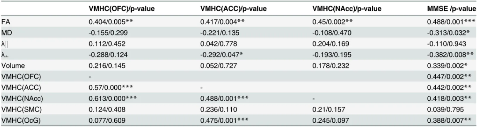
Table 3shows significant positive correlations between VMHC in the anterior brain regions (OFC, ACC, and NAcc) and FA value of the genu of the corpus callosum. Additionally, theλ┴ value of the genu was negatively correlated with VMHC in the ACC.
Fig 2. Cross-cohort comparisons of VMHC. A: The AD subjects showed significantly decreased VMHC in the OFC, ACC, POC, NAcc, putamen, caudate and insula compared to CN.B: The AD subjects showed significantly decreased VMHC in the OFC, putamen, caudate, insula, SMC, and OcG compared to MCI.C: The MCI subjects showed significantly increased VMHC in the SMC compared to CN.D-Hshow the locations of the five anatomical structures where the VMHC was significantly different across the three cohorts. The values in the bar graphs are z-scores transformed from the VMHC values.*represents statistical differences between groups (*,p0.05;***,p0.001).
doi:10.1371/journal.pone.0126310.g002
Table 2. VMHC differences between AD, MCI and CN.
Brain Regions Cluster- size Coordinates
(MNI)
t-score p-value corrected/ uncorrected
x y z
CN>AD OFC/ ACC/ POC/ NAcc putamen/ caudate/ insula 591 -24 39 -21 4 0.000/0.000 MCI>CN SMC (postcentral/ precentral gyrus) 116 -48 3 54 3.54 0.069/0.001
MCI>AD OFC/ putamen/ caudate/ insula 109 -33 39 -9 3.75 0.091/0.001
OcG (middle/ inferior occipital gyrus) 99 -45 -78 12 3.90 0.137/0.002 SMC (postcentral/ precentral gyrus) 218 -60 -18 39 3.58 0.002/0.000
ANOVA, uncorrected, p<0.01, extent threshold = 97. Coordinates are given in the left-posterior-inferior system. For the symmetric results, only the left side structures are listed. Abbreviations: MNI, Montreal Neurological Institute space; x, y, z, coordinates of cluster locations in the MNI space; OFC, orbitofrontal cortex; ACC, anterior cingulate cortex; NAcc, nucleus accumbens; POC, primary olfactory cortex; SMC, sensorimotor cortex; OcG, occipital gyrus.
Fig 3. DTI and volumetric differences among AD, MCI and CN. A:Voxel-based analysis showed significant difference of FA between AD and CN in the genu of the corpus callosum (family-wise error corrected,p<0.05, extent threshold = 10).B-F:Group comparisons revealed the patterns of diffusion parameters and volume changes in the genu of the corpus callosum (FA: AD<MCI<CN; MD: AD>CN / MCI;λ
┴: AD>CN / MCI;λk: no difference; Volume:
AD<CN/MCI).*represents statistical differences between groups (*,p0.05;***,p0.001)
doi:10.1371/journal.pone.0126310.g003
Table 3. Correlation between diffusion parameters, volume of the genu of corpus callosum, VMHC, and MMSE scores of all the subjects.
VMHC(OFC)/p-value VMHC(ACC)/p-value VMHC(NAcc)/p-value MMSE /p-value
FA 0.404/0.005** 0.417/0.004** 0.45/0.002** 0.488/0.001***
MD -0.155/0.299 -0.221/0.135 -0.108/0.470 -0.313/0.032*
λk 0.112/0.452 0.042/0.778 0.204/0.169 -0.110/0.943
λ┴ -0.288/0.124 -0.292/0.047* -0.193/0.195 -0.382/0.008**
Volume 0.216/0.145 0.052/0.727 0.178/0.232 0.339/0.002*
VMHC(OFC) - 0.447/0.002**
VMHC(ACC) 0.57/0.000*** - 0.442/0.002**
VMHC(NAcc) 0.613/0.000*** 0.488/0.001*** - 0.418/0.003**
VMHC(SMC) 0.124/0.408 0.236/0.110 0.21/0.157 0.039/0.795
VMHC(OcG) 0.077/0.609 0.475/0.001*** 0.245/0.097 0.388/0.007**
The values in the table are the correlation coefficients and corresponding p-values (partial correlation with age effect corrected).
*, p0.05;
**, p0.01;
***, p0.001.
Abbreviations: FA, fractional anisotropy; MD, mean diffusivity (×10–3 mm2/s);λk, axial diffusivity (×10–3 mm2/s);λ┴, radial diffusion (×10–3 mm2/s); VMHC, voxel mirror homotopic connectivity; OFC, orbitofrontal cortex; ACC, anterior cingulate cortex; NAcc, nucleus accumbens; SMC, sensorimotor cortex; OcG, occipital gyrus.
Relationship between interhemispheric connectivity and cognitive
performance
There was no significant correlation between either VMHC or the diffusion parameters in the genu of corpus callosum and MMSE within each subject group (r0.451,p0.080). For all the subjects, the DTI parameters including FA, MD,λ┴and volume in the genu of the corpus callosum were closely associated with MMSE scores (Table 3). The values of FA and volume in the genu of the corpus callosum were positively correlated with MMSE, while the values of MD andλ┴were negatively correlated with MMSE scores. Consistent with the structural connectiv-ity, the VMHCs in the brain regions of OFC, ACC, NAcc and OcG were positively correlated with MMSE scores.
Discussion
Major findings
By measuring the VMHC of rs-fMRI signals, we observed decreased VMHC in AD and MCI subjects in anterior brain regions including the prefrontal cortices and subcortical regions with a pattern of AD<MCI<CN. Increased VMHC was observed in the MCI in posterior brain re-gions with patterns of AD/CN<MCI (in SMC) and AD<CN/MCI (in OcG). In addition, DTI analysis among the three cohorts demonstrated that the most significant white matter change located in the genu of corpus callosum, which was revealed by the decreased FA and in-creased MD values in AD and MCI. Further analysis demonstrated that the VMHCs in the pre-frontal cortices and subcortical regions were positively associated with the diffusion
parameters in the genu of corpus callosum which demonstrated the association between struc-tural and functional connectivity. Finally, the VMHCs in the above regions and diffusion pa-rameters in the genu of corpus callosum showed significant correlations with the cognitive performance measured by the MMSE.
Resting-state interhemispheric functional connectivity
The AD and MCI subjects showed decreased VMHC in the prefrontal brain regions (OFC, ACC and POC), subcortical regions (NAcc, putamen, and caudate nucleus) and insula com-pared with the CNs (Fig 2AandTable 2). The significant trend of gradually decreased VMHC, i.e. AD<MCI<CN, indicates a progressive deterioration in interhemispheric connection from MCI to AD.
The prefrontal brain regions are known to be involved in functional deficits at a very early stage of AD. For example, the OFC is involved in AD cognitive functional deficits (spatial con-struction, decision-making, and working memory) [35], and grey matter atrophy in the OFC has frequently been reported in AD patients [36–38]. The ACC plays a key role in cognitive control and emotion [39], which often present dysfunction in AD patients [40,41]. The POC is the key structure for olfaction and also the early site of AD pathology. Olfactory deficits in odor detection, identification, and discrimination are prevalent in the early-stage of AD [42–
The hallmark AD pathology, neurofibrillary tangle and amyloid-βplaque deposition have been found in the OFC [53,54], ACC [55], POC [56,57], insula [58] and subcortical regions [59] at early stages of the disease. This suggests that these regions are preferentially vulnerable to the early attack from the disease, which is consistent with AD pathologic progression pat-terns [60]. Our results extended the findings in previous studies and provided new evidence that VMHC could be a sensitive measure for the interhemispheric functional connectivity for AD studies.
The MCI subjects had significantly stronger VMHC in the SMC than the CN subjects (Fig 2B and 2CandTable 2). The VMHC increase in the SMC of the MCI group may reflect a possi-ble recruitment in hemispheric cooperation as a compensatory mechanism [20,60,61]. It has been shown that older adults commonly recruit more sensorimotor regions to counteract the neurobiological changes due to aging [62]. One previous fMRI study using the VMHC method has demonstrated an increased interhemispheric connectivity in sensorimotor regions during aging [20]. Previous studies have shown that bimanual motor coordination in older adults is associated with increased functional brain connectivity [61–63]. One magnetoencephalography study also showed higher interhemispheric synchronization in MCI subjects than in healthy controls [64]. Our results are in agreement with the literature. Taking together such studies with different modalities, we hypothesize that MCI subjects, challenged by the pathological de-generations in the prefrontal brain areas, would strengthen interhemispheric cooperation of SMC regions to resist the cognitive decline. It may be a similar mechanism happens in the OcG. Before developing to AD, the MCI subjects might be able to recruit additional neural re-sources in the posterior region of OcG that are unaffected or less-affected by the disease, to compensate for interhemispheric disconnections in the disease-affected frontal brain regions. In addition, according to theTable 3, the interhemispheric RSFCs of the OcG were positively correlated with the cognitive performance as measured by MMSE, which suggested that stron-ger inter-hemispheric connectivity of the OcG contributed to the better cognitive performance, supporting the compensatory mechanism. In the study, we didn’t find the significant correla-tion between the SMC regions and the MMSE, we speculated that the sample size may be too small to find a robust association, In the future, to further study the underlying mechanism for this increased VMHC in the MCI, we will perform a longitudinal study of large sample of MCI subjects to provide the more valuable information to further confirm the compensatory mecha-nism in these regions.
It is known that the earliest regions of AD pathology are located in the medial temporal and parietal cortexes, which are involved in the default mode network. It is worthy to mention that the analytic approaches (i.e., VMHC) used in the current study are very different from those previously applied. Previous fMRI studies in AD consistently discussed brain functional con-nectivity (i.e., default mode network) based on whole brain concon-nectivity, i.e., all voxels are cor-related with each other. However, in the current study, we focused on the interhemispheric functional connectivity by measuring the voxel-mirrored homotopic connectivity. For the first time, we applied this method to AD study and provided the spatial pattern of the intrinsic in-terhemispheric connectivity changes across AD, MCI, and CN.
indicate myelin can be directly damaged by oligomerized amyloid-βplaques that are an impor-tant early event in the pathogenesis of AD. Furthermore, homeostatic responses to this myelin breakdown increase intracortical toxicity, which might explain the progressive neuronal dam-age in AD [26,66,67]. The distribution of myelin breakdown is not global. Rather, the regions that are most susceptible to pathological changes in AD are the late myelinated regions, which have fewer oligodendrocytes supporting a greater numbers of axons [26,68,69]. The myelin breakdown process in AD affects the late-myelinated anterior callosal subregions, resulting in a significant atrophy of the genu of the corpus callosum in AD patients [5,70]. Our findings of FA, MD andλ┴abnormalities in the genu of the corpus callosum in AD support the myelin breakdown retrogenesis hypothesis.
It is interesting that the VMHCs in the prefrontal (OFC, ACC) and subcortical regions (NAcc) were positively correlated with the FA values of the genu of the corpus callosum (Table 3). Since the prefrontal regions between the two hemispheres communicate through the genu of the corpus callosum [71], it was not an accident that white matter integrity was im-paired in this region of AD patients, which could disrupt the functional connectivities between prefrontal homotopic regions. Previous studies had provided significant evidence of the robust links between structural and functional connectivity in the human brain [72–74]. A previous study demonstrated a prominent loss of interhemispheric BOLD correlations after complete sectioning of the corpus callosum [75]. Another study confirmed that lack of normal callosal development can lead to deficits in functional connectivity related to cognitive impairments [76]. It is plausible to use the white matter integrity abnormalities in the genu of the corpus cal-losum to explain the reduction of VMHC in the prefrontal regions of AD and MCI. This direct correlation obtained by the two entirely different imaging modalities provided a mutual sup-port of the findings in the functional and structural alterations in AD.
Relationship between interhemispheric connectivity and cognitive
performance
The correlation analysis of all the subjects demonstrated that the interhemispheric RSFCs in specific brain regions and DTI parameters in the genu of corpus callosum were positively cor-related with the cognitive performance as measured by the MMSE. Since we did not observe significant correlation in each subject group, the observed correlation of all the subjects may be dominated by the group differences. Thus the correlation of reduced interhemispheric func-tional and structural connectivity with the cognitive decline in AD needs to be further studied. The brain relies on interhemispheric information transfer for mediating cognition function, which is subserved by the corpus callosum [77]. Previous studies have shown that even subtle degradation of the corpus callosum in neurologically impaired patients can be related to defi-cits in the transfer of information between the hemispheres [78]. Based on the above statement, the bilateral prefrontal regions (OFC and ACC), which are connected by the genu of corpus callosum, played a pivotal role in integrating information and mediating cognitive functions.
Further consideration
between reduced white matter integrity & VMHC. Our findings revealed the close relationship between diffusion changes in the genu of corpus callosum and VMHC changes in the MCI and AD. It would be better to analyze the anatomical connectivity between homotopic voxels to clarify the direct link between VMHC and DTI abnormalities. Future research will further in-vestigate the relationship between these modalities. Finally, the sample size is small and the findings may be altered in large populations. Thus a longitudinal study of large sample of MCI subjects will be beneficial to elucidate the impact of the disease on interhemispheric connectivi-ty, which may in turn provide a valuable imaging marker for the diagnosis of AD.
Conclusions
In this study we demonstrated significant changes of interhemispheric functional connectivity in the AD and MCI using VMHC analysis. The interhemispheric functional connectivity de-cline in the OFC, ACC, and NAcc was significantly correlated with the structural degeneration in the genu of the corpus callosum. These results suggest that VMHC can be used as a biomark-er for the degenbiomark-eration of the intbiomark-erhemisphbiomark-eric connectivity in AD.
Author Contributions
Conceived and designed the experiments: KL QXY. Performed the experiments: ZW. Analyzed the data: JW HZ RM. Contributed reagents/materials/analysis tools: XS HZ. Wrote the paper: ZW JW.
References
1. Petersen RC, Doody R, Kurz A, Mohs RC, Morris JC, Rabins PV, et al. Current concepts in mild cogni-tive impairment. Archives of neurology. 2001; 58:1985–1992. PMID:11735772
2. Lakmache Y, Lassonde M, Gauthier S, Frigon JY, Lepore F. Interhemispheric disconnection syndrome in Alzheimer's disease. Proc Natl Acad Sci U SA.1998; 95: 9042–9046. PMID:9671801
3. Chaim TM,Duran FL,Uchida RR, Périco CA, de Castro CC, Busatto GF, et al. Volumetric reduction of the corpus callosum in Alzheimer's disease in vivo as assessed with voxel-based morphometry. Psy-chiatry Res. 2007; 154:59–68. PMID:17174533
4. Wang PJ, Saykin AJ, Flashman LA, Wishart HA, Rabin LA, Santulli RB, et al. Regionally specific atro-phy of the corpus callosum in AD, MCI and cognitive complaints. Neurobiol Aging. 2006; 27:1613–
1617. PMID:16271806
5. Di Paola M, Di Iulio F, Cherubini A, Blundo C, Casini AR, Sancesario G, et al. When, where, and how the corpus callosum changes in MCI and AD: a multimodal MRI study. Neurology. 2010; 74:1136–
1142. doi:10.1212/WNL.0b013e3181d7d8cbPMID:20368633
6. Preti MG,Baglio F,LaganàMM,Griffanti L, Nemni R, Clerici M, et al. Assessing corpus callosum changes in Alzheimer's disease: comparison between tract-based spatial statistics and atlas-based tractography. PLoS One. 2012; 7: e35856. doi:10.1371/journal.pone.0035856PMID:22545143 7. Xie S,Xiao JX,Gong GL,Zang YF, Wang YH,Wu HK, et al. Voxel-based detection of white matter
abnor-malities in mild Alzheimer disease. Neurology. 2006; 66:1845–1849. PMID:16801648
8. Greicius MD, Supekar K, Menon V, Dougherty RF. Resting-state functional connectivity reflects struc-tural connectivity in the default mode network. Cereb Cortex. 2009; 19:72–78. doi:10.1093/cercor/ bhn059PMID:18403396
9. Wang L, Zang Y, He Y, Liang M, Zhang X, Tian L, et al. Changes in hippocampal connectivity in the early stages of Alzheimer's disease: evidence from resting state fMRI. Neuroimage. 2006; 31:496–504. PMID:16473024
10. Allen G, Barnard H, McColl R, Hester AL, Fields JA, Weiner MF, et al. Reduced hippocampal functional connectivity in Alzheimer disease. Arch Neurol. 2007; 64:1482–1487. PMID:17923631
12. Zhang HY,Wang SJ,Liu B,Ma ZL,Yang M,Zhang ZJ, et al. Resting brain connectivity: changes during the progress of Alzheimer disease. Radiology. 2010; 256:598–606. doi:10.1148/radiol.10091701 PMID:20656843
13. Zhang HY, Wang SJ, Xing J, Liu B, Ma ZL, Yang M, et al. Detection of PCC functional connectivity char-acteristics in resting-state fMRI in mild Alzheimer's disease. Behav Brain Res. 2009; 197:103–108. doi: 10.1016/j.bbr.2008.08.012PMID:18786570
14. Greicius MD, Srivastava G, Reiss AL, Menon V. Default-mode network activity distinguishes Alzhei-mer's disease from healthy aging: evidence from functional MRI. Proc Natl Acad Sci U S A. 2004; 101: 4637–4642. PMID:15070770
15. Sorg C, Riedl V, Mühlau M, Calhoun VD, Eichele T, Laer L, et al. Selective changes of resting-state net-works in individuals at risk for Alzheimer's disease. Proc Natl Acad Sci U S A. 2007; 104:18760–18765. PMID:18003904
16. Biswal B, Yetkin FZ, Haughton VM, Hyde JS. Functional connectivity in the motor cortex of resting human brain using echo-planar MRI. Magn Reson Med.1995; 34: 537–541. PMID:8524021
17. Fox MD,Raichle ME. Spontaneous fluctuations in brain activity observed with functional magnetic reso-nance imaging. Nat Rev Neurosci. 2007; 8:700–711. PMID:17704812
18. Salvador R,Martínez A, Pomarol-Clotet E, Gomar J, Vila F, Sarró S, et al. A simple view of the brain through a frequency-specific functional connectivity measure. Neuroimage.2008; 39:279–289. PMID: 17919927
19. Stark DE,Margulies DS, Shehzad ZE, Reiss P, Kelly AM,Uddin LQ, et al. Regional variation in inter-hemispheric coordination of intrinsic hemodynamic fluctuations. J Neurosci. 2008; 28:13754–13764. doi:10.1523/JNEUROSCI.4544-08.2008PMID:19091966
20. Zuo XN,Kelly C,Di Martino A, Mennes M, Margulies DS, Bangaru S, et al. Growing together and grow-ing apart: regional and sex differences in the lifespan developmental trajectories of functional homo-topy. J Neurosci. 2010; 30:15034–15043. doi:10.1523/JNEUROSCI.2612-10.2010PMID:21068309 21. Anderson JS, Druzgal TJ, Froehlich A, DuBray MB, Lange N, Alexander AL, et al. Decreased
interhemi-spheric functional connectivity in autism. Cereb Cortex. 2011; 21:1134–1146. doi:10.1093/cercor/ bhq190PMID:20943668
22. Kelly C,Zuo XN,Gotimer K,Cox CL,Lynch L, Brock D, et al. Reduced interhemispheric resting state functional connectivity in cocaine addiction. Biol Psychiatry. 2011; 69:684–692. doi:10.1016/j. biopsych.2010.11.022PMID:21251646
23. Hoptman MJ, Zuo XN, D'Angelo D, Mauro CJ, Butler PD, Milham MP, et al. Decreased interhemispher-ic coordination in schizophrenia: a resting state fMRI study. Schizophr Res. 2012; 141:1–7. doi:10. 1016/j.schres.2012.07.027PMID:22910401
24. Zhou Y, Milham M, Zuo XN, Kelly C, Jaggi H, Herbert J, et al. Functional Homotopic Changes in Multi-ple Sclerosis with Resting-State Functional MR Imaging. AJNR Am J Neuroradiol. 2013; 34:1180–
1187. doi:10.3174/ajnr.A3386PMID:23348760
25. Wang L,Li K,Zhang QE,Zeng YW,Jin Z, Dai WJ. Interhemispheric functional connectivity and its rela-tionships with clinical characteristics in major depressive disorder: a resting state fMRI study. PLoS One. 2013; 8:e60191. doi:10.1371/journal.pone.0060191PMID:23555920
26. Bartzokis G, Cummings JL, Sultzer D, Henderson VW, Nuechterlein KH, Mintz J, et al. White matter structural integrity in healthy aging adults and patients with Alzheimer disease: a magnetic resonance imaging study. Arch Neurol. 2003; 60: 393–398. PMID:12633151
27. American Psychiatric Association. Diagnostic and statistical manual of mental disorders (DSM-IV). Washington, DC: American Psychiatric Association Press;1994.
28. McKhann G, Drachman D, Folstein M, Katzman R, Price D, Stadlan EM, et al. Clinical diagnosis of Alz-heimer's disease: report of the NINCDS-ADRDA Work Group under the auspices of Department of Health and Human Services Task Force on Alzheimer's Disease. Neurology.1984; 34:939–944. PMID: 6610841
29. Morris JC. The Clinical Dementia Rating (CDR): current version and scoring rules. Neurology. 1993; 43:2412–2414. PMID:8232972
30. Petersen RC, Smith GE, Waring SC, Ivnik RJ, Tangalos EG, Kokmen E, et al. Mild cognitive im-pairment: clinical characterization and outcome. Arch Neurol.1999; 56: 303–308. PMID:10190820 31. Petersen RC, Stevens JC, Ganguli M, Tangalos EG, Cummings JL, DeKosky ST. Practice parameter:
Early detection of dementia: Mild cognitive impairment (an evidence-based review) Report of the Quali-ty Standards Subcommittee of the American Academy of Neurology. Neurology. 2001; 56:1133–1142. PMID:11342677
Aging-Alzheimer‘s Association workgroups on diagnostic guidelines for Alzheimer’s disease. Alzheimer's & Dementia. 2011; 7:270–279.
33. Yan C, Zang Y. DPARSF: A MATLAB Toolbox for "Pipeline" Data Analysis of Resting-State fMRI. Front Syst Neurosci. 2010; 4:13. doi:10.3389/fnsys.2010.00013PMID:20577591
34. Stadlbauer A, Salomonowitz E, Strunk G, Hammen T, Ganslandt O. Age-related degradation in the central nervous system: assessment with diffusion-tensor imaging and quantitative fiber tracking. Radi-ology.2008; 247:179–188. doi:10.1148/radiol.2471070707PMID:18292477
35. Van Hoesen GW, Parvizi J, Chu CC. Orbito frontal cortex pathology in Alzheimer's disease. Cereb Cor-tex. 2000; 10:243–251. PMID:10731219
36. Plant C,Teipel SJ,Oswald A,Böhm C, Meindl T,Mourao-Miranda J, et al. Automated detection of brain atrophy patterns based on MRI for the prediction of Alzheimer's disease. Neuroimage. 2010; 50:162–
174. doi:10.1016/j.neuroimage.2009.11.046PMID:19961938
37. Wang Z,Yan C,Zhao C,Qi Z,Zhou W, Lu J, et al. Spatial patterns of intrinsic brain activity in mild cogni-tive impairment and alzheimer's disease: A resting-state functional MRI study. Hum Brain Mapp. 2011; 32:1720–1740. doi:10.1002/hbm.21140PMID:21077137
38. Tondelli M,Wilcock GK,Nichelli P,De Jager CA,Jenkinson M, Zamboni G, et al. Structural MRI changes detectable up to ten years before clinical Alzheimer's disease. Neurobiol Aging. 2012; 33:25–36. 39. Bush G, Luu P, Posner MI. Cognitive and emotional influences in anterior cingulate cortex. Trends
Cogn Sci. 2000; 4:215–222. PMID:10827444
40. Agosta F, Pievani M, Geroldi C, Copetti M, Frisoni GB, Filippi M, et al. Resting state fMRI in Alzheimer's disease: beyond the default mode network. Neurobiol Aging. 2012; 33:1564–1578. doi:10.1016/j. neurobiolaging.2011.06.007PMID:21813210
41. Brier MR,Thomas JB,Snyder AZ,Benzinger TL,Zhang D, Raichle ME, et al. Loss of intranetwork and internetwork resting state functional connections with Alzheimer's disease progression. J Neurosci. 2012; 32:8890–8899. doi:10.1523/JNEUROSCI.5698-11.2012PMID:22745490
42. Doty RL, Reyes PF, Gregor T. Presence of both odor identification and detection deficits in Alzheimer's disease. Brain Res Bull.1987; 18:597–600. PMID:3607528
43. Mesholam RI,Moberg PJ,Mahr RN,Doty RL. Olfaction in neurodegenerative disease: a meta-analysis of olfactory functioning in Alzheimer's and Parkinson's diseases. Arch Neurol. 1998; 55:84–90. PMID: 9443714
44. Murphy C. Loss of olfactory function in dementing disease. Physiol Behav.1999; 66:177–182. PMID: 10336141
45. Marigliano V,Gualdi G,Servello A,Marigliano B,Volpe LD, Fioretti A,et al. Olfactory Deficit and Hippo-campal Volume Loss for Early Diagnosis of Alzheimer Disease: A Pilot Study. Alzheimer Dis Assoc Dis-ord. 2014; 28:194–197. doi:10.1097/WAD.0b013e31827bdb9fPMID:23314063
46. Xie C,Bai F,Yu H,Shi Y,Yuan Y, Chen G,et al. Abnormal insula functional network is associated with ep-isodic memory decline in amnestic mild cognitive impairment. Neuroimage. 2012; 63:320–327. doi:10. 1016/j.neuroimage.2012.06.062PMID:22776459
47. González-Burgos I, Feria-Velasco A. Serotonin/dopamine interaction in memory formation. Prog Brain Res.2008; 172: 603–623. doi:10.1016/S0079-6123(08)00928-XPMID:18772052
48. Graybiel AM. Habits, rituals, and the evaluative brain. Annu Rev Neurosci. 2008; 31:359–387. doi:10. 1146/annurev.neuro.29.051605.112851PMID:18558860
49. Guo X, Han Y, Chen K, Wang Y, Yao L. Mapping joint grey and white matter reductions in Alzheimer's disease using joint independent component analysis. Neurosci Lett. 2012; 531:136–141. doi:10.1016/ j.neulet.2012.10.038PMID:23123779
50. de Jong LW, Ferrarini L, van der Grond J, Milles JR, Reiber JH, Westendorp RG, et al. Shape abnor-malities of the striatum in Alzheimer's disease. J Alzheimers Dis. 2011; 23:49–59. doi: 10.3233/JAD-2010-101026PMID:20930298
51. Hall AM, Moore RY, Lopez OL, Kuller L, Becker JT. Basal forebrain atrophy is a presymptomatic marker for Alzheimer's disease. Alzheimers Dement. 2008; 4: 271–279. doi:10.1016/j.jalz.2008.04.005PMID: 18631978
52. Teipel SJ, Flatz WH, Heinsen H, Bokde AL, Schoenberg SO, Stockel S, et al. Measurement of basal forebrain atrophy in Alzheimer's disease using MRI. Brain. 2005; 128:2626–2644 PMID:16014654 53. Resnick SM, Lamar M, Driscoll I. Vulnerability of the orbitofrontal cortex to age-associated structural
and functional brain changes. Ann N Y Acad Sci. 2007; 1121: 562–575. PMID:17846159
55. Van Hoesen GW, Parvizi J, Chu CC. Orbitofrontal cortex pathology in Alzheimer's disease. Cereb Cor-tex. 2000; 10:243–251. PMID:10731219
56. Attems J, Jellinger KA. Olfactory tau pathology in Alzheimer disease and mild cognitive impairment. Clin Neuropathol. 2006; 25:265–271. PMID:17140156
57. Wesson DW, Levy E, Nixon RA, Wilson DA. Olfactory dysfunction correlates with amyloid-beta burden in an Alzheimer's disease mouse model. J Neurosci. 2010; 30: 505–514. doi:10.1523/JNEUROSCI. 4622-09.2010PMID:20071513
58. Villemagne VL,Burnham S,Bourgeat P,Brown B,Ellis KA, Salvado O,et al. Amyloid beta deposition, neurodegeneration, and cognitive decline in sporadic Alzheimer's disease: a prospective cohort study. Lancet Neurol. 2013; 12: 357–367. doi:10.1016/S1474-4422(13)70044-9PMID:23477989
59. Selden N, Mesulam MM, Geula C. Human striatum: the distribution of neurofibrillary tangles in Alzhei-mer's disease. Brain Res.1994; 648:327–331. PMID:7922549
60. Braak H, Braak E. Neuropathological stageing of Alzheimer-related changes. Acta Neuropathol.1991; 82:239–259. PMID:1759558
61. Donchin O,Gribova A,Steinberg O,Bergman H,Vaadia E. Primary motor cortex is involved in bimanual coordination. Nature.1998; 395:274–278. PMID:9751054
62. Marion SD,Kilian SC,Naramor TL,Brown WS. Normal development of bimanual coordination: visuomo-tor and interhemispheric contributions. Dev Neuropsychol. 2003; 23:399–421. PMID:12740193 63. Heitger MH,Goble DJ,Dhollander T,Dupont P,Caeyenberghs K, Leemans A, et al. Bimanual motor
co-ordination in older adults is associated with increased functional brain connectivity—a graph-theoretical analysis. PLoS One. 2013; 8: e62133. doi:10.1371/journal.pone.0062133PMID:23637982
64. Bajo R,Maestú F,Nevado A,Sancho M, érrez R,Campo P, et al. Functional connectivity in mild cognitive impairment during a memory task: implications for the disconnection hypothesis. J Alzheimers Dis. 2010; 22:183–193. doi:10.3233/JAD-2010-100177PMID:20847450
65. Wang Z, Guo X, Qi Z, Yao L, Li K. Whole-brain voxel-based morphometry of white matter in mild cogni-tive impairment. Eur J Radiol. 2010; 75:129–133. doi:10.1016/j.ejrad.2009.04.041PMID:19443157 66. Bartzokis G, Sultzer D, Lu PH, Nuechterlein KH, Mintz J, Cummings JL, et al. Heterogeneous
age-relat-ed breakdown of white matter structural integrity: implications for cortical "disconnection" in aging and Alzheimer's disease. Neurobiol Aging. 2004; 25:843–851. PMID:15212838
67. Bartzokis G,Lu PH,Geschwind DH,Tingus K, Huang D, Mendez MF, et al. Apolipoprotein E affects both myelin breakdown and cognition: implications for age-related trajectories of decline into dementia. Biol Psychiatry. 2007; 62:1380–1387. PMID:17659264
68. Braak H, Braak E. Development of Alzheimer-related neurofibrillary changes in the neocortex inversely recapitulates cortical myelogenesis. Acta Neuropathol. 1996; 92:197–201. PMID:8841666
69. Braak H, Del Tredici K, Schultz C, Braak E. Vulnerability of select neuronal types to Alzheimer's dis-ease. Ann N Y Acad Sci. 2000; 924:53–61. PMID:11193802
70. Di Paola M, Spalletta G, Caltagirone C. In vivo structural neuroanatomy of corpus callosum in Alzhei-mer's disease and mild cognitive impairment using different MRI techniques: a review. J Alzheimers Dis. 2010; 20:67–95. doi:10.3233/JAD-2010-1370PMID:20164572
71. van der Knaap LJ, van der Ham IJ. How does the corpus callosum mediate interhemispheric transfer? A review. Behav Brain Res. 2011; 223:211–221. doi:10.1016/j.bbr.2011.04.018PMID:21530590 72. Hermundstad AM,Bassett DS,Brown KS,Aminoff EM,Clewett D,Freeman S, et al. Structural
founda-tions of resting-state and task-based functional connectivity in the human brain. Proc Natl Acad Sci U S A. 2013; 110:6169–6174. doi:10.1073/pnas.1219562110PMID:23530246
73. van den Heuvel MP, Mandl RC, Kahn RS, Hulshoff Pol HE. Functionally linked resting-state networks reflect the underlying structural connectivity architecture of the human brain. Hum Brain Mapp. 2009; 30:3127–3141. doi:10.1002/hbm.20737PMID:19235882
74. Greicius MD, Supekar K, Menon V, Dougherty RF. Resting-state functional connectivity reflects struc-tural connectivity in the default mode network. Cereb Cortex. 2009; 19:72–78. doi:10.1093/cercor/ bhn059PMID:18403396
75. Johnston JM,Vaishnavi SN,Smyth MD,Zhang D,He BJ, Zempel JM,et al. Loss of resting interhemi-spheric functional connectivity after complete section of the corpus callosum. J Neurosci. 2008; 28:6453–6458. doi:10.1523/JNEUROSCI.0573-08.2008PMID:18562616
77. Doron KW, Gazzaniga MS. Neuroimaging techniques offer new perspectives on callosal transfer and interhemispheric communication. Cortex. 2008; 44: 1023–1129. doi:10.1016/j.cortex.2008.03.007 PMID:18672233