60
https://doi.org/10.1590/0004-282X20170169
IMAGES IN NEUROLOGY
Delayed hemiparkinsonism after closed
head injury
Hemiparkinsonismo tardio após traumatismo craniano fechado
Monalisa da Silveira Dias
1, Pedro Renato Brandão
1,2, Talyta Grippe
1,3, Cassio Jovem
4,5, Marcelo Gomes
6,
Flávio Faria Pereira
11Hospital de Base do Distrito Federal, Unidade de Neurologia, Centro de Distúrbios do Movimento e Doença de Parkinson, Brasília DF, Brasil;
2Congresso Nacional, Câmara dos Deputados, Departamento Médico, Brasília DF, Brasil;
3Universidade de Brasília, Faculdade de Medicina, Programa de Pós Graduação, Brasília DF, Brasil;
4Clínica Villa Rica, Brasília DF, Brasil;
5Hospital de Base do Distrito Federal, Departamento de Radiologia, Brasília DF, Brasil;
6Núcleos Medicina Nuclear, Brasília DF, Brasil.
Correspondence: Talyta Cortez Grippe; Faculdade de Medicina da Universidade de Brasília; Campus Universitário Darcy Ribeiro, Asa Norte; 70904-970 Brasília DF, Brasil; E-mail: [email protected]
Conflict of interest: There is no conflict of interest to declare.
Received 18 November 2016; Received in final form 29 August 2017; Accepted 05 September 2017.
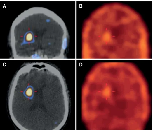
A 40-year-old man complained of insidiously-reduced
right arm dexterity, which started three years previously.
He had had a severe closed head injury 19 years before.
At the examination, he presented with rigidity, akinesia, and
dystonia uniquely over the right side. According to Crouzon
and Justin-Besancon, the criteria for traumatic secondary
parkinsonism are severe trauma, brain concussion, and
a temporal relationship between the trauma and
symp-toms
1. In this patient, presynaptic dopaminergic imaging
corroborated nigrostriatal denervation induced,
presum-ably, by a previous traumatic hemorrhage. As a result,
neu-roimaging (Figures 1 and 2) showed speciic features that
validated the diagnosis of parkinsonism secondary to a
traumatic etiology
2,3.
Figure 1.
Magnetic resonance imaging, with T2 (A), and susceptibility weighted imaging (B), discloses, in detail, a focal lesion with
hemosiderin deposits, over the left cerebral peduncle and substantia nigra.
61
Dias MS. Delayed hemiparkinsonism imagingReferences
1. Crouzon O, Justin-Besancon L-E. Post-traumatic parkinsonism. Presse Med. 1929;37:1325-7. https://doi.org/10.1016/j.lpm.2017.01.018
2. Loher TJ, Krauss JK. Dystonia associated with
pontomesencephalic lesions. Mov Disord. 2009;24(2):157-67. https://doi.org/10.1002/mds.22196
3. Krauss JK, Jankovic J. Head injury and posttraumatic movement disorders. Neurosurgery. 2002;50(5):927-39. https://doi.org/10.1097/00006123-200205000-00003