Article
J. Braz. Chem. Soc., Vol. 24, No. 8, 1314-1321, 2013. Printed in Brazil - ©2013 Sociedade Brasileira de Química 0103 - 5053 $6.00+0.00
A
*e-mail: [email protected]
Clusiaxanthone and Tocotrienol Series from
Clusia pernambucensis
and
their Antileishmanial Activity
Everton M. Silva,a Renata M. Araújo,b Lindomar G. Freire-Filha,a Edilberto R. Silveira,c
Norberto P. Lopes,d José Elias de Paula,e Raimundo Braz-Filhof and Laila S. Espindola*,a
aLaboratório de Farmacognosia, Universidade de Brasília, 70910-900 Brasília-DF Brazil
bInstituto de Química, Universidade Federal do Rio Grande do Norte, 59072-970 Natal-RN, Brazil
cDepartamento de Química Orgânica, Universidade Federal do Ceará, 60451-970 Fortaleza-CE, Brazil
dFaculdade de Ciências Farmacêuticas de Ribeirão Preto, Universidade de São Paulo,
14040-903 Ribeirão Preto-SP, Brazil
eLaboratório de Anatomia Vegetal, Universidade de Brasília, 70910-900 Brasília-DF, Brazil
fLaboratório de Ciências Químicas, Universidade Estadual do Norte Fluminense,
28013-602 Campos dos Goytacazes-RJ and Departamento de Química, Universidade Rural do Rio de Janeiro, 23890-000 Seropédica-RJ, Brazil
A análise fitoquímica do extrato acetato de etila da casca do caule de Clusia pernambucensis
G. Mariz, Clusiaceae, uma espécie do Cerrado brasileiro, conduziu ao isolamento e caracterização completa de uma nova xantona, 1,7-dihidróxi-2-(3-metil-2-butenil)-6’,6’-dimetilpirano(2’,3’:3,4) xantona, denominada clusiaxantona. Quatro tocotrienóis ainda não relatados nesta espécie também foram isolados. Um derivado foi obtido a partir da clusiaxantona, 1-hidróxi,7-metóxi-2-(3-metil-2-butenil)-6’,6’-dimetilpirano(2’,3’:3,4)xantona (7-O-metil-clusiaxantona), e um segundo derivado foi obtido a partir do ácido Z-δ-tocotrienolóico. As estruturas foram estabelecidas com base em dados de ressonância magnética nuclear de 1H e 13C (NMR 1D e 2D), espectrometria de massa com
ionização por electrospray de alta resolução (HRESIMS) e espectroscopia no infravermelho. No controle da infecção de macrófagos com amastigotas de Leishmania (Leishmania) amazonensis, os compostos ativos foram clusiaxantona e seu derivado (CI50 = 66,9 e 57,4 µM, respectivamente).
A citotoxicidade dos compostos foi determinada em macrófagos peritoneais de camundongos BALB/c.
Phytochemical analysis of the ethyl acetate extract from the stem bark of Clusia pernambucensis
G. Mariz, Clusiaceae, a Brazilian Cerrado species, led to the isolation and full characterization of a new xanthone, 1,7-dihydroxy-2-(3-methyl-2-butenyl)-6’,6’-dimethylpyrano(2’,3’:3,4)xanthone, namely clusiaxanthone. Four previously unreported tocotrienols from this species were also isolated. A derivative was obtained from clusiaxanthone, 1-hydroxy,7-methoxy-2-(3-methyl-2-butenyl)-6’,6’-dimethylpyrano(2’,3’:3,4)xanthone (7-O-methylclusiaxanthone), and an additional derivative was obtained from Z-δ-tocotrienoloic acid. The structures of these compounds were established based on data from 1H and 13C nuclear magnetic resonance (1D and 2D NMR), high
resolution electrospray ionization mass spectrometry (HRESIMS) and infrared spectroscopy. The clusiaxanthone and its derivative were able to control macrophage infection by Leishmania
(Leishmania) amazonensis amastigotes (IC50 = 66.9 and 57.4 µM, respectively). The cytotoxicity
of the compounds was assessed in BALB/c mouse peritoneal macrophages.
Keywords: Clusia pernambucensis, Clusiaceae, xanthone, tocotrienol series, Leishmania
Introduction
Cutaneous leishmaniasis is a worldwide endemic disease that causes skin lesions usually persisting for several months or years.1 Patients also suffer because of the toxicity
induced by prolonged use of antileishmanial drugs. The clinical symptoms of leishmaniasis are determined by the involved species of Leishmania, the parasite virulence and host immune response.2 On account of the limited treatment
options,3 new therapeutic solutions have been proposed,
such as the use of miltefosine and paromomycin4 for the
past twenty years.
Despite these efforts, the need for new drugs remains. However, progress in the development of new treatments has been slow.2,5-7 As previously reported, the screening
for compounds from natural product libraries is a key strategy because many plants contain chemical compounds with powerful leishmanicidal activity.8,9 In addition,
approximately 80% of the population in the Americas use plants to treat various diseases,10 including leishmaniasis.11,12
Benzophenones, triterpenes and several flavonoids have been isolated from Clusia pernambucensis, C. columnaris,
C. grandiflora and C. spirictu-sanctensis.13,14 These
compounds have presented anti-inflammatory, antifungal, antibacterial,15 anti-HIV,16 antioxidant and antitumor
activities.17-19 Peraza-Sanchez et al.20 demonstrated the
activity of the methanol extract of C. flava leaves, popularly used for treating wounds and syphilis, in promastigotes of Leishmania major. A Peru based research team supported the use of traditional medicine for the treatment of cutaneous leishmaniasis.11 The group observed the
activity of C. amazonica extracts in L. amazonensis axenic amastigotes.11
I n t h i s s t u d y, w e eva l u a t e d t h e p o t e n t i a l antileishmanial effects of compounds and derivatives from Clusiapernambucensis G. Mariz, Clusiaceae against
Leishmania (Leishmania) amazonensis, a parasite species that causes cutaneous leishmaniasis.
Results and Discussion
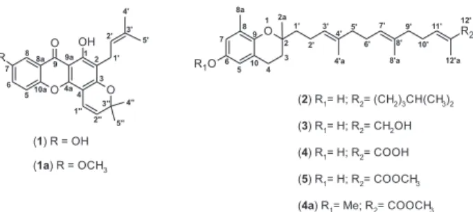
The fractionation of the ethyl acetate extract from the stem bark of Clusia pernambucensis led to the isolation of a previously unreported compound, clusiaxanthone (1), and six known compounds (2-7), δ-tocotrienol (2),21δ-tocotrienolic
alcohol (3),22Z-δ-tocotrienoloic acid (4) and δ-tocotrienol
methyl ester (5),23 betulinic acid (6),24,25 and β-sitosterol (7)26
(Figure 1). Compounds 2 and 5 were isolated from a natural source for the first time. For Z-δ-tocotrienoloic acid (4), the chemical shift of the methyl 12a’ carbon (d 12.21 ppm) under the protective effect of the γ-methylenic carbon 10’
(d 27.78 ppm) contributed to the stereochemical identification of the carboxyl group. The structures of compounds 2-7 were determined by comparing the experimental data from the
1H and 13C nuclear magnetic resonance (NMR) spectra and
high resolution electrospray ionization mass spectrometric (HRESIMS) analyses with previously reported data. This was the first time compounds 2 to 5 were isolated from
C.pernambucensis.
The methylation of clusiaxanthone and Z-δ-tocotrienolic acid resulted in the derivatives 1a, 1-hydroxy,7-methoxy-2-(3-methyl-2-butenyl)-6’,6’-dimethylpyrano(2’,3’:3,4) xanthone (7-O-methylclusiaxanthone), and 4a, (2E,6E,10E )-methyl 13-(3,4-dihydro-6-methoxy-2,8-di)-methyl- 2H-chromen-2-yl)-2,6,10-trimethyltrideca-2,6,10-trienoate, respectively.
Clusiaxanthone (1) was isolated as a yellow solid with [α]D −57.4 (MeOH, c 0.11). The molecular formula for
clusiaxanthone was determined as C23H22O5 based on the
quasi-molecular ion at m/z 379.1540 [M + H]+ (1 ppm
error) in the HRESIMS spectrum. The vibrational bands at 3325 and 1647 cm-1 in the IR spectrum were consistent
with hydroxyl and carbonyl groups, respectively.
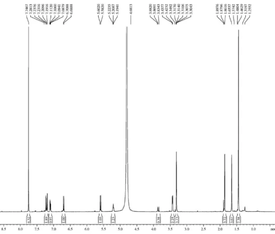
The 1H NMR spectrum of clusiaxanthone (500 MHz,
C5D5N) showed signals at dH 1.74 (s, 3H-4’), 1.96 (s,
3H-5’), 3.62 (d, J 7.5 Hz, 2H-1’) and 5.49 (t’, J 7.5 Hz, H-2), and correlations with the carbon signals (1J
CH) at
dC 18.5 (CH3-5’), 26.2 (CH3-4’), 22.3 (CH2-1’) and 123.4
(CH-2’) in the HSQC spectrum, respectively, used to characterize the presence of a prenyl moiety. The presence of a 2,2-dimethyl-3,4-dehydropyran was also identified by the signals at dH 1.49 (s, 6H-3H4”/3H5’’), 5.65 (d, H-2”,
10.0 Hz) and 6.97 (d, H-1’’, 10.0 Hz), which showed correlations (1J
CH) with the signals corresponding to carbons
C-4”/C-5” (d 28.6), C-2" (d 116.5) and C-1" (d 128.2) in the HSQC spectrum, respectively. The signal at dH 13.86
(s) in the 1H NMR spectrum suggested the presence of a
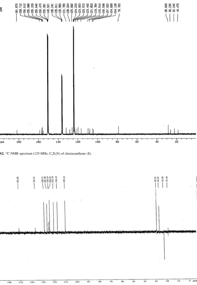
chelatogenic hydroxyl group (HO-1) (Table 1). The 13C NMR spectrum (125 MHz, C
5D5N) of
compound 1, which was supported by the data from the
13C DEPT (distortionless enhancement by polarization)
135 analysis, revealed 23 signals corresponding to twelve non-hydrogenated, six methine (all sp2), one methylene
(sp3) and four methyl carbon atoms. Therefore, the
expanded molecular formula C23H22O7 was determined
for compound 1 based on these data, and this formula is consistent with the chemical formula of C23H22O5, which
was determined by HRESIMS, with the presence of two ether functions.
For compound 1, a signal at dC 181.9 was assigned
to a carbonyl carbon (C-9), three signals were associated with mono-hydrogenated aromatic carbons at dC 109.2
(C-8), 119.8 (C-5) and 125.7 (C-6), and eight signals were correlated with non-hydrogenated aromatic carbons at d 104.1 (C-9a), 104.7 (C-4a), 107.9 (C-2), 121.8 (C-8a), 155.2 (C-1), 156.6 (C-4a), 156.0 (C-7) and 158.7 (C-3).
The last four carbons represent oxygenated carbons. It was also possible to observe the presence of two olefin carbons at dC 123.4 (C-2’) and 131.8 (C-3’) associated with a
tri-substituted double bond in the prenyl side chain, and two additional carbons at dC 116.5 (C-1”) and 128.2 (C-2”) of
the pyran substituent. The location of the carbons in the xanthone skeleton and all the unequivocal chemical shift assignments were based on the heteronuclear long-range coupling (2J
CH and 3JCH) between the hydrogen and carbon
atoms as determined by the 2D HMBC spectrum. The location of the prenyl moiety at C-2 was indicated by the HMBC correlations between the methylene protons 2H-1’ (dH 3.62) with both C-2’ (dC 123.4, 2JCH) and C-2 (dC 107.9, 2J
CH) carbons. The location of the prenyl moiety was also
confirmed by correlations with both the C-1 (dC 155.2,
Table 1. 1H and 13C NMR data of compounds 1 and 1a, including the results of the HSQC and HMBC analysesa
1, C5D5N 1a, CDCl3 + CD3OD
HSQC HMBC HSQC
dC / ppm dH / ppm 2JCH / Hz 3JCH / Hz dC / ppm dH / ppm
1 155.2 − 2H-1’ 156.0 −
2 107.9 − 2H-1’ H-2’ 107.5 −
3 158.7 − H-1”; 2H-1’ 158.5 −
4 104.9 − H-1” H-2” 104.8 −
4a 156.6 − H-1” 157.2 −
5 119.8 7.53 (d, 9.0) 119.7 7.23 (d, 3.0)
6 125.7 7.58 (dd, 9.0, 3.0) H-8 129.9 7.19 (d, 9.0)
7 156.0 − H-6; H-8 H-5 165.2 −
8 109.2 8.04 (d, 3.0) 110.7 7.10 (dd. 9.0, 3.0)
8a 121.8 − H-8 H-5 122.2 −
9 181.9 − H-8 183.0 −
9a 104.1 − 104.0 −
10a 150.5 − H-5 H-6; H-8 148.6 −
1’ 22.3 3.62 (d, 7.5) 22.2 3.43 (d, 7.4)
2’ 123.4 5.49 (t, 7.5) 2H-1’ 3H-4’; 3H-5’ 123.7 5.23 (t, 7.4)
3’ 131.8 − H-2’; 3H-4’/3H-5’ 2H-1’ 131.2 −
4’ 26.2 1.74 (brs) H-2’ 26.1 1.65 (brs)
5’ 18.5 1.96 (brs) H-2’ 18.3 1.86 (brs)
1” 116.5 6.97 (d, 10.0) H-2” 117.0 6.69 (d, 10.0)
2” 128.2 5.65 (d, 10.0) H-1” 3H-4”/3H-5” 127.8 5.59 (d, 10.0)
3” 78.8 − 3H-4”; 3H-5” H-1” 78.0 −
4” 28.6 1.49 (s) H-2”; 3H-5” 28.7 1.48 (s)
5” 28.6 1.49 (s) H-2”; 3H-4” 28.7 1.48 (s)
MeO − − − − 49.2 3.31 (s)
HO-1 − 13.86 (s)
aThe number of hydrogens bound to carbon atoms was determined by the comparative analysis of 1H and DEPT 13C NMR spectra. The chemical shifts and
3J
CH) and C-3 (dC 158.7, 3JCH) carbons. The doublet at d 6.97
(H-1'') showed an HMBC cross-peak to the aromatic C-4 (dC 104.9, 2JCH), C-4a (dC 156.6, 3JCH) and C-3 (dC 158.7, 3J
CH) carbons, thus establishing the connectivity of C-1”
with C-4. Other similar interactions are summarized in Table 1.
The methylation of compound 1 with an ethereal solution of diazomethane yielded the mono methyl ether 1a and confirmed the presence of a hydroxyl group at C-7, which formed the methyl ether 7-O-Me derivative only. This was determined by its corresponding signal at dC 165.2 (Table 1). A singlet at dH 13.86 (vide supra) in the 1H NMR spectrum of 1 (Table 1) indicated the presence of
another hydroxyl group at C-1 that chelated the carbonyl carbon at C-9 and interfered with the methylation by diazomethane. Derivative 1a had a molecular formula of C24H25O5 determined by the protonated peak at m/z
393.1699 ([M + H]+ calculated m/z 393.1702) in the
HRESIMS spectrum.
Therefore, the structure of the new xanthone isolated from
Clusia pernambucensiswas determined as 1,7-hydroxy-2-(3-methyl-2-butenyl)-6’,6’-dimethylpyrano(2’,3’:3,4) xanthone. This compound was named clusiaxanthone (1). Unlike other reported related xanthones bearing two hydroxyl groups in the aromatic ring, this compound has one OH group less, similar to that identified in Garcinia nigrolineata.27-30
The basic core of this chemical class is a tricycle with a symmetrical skeleton. Ring A is formed from the acetate pathway, involving 1-4 carbon atoms, whereas ring B is composed of the 5-8 carbon atoms and is derived from the shikimic acid pathway.31
T h e E t OA c ex t r a c t f r o m t h e s t e m b a r k o f
C.pernambucensis exhibited an IC50 = 65.0 µg mL
-1 against L. (L.) amazonensis promastigotes. The antileishmanial activity of compounds 1-6 in peritoneal macrophages infected with amastigotes was also determined and indicated that clusiaxanthone (1) displayed the most significant antileishmanial activity with an IC50 = 66.9 µM
(Table 2). The antileishmanial activity of derivative 1a was similar to that of 1 with an IC50 = 57.4 µM.
Azebaze et al.32 showed that the activity of synthetic
xanthones against axenic L. amazonensis amastigotes was increased in the presence of prenyl groups at positions 2 and 4, similar to the activity of the 2-substituted xanthone observed in this study. Hydroxylated and methoxylated xanthones (with structures similar to compound 1a), which were isolated from Andrographis paniculata
(Acanthaceae), showed activity against L. infantum
amastigotes inside peritoneal macrophages.33 Lipophilic
groups play an important role in facilitating the compound
internalization by macrophages, thereby increasing its availability.34,35
According to the literature, the hydroxy-substituted xanthone nucleus shows potential for the development of active derivatives against Leishmania sp., which was observed for the 3,6-bis-ω-diethylaminoalkoxyxanthones tested in L. mexicana.36 The results of this study confirm
the data obtained in our study.
Monzote et al.37 suggested that in Leishmania sp.
amastigotes, mitochondrial complex III can be a target for compounds, such as tocotrienols, that contain chromanol groups. Among the tocotrienols studied here, only
Z-δ-tocotrienoloic acid (4) showed an IC50 = 181.0 µM
against L. (L.) amazonensis amastigotes. The derivative compound 4a did not show increased antileishmanial activity. Therefore, it is possible that the presence of the carboxylic group is beneficial to the antileishmanial activity because the change in substituent groups at C12’ in compounds 2, 3 and 5 failed to improve the antileishmanial activity of tocotrienol.
Conclusions
We isolated and extensively characterized one unreported xanthone (1) and a series of four known tocotrienols, δ-tocotrienol (2), δ-tocotrienolic alcohol (3), Z-δ-tocotrienoloic acid (4) and δ-tocotrienol methyl ester (5), which were previously undescribed in
C. pernambucensis. Compounds 2 and 5 were isolated from natural sources for the first time, and two compounds, betulinic acid (6) and β-sitosterol (7), are ubiquitous in plants. Clusiaxanthone (1) showed moderate activity against Leishmania (Leishmania) amazonensis.
Table 2. Compound activities against L. (L.) amazonensis amastigotes IC50, and cytotoxicity in murine macrophages CC50
Compound
IC50 / µM amastigotes
L. (L.) amazonensis
CC50 / µM murine macrophages
Clusiaxanthone (1) 66.9 ± 6.1 45.5 ± 7.7
Compound 1a 57.4 ± 4.1 83.2 ± 8.4
δ-Tocotrienol (2) > 200.0 > 400.0
δ-Tocotrienolic alcohol (3) > 200.0 400.0
Z-δ-Tocotrienoloic acid (4) 181.0 ± 8.0 > 400.0
Compound 4a > 200.0 > 400.0
δ-Tocotrienol-methyl-ester (5) > 200.0 177.0 ± 16.1
Betulinic acid (6) > 200.0 > 400.0
Amphotericin B 0.2 ± 0.1 −
Meglumine antimoniate 25.0 ± 2.2 −
Chemical studies to identify derivatives and analogs with lower cytotoxicity and improved antileishmanial activity are desirable. These compounds can be evaluated using an amastigote intramacrophage system. This system is advantageous since it takes into account the pharmacokinetic barriers that all compounds must overcome to reach the target parasite.38,39
Our laboratory is focused on the development of antileishmanial candidates and is currently developing new xanthone derivatives that contain the original hydroxylated xanthone nucleus.
Experimental
General experimental procedures
Optical rotation data were collected on a DIP-370 polarimeter. For infrared analysis, the samples were embedded in KBr pellets under 10,000 kgf, and the experiment was performed with an infrared spectrophotometer (Perkin-Elmer RX-1). Fractionation was performed by column chromatography (7 × 100 cm) on silica gel (0.04-0.063 mm or 230-400 mesh). Thin layer chromatography (TLC) was performed in silica gel plates (60 F254 aluminum, Merck).
1H and 13C NMR spectra were recorded at room
temperature on a Bruker Avance DRX 500 (500.13 and 125.77 MHz) spectrometer using an inverse detection probe. The chemical shifts are expressed in parts per million (ppm) relative to tetramethylsilane (TMS), and the coupling constants (J values) are shown in hertz (Hz).
HRESIMS spectra were recorded on Bruker UltrOTOF and MicrOTOF spectrometers. Electrospray ionization was performed using a time of flight analyzer (ESI-TOF-MS) in both the positive and negative modes.
Analytical high-performance liquid chromatography (HPLC) of the samples was performed on a Waters model 1525 coupled to a UV photodiode array detector (190-800 nm), series 2996, thermostatic oven and Phenomenex silica column (4.6 × 250 mm, 5 µm). The mobile phase consisted of hexane-ethyl acetate pre-filtered through 0.45 µm nylon membranes, vacuum degassed and sonicated for 15 min. Reversed phase-HPLC was performed on a Shimadzu SL6Vp equipment coupled to a UV photodiode array detector (190-800 nm) equipped with a degasser. A Shimadzu octadecylsilane column (4.6 × 250 mm, 5 µm) with an injection volume of 20 µL (2 mg mL-1) and flow rate of
1 mL min-1 was used in the analytical mode. The separation
was achieved on a Shimadzu preparative octadecylsilane (20 × 250 mm, 10 µm) column with an injection volume of 1 mL (20 mg mL-1) and a flow rate of 9 mL min-1. The
compound detection was monitored at 254 nm.
Plant material
The stem bark of Clusia pernambucensis G. Mariz, Clusiaceae, was collected from the gallery forest of the Cerrado biome, Santo Antônio do Descoberto, Goiás State, at an altitude of 1055 metres; south latitude 15º48’35.0”; south longitude 48º20’12.0”. The identification of the plant was confirmed by Professor Dr. José Elias de Paula, and a voucher specimen was deposited in the Herbarium of the University of Brasília under the accession number (UB) 3771.
Extraction and isolation
C. pernambucensis extract (22 g) was obtained by macerating 197.11 g of C. pernambucensis stem bark with ethyl acetate. From this extract, 14.15 g were fractionated into 312 fractions of 100 mL by column chromatography with a cyclohexane-EtOAc polarity gradient (100:0; 99:1; 98:2; 97:3; 95:5; 93:7; 90:10; 85:15; 80:20; 75:25; 70:30; 60:40: 50:50; 0:100), and sequentially with an EtOAc-MeOH gradient (100:0; 90:10; 85:15; 80:20; 75:25; 70:30; 60:40; 50:50; 0:100). Based on the TLC profile, the fractions were assembled in 21 fractions (CP1-CP21). Fraction CP15 was purified on a silica column to obtain compound 1 (36.2 mg, 0.2 %). The compound detection was monitored at 254 nm. The purification of fraction CP19 was performed using an HPLC Shimadzu, model SL6Vp equipped with an octadecilsilane-C18 (20 × 250 mm, 10 µm)
preparative column. The eluent consisted of an isocratic ternary mixture of H2O:MeOH:MeCN (8:32:60). The
samples were injected in a volume of 1 mL (20 mg mL-1)
with a flow rate of 9 mL min-1, and the compound detection
was monitored at 190, 209 and 254 nm. At the end of this process, four compounds were isolated, δ-tocotrienol (2, 1.4 mg, 0.009 %), δ-tocotrienolic alcohol (3, 10.2 mg, 0.07%), Z-δ-tocotrienoloic acid (4, 85.3 mg, 0.6%) and
δ-tocotrienol-methyl ester (5, 0.6 mg, 0.004%). Betulinic acid (6, 100 mg, 0.7%) and β-sitosterol (7, 20.2 mg, 0.1%) were also isolated.
A methyl ether derivative (1a) of clusiaxanthone (1) was produced by methylation of 1 with diazomethane. Compound 4a was obtained by the methylation of a hydroxyl and terminal carboxyl of Z-δ-tocotrienoloic acid (4).
1,7-Dihydroxy-2-(3-methyl-2-butenyl)-6’,6’-dimethyl-pyrano(2’,3’:3,4)xanthone (1): yellow solid; [α]D −57.4
(MeOH, c 0.11); IR ν/cm-1 3324, 2983, 2962, 2909,
2857, 1700, 1647, 1480, 1465, 1357; 1H and 13C NMR
(500.13 and 125.77 MHz, C5D5N, d/ppm) see Table 1;
HRESIMS (positive mode) m/z found: 379.1540 [M + H]+;
1 H y d r ox y, 7 m e t h ox y 2 ( 3 m e t hy l 2 bu t e ny l ) -6’,6’-dimethyl pyrano(2’,3’:3,4)xanthone (1a): yellow solid. 1H and 13C NMR (500.13 and 125.77 MHz, C
5D5N,
d/ppm) see Table 1. HRESIMS (positive mode) m/z found: 393.1699 [M + H]+; calc m/z for [C
24H25O5]+ = 393.1702.
Leishmania (Leishmania) amazonensis promastigote
culture
C57BL/6 mice were infected with Leishmania (Leishmania) amazonensis (strain L(L)a)-MHOM/BR/PH8) and maintained in the biotherium at the Faculty of Health Science/Medicine (University of Brasília, Brazil). Blood was collected by puncture of the hind paw of infected animals. The parasites were incubated in McNeal, Novy and Nicolle (NNN) culture medium pH 7.2 at 22 °C for seven days, and subsequently in Schneider’s medium supplemented with 20% fetal bovine serum and 0.25% gentamicin pH 7.2 at 22 °C.
Compound evaluation in L. (L.) amazonensis promastigotes
Sterile Schneider’s medium (Sigma-Aldrich) was added to each well of a 96 well cell culture plate. Promastigotes (105) were then added to the wells and
counted in a Neubauer chamber. Serial dilution of the compounds was performed starting at an initial concentration of 300 µmol L-1, with 0.1% DMSO
(dimethyl sulfoxide) as a negative control. The cell culture plate was incubated for 48 h at 22-26 °C. Next, 100 µg per
well of a solution of 5 mg mL-1
3-(4,5-dimethylthiazol-2-yl)-2,5-diphenyltetrazolium bromide (MTT) in PBS (phosphate buffered saline, Sigma-Aldrich) was added to the wells for 4 h at 37 °C. Viability measurements were performed after the addition of 100 µL DMSO to solubilize the formazan crystals.40-42 In this experiment,
we used promastigotes in log phase growth to optimize the spectrophotometer reading at 570 nm. The experiments were performed in triplicate, and the IC50 values were
calculated using Microsoft Excel.
Compound evaluation in L. (L.) amazonensis amastigotes
Peritoneal macrophages from BALB/c mice were collected and centrifuged in RPMI 1640 (Gibco) medium at 1190 rpm or 300 g. The pellets were recovered and resuspended to count the viable cells by trypan blue (Sigma) staining in a Neubauer chamber. Glass coverslips (13 mm) were placed into a 24 well plate with 4 × 10⁵
macrophages per well for 24 h at 37 °C in a 5% CO2
atmosphere. After the incubation period, the non-adherent
cells were removed and the macrophages were incubated with L. (L.) amazonensis promastigotes in stationary phase at a ratio of 10:1 for 4 h at 37 °C in a 5% CO2
atmosphere. The promastigote-free supernatant was removed, and the infected macrophages were treated with the compounds at initial concentrations of 300 µmol L-1
for 48 h. The cells were subsequently harvested, fixed with methanol and stained with Giemsa stain. Miltefosine (Sigma-Aldrich), amphotericin B (Sigma-Aldrich), and meglumine antimoniate (Glucantime®, Aventis Pharma
Brazil; 300 mg mL-1, 81 mg mL-1 Sb (V)) at concentrations
of 30.0 to 0.056 µmol L-1 were used as positive controls, and
0.1% DMSO in a culture medium with amastigotes as a negative control. For each concentration of compound, 200 macrophages were counted using an optical microscope (Olympus). The number of amastigote-infected cells was calculated compared to the negative control.43,44 The tests
were performed in triplicate.
Evaluation of compound cytotoxicity in murine macrophages
After the peritoneal macrophages were isolated from BALB/c mice, 106 cells were placed in each well of a
96 well microplate. The cells were incubated for 12 h at 37 °C in a 5% CO2 atmosphere. The culture medium was
then replaced with the diluted compounds in new culture medium, and the cells were then incubated at 37 °C in a 5% CO2 atmosphere. A solution of 0.1% DMSO was used as the
negative control. After 24 h, cell viability was determined by adding 100 µg per well MTT in PBS (5 mg mL-1) for 4 h.
The culture medium was removed and 100 µL DMSO was added. The formazan was quantified in a spectrophotometer at 570 nm.45,46
Supplementary Information
1H and 13C NMR, HSQC, HMBC and 1H-1H COSY
spectra and HRESIMS spectra of clusiaxanthone and derivative 1a are available free of charge at http://jbcs.sbq.org.br as PDF file.
Acknowledgments
The authors gratefully acknowledge the CNPq, CAPES and FAPDF for financial support.
References
3. Siqueira-Neto, J. L.; Song, O. R.; Oh, H.; Sohn, J. H.; Yang, G.; Nam, J.; Jang, J.; Cechetto, J.; Lee, C. B.; Moon, S.; Genovesio, A.; Chatelain, E.; Christophe, T.; Freitas-Junior, L. H.; PLoS Negl. Trop. Dis.2010, 4, e675.
4. Olliaro, P. L.; Curr. Opin. Infect. Dis.2010, 23, 595. 5. Croft, S. L.; Olliaro, P.; Clin. Microbiol. Infect.2011, 17,
1478.
6. Grogl, M.; Hickman, M.; Ellis, W.; Hudson, T.; Lazo, J. S.; Sharlow, E. R.; Johnson, J.; Berman, J; Sciotti, R. J.; Am. J. Trop. Med. Hyg. 2013, 88, 216.
7. Salah, A. B.; Messaoud, N. B.; Guedri, E.; Zaatour, A.; Alaya, N. B.; Bettaieb, J.; Gharbi, A.; Hamida, N. B.; Boukthir, A.; Chlif, S.; Abdelhamid, K.; Ahmadi, Z. E.; Louzir, H.; Mokni, M.; Morizot, G.; Buffet, P.; Smith, P. L.; Kopydlowski, K. M.; Kreishman-Deitrick, M.; Smith, K. S.; Nielsen, C. J.; Ullman, D. R.; Norwood, J. A.; Thorne, G. D.; McCarthy, W. F.; Adams, R. C.; Rice, R. M.; Tang, D.; Berman, J.; Ransom, J.; Magill, A. J.; Grogl, M.; N. Engl. J. Med. 2013,
368, 524.
8. Sen, R.; Chatterjee, M.; Phytomedicine2011, 18, 1056. 9. Vieira, N. C.; Herrenknecht, C.; Vacus, J.; Fournet, A.;
Bories, C.; Figadère, B.; Espindola, L. S.; Loiseau, P. M.;
Biomed. Pharmacother.2008, 62, 684.
10. Newman, D. J.; Cragg, G. M.; J. Nat. Prod.2007, 70, 461. 11. Odonne, G.; Bourdy, G.; Castillo, D.; Estevez, Y.;
Lancha-Tangoa, A.; Alban-Castillo, J.; Deharo, E.; Rojas, R.; Stien, D.; Sauvain, M.; J. Ethnopharmacol.2009, 126, 149. 12. Odonne, G.; Berger, F.; Stien, D.; Grenand, P.; Bourdy, G.;
J.Ethnopharmacol.2011, 137, 1228.
13. Porto, A. L. M.; Machado, S. M. F.; De Oliveira, C. M. A.; Bittrich, V.; Amaral, M. D. E.; Marsaioli, A. J.; Phytochemistry
2000, 55, 755.
14. Compagnone R. S.; Suarez, A. C.; Leitão, S. G.; Delle Monache, F.;
Braz. J. Pharmacogn.2008, 18, 6.
15. Moura, A. C. G.; Perazzo, F. F.; Maistro, E. L.; Genet. Mol. Res.2008, 7, 1360.
16. Huerta-Reyes, M.; Basualdo, M. D.; Lozada, L.; Jimenez-Estrada, M.; Soler, C.; Reyes-Chilpa, R.; Biol. Pharm. Bull.2004, 27, 916.
17. Díaz-Carballo, D.; Malak, S.; Bardenheuer, W.; Frestuehler, M.; Reusch, H. P.; J. Cel. Mol. Med.2008, 12, 2598.
18. Díaz-Carballo, D.; Ueberla, K.; Kleff, V.; Ergun, S.; Malak, S.; Frestuehler, M.; Somogyi, S.; Kucherer, C.; Bardenheuer, W.; Strumberg, D.; Int. J. Clin. Pharmacol. Ther.2010, 48, 670. 19. Chaturvedula, V. S.; Schilling, J. K.; Kingston, D. G.; J. Nat.
Prod.2002, 65, 965.
20. Peraza-Sánchez, S. R.; Cen-Pacheco, F.; Noh-Chimal, A.; May-Pat, F.; Simá-Polanco P.; Dumonteil, E.; García-Miss, M. R.; Mut-Martín, M.; Fitoterapia2007, 78, 315.
21. Yamamoto, I.; Takigawa, T.; Kenmochi, K.; Mizuno, M.; JP 59219280 1984.
22. Terashima, K.; Takaya, Y.; Niwa, M.; Bioorg. Med. Chem.2002,
1, 1619.
23. Monache, F. D.; Marta, M.; Mac-Quhae, M. M.; Nicoletti, M.;
Gazz. Chim. Ital.1984, 114, 135.
24. Bruckner, V.; Kovacs, J.; Koczka, I.; J. Chem. Soc.1948, 1, 948.
25. Pancharoen, O.; Tuntiwachwuttikul, P.; Taylor, W. C.; Picker, K.; Phytochemistry1994, 35, 987.
26. Jares, E. A.; Tettamanzi, M. C.; Pomilio, A. B.; Phytochemistry
1990,29, 340.
27. Bayma, J. C.; Arruda, M. S. P.; Neto, M. S.; Phytochemistry
1998, 49, 1159.
28. Rukachaisirikul, V.; Ritthiwigroma, A. P.; Sawangchoteb, P.; Taylor, W. C.; Phytochemistry2003, 64, 1149.
29. Rukachaisirikul, V.; Kamkaew, M.; Sukavisit, D.; Phongpaichit, S.; Sawangchoteb, P.; Taylor, W. C.; J. Nat. Prod.2003, 66, 1531.
30. Chen, Y.; Yang, G.; Zhong, F.; He, H.; Bull. Korean Chem. Soc.
2010, 31, 3418.
31. El-Seedi, H. R.; El-Barbary, M. A.; El-Ghorab, D. M. H.; Bohlin, L.; Borg-Karlson, A.; Goransson, U.; Verpoorte, R.;
Curr. Med. Chem. 2010, 17, 854.
32. Azebaze, A. G. B.; Ouahouo, B. M. W.; Vardamides, J. C.; Valentin, A.; Kuete, V.; Acebey, L.; Beng, V. P.; Nkengfack, A. E.; Meyer, M.; Chem. Nat. Compd.2008, 44, 582. 33. Dua, V. K.; Verma, G.; Dash, A. P.; Phytother. Res.2009, 23, 126. 34. Stec, J.; Huang, Q.; Pieroni, M.; Kaiser, M.; Fomovska, A.;
Mui, E.; Witola, W. H.; Bettis, S.; McLeod, R.; Brun, R.; Kozikowski, A. P.; J. Med. Chem.2012, 55, 3088.
35. Audisio, D.; Messaoudi, S.; Cojean, S.; Peyrat, J. F.; Brion, J. D.; Bories, C.; Huteau, F.; Loiseau, P. M.; Alami, M.; Eur. J. Med. Chem.2012,52, 44.
36. Kelly, J. X.; Ignatushchenko, M. V.; Bouwer, H. G.; Peyton, D. H.; Hinrichs, D. J.; Winter, R. W.; Riscoe, M.; Mol. Biochem. Parasitol.2003, 126, 43.
37. Monzote, L.; Stamberg, W.; Patel, A.; Rosenau, T.; Maes, L.; Cos, P.; Gille, L.; Chem. Res. Toxicol.2011, 24, 1678. 38. Dutta, S.; Ongarora, B. G.; Li, H.; Vicente, M. G. H.; Kolli,
B. K.; Chang, K. P.; PLoS One2011, 6, e20786.
39. Loiseau, P. M.; Gupta, S.; Verma, A.; Srivastava, S.; Puri, S. K.; Sliman, F.; Normand-Bayle, M.; Desmaele, D.; Antimicrob. Agents Chemother.2011, 55, 1777.
40. Berg, K.; Zhai, L.; Chen, M.; Kharazmi, A.; Owen, T. C.;
Parasitol. Res.1994, 80, 235.
41. Dutta, A.; Bandyopadhyay, S.; Mandal, C.; Chatterjee, M.;
Parasitol. Int.2005, 54, 119.
42. Albernaz, L. C.; De Paula, J. E.; Romero, G. A. S.; Silva, M. R. R.; Grellier, P.; Mambu, L.; Espindola, L. S.; J.Ethnopharmacol.
2010, 131, 116.
44. Tiuman, T. S.; Ueda-Nakamura, T.; Cortez, D. A. G.; Dias-Filho, B. P.; Morgado-Diaz, J. A.; Souza, W.; Nakamura C. V.;
Antimicrob. Agents Chemother.2005, 49, 176.
45. Albernaz, L. C.; Deville, A.; Dubost, L.; De Paula, J. E.; Bodo, B.; Grellier, P.; Espindola, L. C.; Mambu, L.; Planta Med.2011,
78, 1.
46. Mosmann, T.; J. Immunol Methods 1983, 65, 55.
Submitted: March 12, 2013 Published online: July 10, 2013
Supplementary Information
Printed in Brazil - ©2013 Sociedade Brasileira de Química0103 - 5053 $6.00+0.00S
I
*e-mail: [email protected]
Clusiaxanthone and Tocotrienol Series from
Clusia pernambucensis
and
their Antileishmanial Activity
Everton M. Silva,a Renata M. Araújo,b Lindomar G. Freire-Filha,a Edilberto R. Silveira,c
Norberto P. Lopes,d José Elias de Paula,e Raimundo Braz-Filhof and Laila S. Espindola*,a
aLaboratório de Farmacognosia, Universidade de Brasília, 70910-900 Brasília-DF Brazil bInstituto de Química, Universidade Federal do Rio Grande do Norte, 59072-970 Natal-RN, Brazil cDepartamento de Química Orgânica, Universidade Federal do Ceará, 60451-970 Fortaleza-CE, Brazil
dFaculdade de Ciências Farmacêuticas de Ribeirão Preto, Universidade de São Paulo,
14040-903 Ribeirão Preto-SP, Brazil
eLaboratório de Anatomia Vegetal, Universidade de Brasília, 70910-900 Brasília-DF, Brazil fLaboratório de Ciências Químicas, Universidade Estadual do Norte Fluminense, 28013-602
Campos dos Goytacazes-RJ and Departamento de Química, Universidade Rural do Rio de Janeiro, 23890-000 Seropédica-RJ, Brazil
Figure S1.1H NMR spectrum (500 MHz, C
Figure S3.13C DEPT 135 NMR spectrum (125 MHz, C
5D5N) of clusiaxanthone (1).
Figure S2. 13C NMR spectrum (125 MHz, C
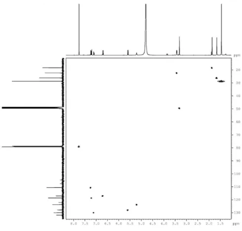
Figure S5. HMBC spectrum (C5D5N) of clusiaxanthone (1).
Figure S7. HRESIMS spectrum of clusiaxanthone (1) in the positive mode [M + H]+.
Figure S9. 13C NMR spectrum (125 MHz, CDCl
3 + CD3OD) of the derivative 1a, obtained from clusiaxanthone (1).
Figure S8.1H NMR spectrum (500 MHz, CDCl
Figure S11. HMBC spectrum (CDCl3 + CD3OD) of the derivative 1a, obtained from clusiaxanthone (1).


![Figure S7. HRESIMS spectrum of clusiaxanthone (1) in the positive mode [M + H] + .Figure S6](https://thumb-eu.123doks.com/thumbv2/123dok_br/18997245.462579/12.892.190.674.112.603/figure-s-hresims-spectrum-clusiaxanthone-positive-mode-figure.webp)
![Figure S12. HRESIMS spectrum of the derivative 1a, obtained from clusiaxanthone (1) in the positive mode [M + H] + .](https://thumb-eu.123doks.com/thumbv2/123dok_br/18997245.462579/15.892.131.787.122.684/figure-hresims-spectrum-derivative-obtained-clusiaxanthone-positive-mode.webp)
